. 2012;7(6):e39104.
doi: 10.1371/journal.pone.0039104. Epub 2012 Jun 18.

Evaluation of 3-(3-chloro-phenyl)-5-(4-pyridyl)-4,5-dihydroisoxazole as a novel anti-inflammatory drug candidate

Affiliations

Evaluation of 3-(3-chloro-phenyl)-5-(4-pyridyl)-4,5-dihydroisoxazole as a novel anti-inflammatory drug candidate

Amanda Roberta Revoredo Vicentino et al. PLoS One. 2012.

Abstract

Background: 3-(3-chloro-phenyl)-5-(4-pyridyl)-4,5-dihydroisoxazole (DIC) is a five-membered heterocyclic compound containing a N-O bond. The anti-inflammatory effects of this compound were studied both in vitro and in vivo.

Principal findings: DIC effectively decreased TNF-α and IL-6 release from LPS-stimulated macrophages in a dose dependent manner. DIC diminished the levels of COX-2 with subsequent inhibition of PGE(2) production. DIC also compromised HMGB1 translocation from the nucleus to the cytoplasm. Moreover, DIC prevented the nuclear translocation of NF-κB and inhibited the MAPK pathway. In vivo, DIC inhibited migration of neutrophils to the peritoneal cavity of mice.

Conclusions: This study presents the potential utilization of a synthetic compound, as a lead for the development of novel anti-inflammatory drugs.


Conflict of interest statement

Competing Interests: The authors have declared that no competing interests exist.

Figures

Figure 1
Figure 1. Chemical structure of 3-(3-chloro-phenyl)-5-(4-pyridyl)-4,5-dihydroisoxazole, DIC.
Figure 2
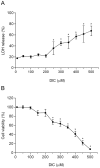
Figure 2. Effect of DIC on macrophage viability.
RAW 264.7 macrophages were treated with DIC (from 10 µM to 500 µM) for 24 h. Cell viabilities were determined by LDH release (A) and MTT assay (B). Values represent means ± SD of three independent experiments. * Significant differences (p>0.05) between treated and untreated cells (250–500 µM), using unpaired t-test.
Figure 3
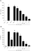
Figure 3. Effect of DIC on LPS-induced TNF-α and IL-6 production.
A and B, following pretreatment with Polymyxin B (Pol B, 15 µg/mL), vehicle (DMSO 0.25%) or DIC (10−200 µM) for 2 h, the cells were treated with LPS (100 ng/mL) for 4 h (A) or 24 h (B). Negative control (CTRL −): cell medium only; Positive control (CTRL +): cells stimulated with LPS, only. TNF-α and IL-6 levels were assayed by ELISA. Values represent means ± SD of three independent experiments. NS, non-significant vs CTRL +; * p<0.05 vs vehicle; ** non-significant vs vehicle. Significances between treated groups were determined using unpaired t-test.
Figure 4
Figure 4. Effect of DIC on nuclear translocation of HMGB1.
RAW 264.7 macrophages were pretreated with DIC 200 µM for 2 h prior to addition of LPS (1 µg/mL) for 24 h. Intracellular HMGB1 was visualized with green immunofluorescent FITC-staining. Untreated cells (UT); LPS-stimulated cells (LPS); DIC-treated cells stimulated with LPS (LPS + DIC).
Figure 5
Figure 5. Effect of DIC on LPS-induced PGE2 production.
(A) RAW 264.7 macrophages were pretreated with DIC 200 µM for 2 h prior to addition of LPS (1 µg/mL) for 24 h and then PGE2 levels were determined by EIA. The values shown are means ± SD of three independent experiments. NS, non-significant vs CTRL+; * p<0.05 vs vehicle; ** non-significant vs vehicle. Significances between treated groups were determined using unpaired t-test. (B) Protein levels of COX-2 were determined by western blot analysis of cellular protein extract (upper panel). A representative immunoblot out of three independent experiments were shown.
Figure 6
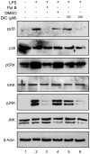
Figure 6. Effect of DIC on the MAPK pathway.
RAW 264.7 macrophages were pretreated with 200 µM of DIC for 2 h prior to addition of LPS (1 µg/mL) for 15 min, and then the whole cell lysate was analyzed by western blot using antibodies against the phosphorylated (activated) and unphosphorylated MAPK. The data shown are representative of three independent experiments.
Figure 7
Figure 7. Effect of DIC on the activation of NF-κB signaling pathway.
(A) RAW 264.7 macrophages were grown on coverslips, pretreated with DIC at a concentration of 200 µM for 2 h and stimulated with LPS (1 µg/mL) for 1 h. FITC-immunostained for NF-κB/p65 viewed under a fluorescence microscope in the ApoTome mode. Untreated cells (NT); LPS-stimulated cells (LPS); DIC-treated cells stimulated with LPS (LPS + DIC). Scale bar: 10 µm. (B) EMSA analysis: nuclear extracts were prepared from unstimulated cells (lane 2); cells stimulated with LPS (1 µg/mL - lane 3); cells pretreated with Polymyxin B (15 µg/mL - lane 4), cells treated with vehicle only (DMSO 0.25% - lane 5); cells treated with increasing concentrations of DIC (lanes 6 and 7) for 2 h and stimulated with LPS for 1 h. The analysis was based on the DNA binding by the active NF-ΚB heterodimer p50–p65. DNA binding inhibition of the p50–p65 heterodimers by DIC is observed (compare lanes 3 and 5 with lanes 6 and 7; the intensities of the upper bands [p50–p65 arrow] in lanes 6 and 7 are significantly lower).
Figure 8
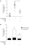
Figure 8. Effect of DIC on cell migration in thioglycollate-induced peritonitis in mice.
(A) DIC (5 mg/kg) or vehicle (DMSO 2.4%) was administered intraperitoneally 30 min before the thioglycollate administration. Mice were sacrificed after 4 h of thioglycollate-induced peritonitis. Total cell migration was counted using a Neubauer chamber. (B) Differential cell count was evaluated by Cytospin. Data represent mean ± S.D. from at least 8 animals per group. * P<0.05 (significances between treated groups were determined using unparied t-test).

Similar articles

Cited by

  • HMGB1 in health and disease.
    Kang R, Chen R, Zhang Q, Hou W, Wu S, Cao L, Huang J, Yu Y, Fan XG, Yan Z, Sun X, Wang H, Wang Q, Tsung A, Billiar TR, Zeh HJ 3rd, Lotze MT, Tang D. Kang R, et al. Mol Aspects Med. 2014 Dec;40:1-116. doi: 10.1016/j.mam.2014.05.001. Epub 2014 Jul 8. Mol Aspects Med. 2014. PMID: 25010388 Free PMC article. Review.
  • Emerging Role of HMGB1 in the Pathogenesis of Schistosomiasis Liver Fibrosis.
    Vicentino ARR, Carneiro VC, Allonso D, Guilherme RF, Benjamim CF, Dos Santos HAM, Xavier F, Pyrrho ADS, Gomes JAS, Fonseca MC, de Oliveira RC, Pereira TA, Ladislau L, Lambertucci JR, Fantappié MR. Vicentino ARR, et al. Front Immunol. 2018 Sep 12;9:1979. doi: 10.3389/fimmu.2018.01979. eCollection 2018. Front Immunol. 2018. PMID: 30258438 Free PMC article.

References

    1. Choy EH, Panayi GS. Cytokine pathways and joint inflammation in rheumatoid arthritis. N Engl J Med. 2001;344:907–916. - PubMed
    1. Wagner G, Laufer S. Small molecular anti-cytokine agents. Med Res Rev. 2006;26:1–62. - PubMed
    1. Kumar S, Boehm J, Lee JC. p38 MAP kinases: key signalling molecules as therapeutic targets for inflammatory diseases. Nat Rev Drug Discov. 2003;2:717–726. - PubMed
    1. Penzo M, Molteni R, Suda T, Samaniego S, Raucci A, et al. Inhibitor of NF-kappa B kinases alpha and beta are both essential for high mobility group box 1-mediated chemotaxis [corrected]. J Immunol. 2010;184:4497–4509. - PMC - PubMed
    1. Andersson U, Tracey KJ. HMGB1 is a therapeutic target for sterile inflammation and infection. Annu Rev Immunol. 2011;29:139–162. - PMC - PubMed

Publication types

MeSH terms