. 2012;7(6):e39255.
doi: 10.1371/journal.pone.0039255. Epub 2012 Jun 19.

A two-stage model for lipid modulation of the activity of integral membrane proteins

Affiliations

A two-stage model for lipid modulation of the activity of integral membrane proteins

Martín M Dodes Traian et al. PLoS One. 2012.

Abstract

Lipid-protein interactions play an essential role in the regulation of biological function of integral membrane proteins; however, the underlying molecular mechanisms are not fully understood. Here we explore the modulation by phospholipids of the enzymatic activity of the plasma membrane calcium pump reconstituted in detergent-phospholipid mixed micelles of variable composition. The presence of increasing quantities of phospholipids in the micelles produced a cooperative increase in the ATPase activity of the enzyme. This activation effect was reversible and depended on the phospholipid/detergent ratio and not on the total lipid concentration. Enzyme activation was accompanied by a small structural change at the transmembrane domain reported by 1-aniline-8-naphtalenesulfonate fluorescence. In addition, the composition of the amphipilic environment sensed by the protein was evaluated by measuring the relative affinity of the assayed phospholipid for the transmembrane surface of the protein. The obtained results allow us to postulate a two-stage mechanistic model explaining the modulation of protein activity based on the exchange among non-structural amphiphiles at the hydrophobic transmembrane surface, and a lipid-induced conformational change. The model allowed to obtain a cooperativity coefficient reporting on the efficiency of the transduction step between lipid adsorption and catalytic site activation. This model can be easily applied to other phospholipid/detergent mixtures as well to other membrane proteins. The systematic quantitative evaluation of these systems could contribute to gain insight into the structure-activity relationships between proteins and lipids in biological membranes.


Conflict of interest statement

Competing Interests: The authors have declared that no competing interests exist.

Figures

Figure 1
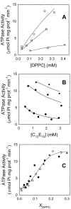
Figure 1. Effects of phospholipids and detergent on the ATPase activity of PMCA.
(A) Purified PMCA, reconstituted in (○) 1.20, (□) 2.75 and (▵) 7.50 mM C12E10 micelles was supplemented with increasing concentrations of DPPC before measuring the ATPase activity. (B) Alternatively, C12E10 was added to purified PMCA already supplemented with (•) 0.06, (▪) 0.22 and (▴) 0.53 mM DPPC before measuring the ATPase activity. Solid lines in A and B are a guide to the eye. (C) Data from panels A and B were plotted as a function of the phospholipid mole fraction in the micellar phase. The continuous line in C corresponds to the graphical representation of equation 8 with the best fitting parameter values shown in Table 1.
Figure 2
Figure 2. PMCA structural changes upon activation by DPPC.
Purified PMCA was supplemented with C12E10 up to 1.2 mM (orange dashed lines) or up to 1.7 mM DPPC/C12E10 and a final DPPC mole fraction of 0.3 ([C12E10] = 1.2 mM) (continuous green lines). After 10 minutes of incubation at 25°C, (A) far UV circular dichroism, and (B) Trp fluorescence were registered. ANS and Trp fluorescence were registered after adding 3 µM ANS to both samples and exciting at 380 nm (ANS, C) and at 295 nm (PMCA-ANS FRET, D). The emission spectrum of PMCA in the absence of ANS is shown as blue dash dotted line. Apparent energy transfer efficiencies were 0.14 in the presence of lipids and 0.07 in the absence of lipids. The final volume and protein concentration in all the samples was identical in order to avoid dilution corrections.
Figure 3
Figure 3. The two stage model for lipid modulation of the enzyme activity.
The scheme shows the transition between low and high activity states of PMCA. In the first stage the enzyme selects a particular lipidic microenvironment among the available amphiphiles according to their relative affinities. The interaction of the protein with specific phospholipids induces, in a second stage, a conformational change at the transmembrane region which is further propagated towards the catalytic domain.
Figure 4
Figure 4. Determination of the exchange constant between DPPC and C12E10.
(A) Lipid free purified PMCA (blue dash dotted line), PMCA with HPPC (orange dashed line) and PMCA-HPPC with DPPC/C12E10 up to a final DPPC mole fraction of 0.3 (continuous green line) were excited at 295 nm and fluorescence emission spectra were registered. (B) PMCA samples were supplemented with DPPC up to mole fractions of: 0.34 (▵), 0.36 (•), 0.41(▪), 0.44 (□), 0.48 (○). PMCA emission intensity was measured after adding increasing quantities of HPPC and mixing for 1 min. Total intensity values were corrected for the dilution (<7%) caused by the addition of the probe. The 2D projection surface is the graphical representation of equation 4 with the best fitting parameter values shown in Table 1.

Similar articles

Cited by

References

    1. Singer SJ, Nicolson GL. The fluid mosaic model of the structure of cell membranes. Science. 1972;175:720–731. - PubMed
    1. Phillips R, Ursell T, Wiggins P, Sens P. Emerging roles for lipids in shaping membrane-protein function. Nature. 2009;459:379–385. - PMC - PubMed
    1. Lee AG. Biological membranes: the importance of molecular detail. Trends in Biochemical Sciences. 2011;36:493–500. - PubMed
    1. Adamian L, Naveed H, Liang J. Lipid-binding surfaces of membrane proteins: Evidence from evolutionary and structural analysis. Biochim Biophys Acta. 2011;1808:1092–1102. - PMC - PubMed
    1. Hunte C, Richers S. Lipids and membrane protein structures. Curr Op Struct Biol. 2008;18:406–411. - PubMed

Publication types