

Hollow spherical nucleic acids for intracellular gene regulation based upon biocompatible silica shells
- PMID: 22725653
- PMCID: PMC3397824
- DOI: 10.1021/nl3020846
Hollow spherical nucleic acids for intracellular gene regulation based upon biocompatible silica shells
Abstract
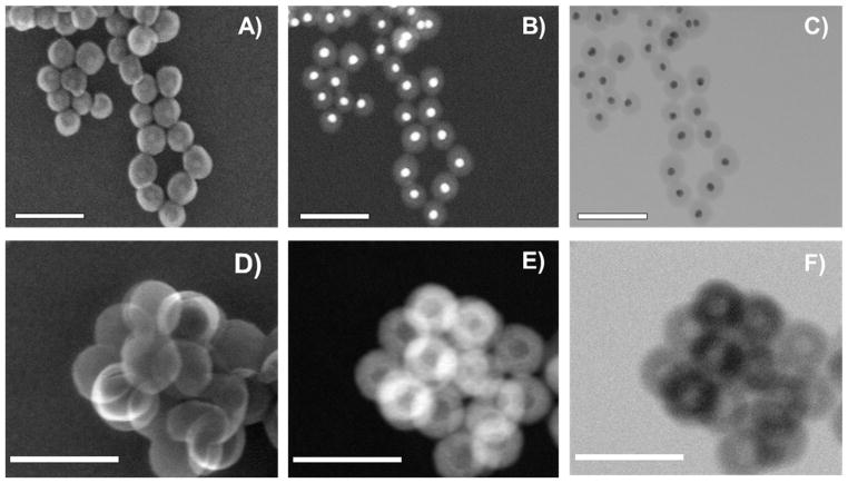
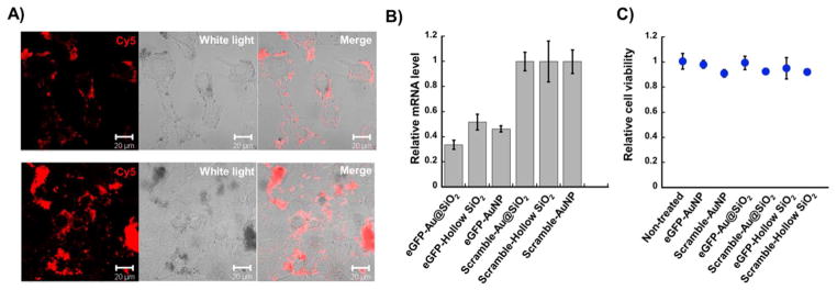
Cellular transfection of nucleic acids is necessary for regulating gene expression through antisense or RNAi pathways. The development of spherical nucleic acids (SNAs, originally gold nanoparticles functionalized with synthetic oligonucleotides) has resulted in a powerful set of constructs that are able to efficiently transfect cells and regulate gene expression without the use of auxiliary cationic cocarriers. The gold core in such structures is primarily used as a template to arrange the nucleic acids into a densely packed and highly oriented form. In this work, we have developed methodology for coating the gold particle with a shell of silica, modifying the silica with a layer of oligonucleotides, and subsequently oxidatively dissolving the gold core with I(2). The resulting hollow silica-based SNAs exhibit cooperative binding behavior with respect to complementary oligonucleotides and cellular uptake properties comparable to their gold-core SNA counterparts. Importantly, they exhibit no cytotoxicity and have been used to effectively silence the eGFP gene in mouse endothelial cells through an antisense approach.
Conflict of interest statement
The authors declare no competing financial interest.
Figures


References
-
- Rosi NL, Giljohann DA, Thaxton CS, Lytton-Jean AKR, Han MS, Mirkin CA. Science. 2006;312:1027–1030. - PubMed
-
- Cutler JI, Auyeung E, Mirkin CA. J Am Chem Soc. 2012;134:1376–1391. - PubMed
-
- Sokolova V, Epple M. Angew Chem, Int Ed. 2008;47:1382–1395. - PubMed
-
- Lv H, Zhang S, Wang B, Cui S, Yan J. J Controlled Release. 2006;114:100–109. - PubMed
-
- Dokka S, Toledo D, Shi X, Castranova V, Rojanasakul Y. Pharm Res. 2000;17:521–525. - PubMed
Publication types
MeSH terms
Substances
Grants and funding
LinkOut - more resources
Full Text Sources
Other Literature Sources
Research Materials
