Review
. 2012 Jul;248(1):188-204.
doi: 10.1111/j.1600-065X.2012.01138.x.

Rethinking inflammation: neural circuits in the regulation of immunity

Affiliations
Review

Rethinking inflammation: neural circuits in the regulation of immunity

Peder S Olofsson et al. Immunol Rev. 2012 Jul.

Abstract

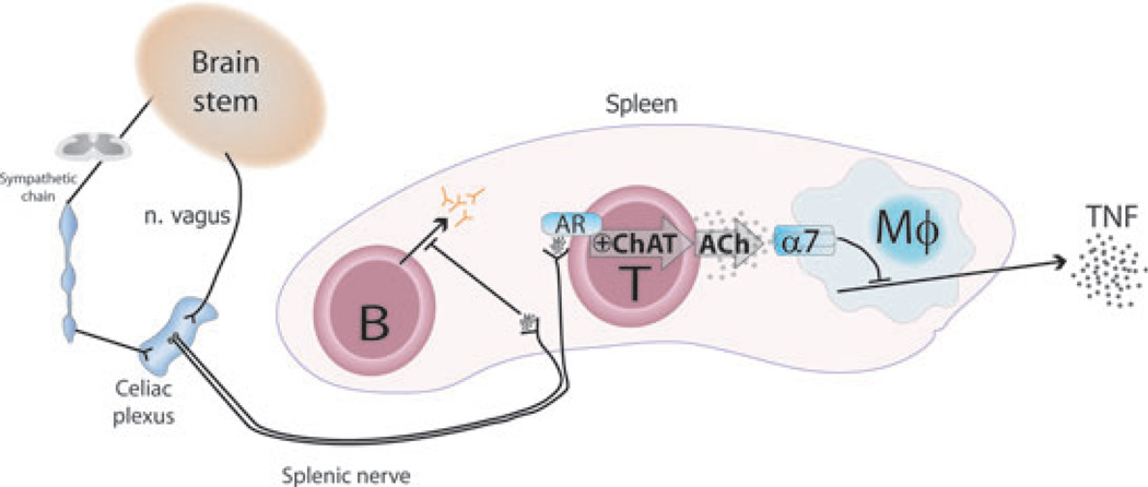
Neural reflex circuits regulate cytokine release to prevent potentially damaging inflammation and maintain homeostasis. In the inflammatory reflex, sensory input elicited by infection or injury travels through the afferent vagus nerve to integrative regions in the brainstem, and efferent nerves carry outbound signals that terminate in the spleen and other tissues. Neurotransmitters from peripheral autonomic nerves subsequently promote acetylcholine-release from a subset of CD4(+) T cells that relay the neural signal to other immune cells, e.g. through activation of α7 nicotinic acetylcholine receptors on macrophages. Here, we review recent progress in the understanding of the inflammatory reflex and discuss potential therapeutic implications of current findings in this evolving field.


Conflict of interest statement

M.R.B and P.S.O. have no conflicts of interest to declare.

Figures

Fig. 1
Fig. 1. The inflammatory reflex
Immune responses are regulated by neural reflex circuits that sense peripheral inflammation and provide regulatory feedback through specific nervous signals and humoral factors. Sensory vagus fibers innervate a multitude of organs, for example the intestine and glomus caroticum. They are activated by cytokines induced by tissue damage or PAMPs in the periphery and transmit signals to the nucleus tractus solitarius (NTS) in the brainstem. Polysynaptic relays connect to the vagal motor neurons in the dorsal vagal motor nucleus and nucleus ambiguus and sympathoexcitatory neurons in the rostral ventrolateral medulla. Efferent vagus nerve signals travel to the celiac plexus and also directly to target organs and suppress innate immune responses. Activation of afferent vagus signals also triggers a ‘sickness response’ and activates the hypothalamic-pituitary-adrenal (HPA) axis, which promotes glucocorticoid release from the adrenal glands.
Fig. 2
Fig. 2. Current model of the efferent arc of the inflammatory reflex
Efferent signals from the brain stem travel through the efferent vagus nerve to the celiac plexus, which also receives input from the sympathetic trunk. The catecholaminergic splenic nerve arises in the celiac plexus and projects to the spleen. Choline acetyltransferase+ (ChAT) T cells and B cells are found in close proximity of splenic nerve fibers. Efferent, outgoing signals in the vagus nerve activate the splenic nerve, which releases its neurotransmitters, including norepinephrine, in the spleen. Activation of choline acetyltransferase-expressing T cells, possibly through adrenergic receptors (AR), promotes production and release of T cell-derived acetylcholine (ACh). This acetylcholine then acts on the α7 nicotinic acetylcholine receptors (α7) on macrophages and other immune cells and suppresses endotoxin-induced release of TNF. Activation of the splenic nerve also arrests B-cell migration and inhibits antibody production.
Fig. 3
Fig. 3. Putative mechanism of acetylcholine synthesis and release in lymphocytes
Acetylcholine (ACh) synthesis is catalyzed by choline acetyltransferase (ChAT) that utilizes acetyl-coenzyme A (Acetyl-CoA) and choline as substrates. Acetyl-CoA can derive from glucose and fatty acid oxidation through glycolysis and beta-oxidation, respectively. Choline can be synthesized de novo, obtained from membrane phospholipid, or taken up through choline transporter 1 (ChT1) after hydrolysis of acetylcholine by acetylcholinesterase (AChE). In T cells, cytosolic acetylcholine is released through mediatophore; however, release of vesicular acetylcholine has not been ruled out. Acetylcholine is released by T cells through unknown mechanisms upon polyclonal stimulation or incubation with norepinephrine. An intriguing question is whether acetylcholine is released in a quantal or non-quantal fashion. Blue arrows indicate unknown processes.

References

    1. Backhed F, Ley RE, Sonnenburg JL, Peterson DA, Gordon JI. Host-bacterial mutualism in the human intestine. Science. 2005;307:1915–1920. - PubMed
    1. Lee YK, Mazmanian SK. Has the microbiota played a critical role in the evolution of the adaptive immune system? Science. 2010;330:1768–1773. - PMC - PubMed
    1. Tracey KJ. Reflex control of immunity. Nat Rev Immunol. 2009;9:418–428. - PMC - PubMed
    1. Rittirsch D, Flierl MA, Ward PA. Harmful molecular mechanisms in sepsis. Nat Rev Immunol. 2008;8:776–787. - PMC - PubMed
    1. Serhan CN, et al. Resolvins: a family of bioactive products of omega-3 fatty acid transformation circuits initiated by aspirin treatment that counter proinflammation signals. J Exp Med. 2002;196:1025–1037. - PMC - PubMed

Publication types