

Review
doi: 10.1146/annurev-micro-092611-150051.
Epub 2012 Jun 15.
Herpesvirus transport to the nervous system and back again
Affiliations
- PMID: 22726218
- PMCID: PMC3882149
- DOI: 10.1146/annurev-micro-092611-150051
Item in Clipboard
Review
Herpesvirus transport to the nervous system and back again
Annu Rev Microbiol.
2012.
Abstract
Herpes simplex virus, varicella zoster virus, and pseudorabies virus are neurotropic pathogens of the Alphaherpesvirinae subfamily of the Herpesviridae. These viruses efficiently invade the peripheral nervous system and establish lifelong latency in neurons resident in peripheral ganglia. Primary and recurrent infections cycle virus particles between neurons and the peripheral tissues they innervate. This remarkable cycle of infection is the topic of this review. In addition, some of the distinguishing hallmarks of the infections caused by these viruses are evaluated in terms of their underlying similarities.
Figures
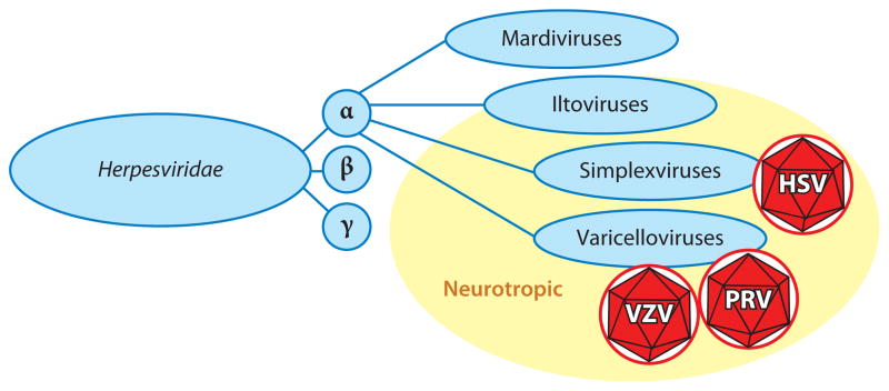
The Alphaherpesvirinae subfamily consists of four genera. Two of the genera, the Simplexvirus and Varicellovirus, consist of human and veterinary pathogens that establish latency in peripheral neurons. Herpes simplex virus (HSV) is a simplexvirus, while varicella zoster virus (VZV) and pseudorabies virus (PRV) are varicelloviruses. At least one member of the Iltovirus genus is also proposed to establish latent infections in peripheral neurons (177), but the Mardivirus genus lacks neurotropism.
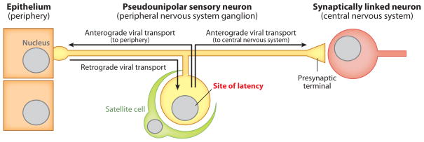
Fundamentals of a neurotropic herpesvirus niche. Although the herpesviruses discussed in this article are referred to as neurotropic, these agents are more accurately described as dual tropic or multitropic. Primary infections begin in exposed tissues such as mucosal epithelia (left). Subsequent spread is normally restricted to innervating neurons resident in peripheral ganglia, where latent infections are maintained, following a single round of retrograde axon transport (middle). Reactivation results in the production of new viruses and the return to peripheral tissues (anterograde axon transport to epithelia). Severe disease associated with invasion of second-order neurons (anterograde axon transport to CNS) occurs only rarely in the natural host but may be frequent in secondary hosts (right). Anterograde and retrograde spread is dictated by the orientation of axonal microtubules and should not be confused with the direction of action potential propagation in the pseudounipolar neuron. Neurotropic herpesviruses can also infect cells of the circulatory system to varying degrees (not illustrated).
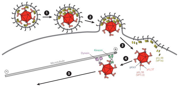
Early events in herpesvirus infection. ❶ Virion contacts plasma membrane of a somatic cell or the axon membrane of a neuron. Tegument proteins redistribute and the virion orients with the bulk of its mass away from the cell. ❷ Fusion between the virion envelope and the cell membrane deposits the capsid and tegument proteins into the cytosol. ❸ The capsid releases from the majority of tegument and envelope proteins (including VP16), but a subset of inner tegument proteins (including pUs3, VP1/2, pUL37) remain capsid bound and together compose the retrograde transport complex. VP16, which enters the nucleus in somatic cells and promotes productive infection, may be lost upon entering a neuron owing to an inability to efficiently participate in retrograde axon transport. ❹ The retrograde transport complex traverses cortical actin (not illustrated) and associates with dynein and kinesin motors that in turn bind microtubules. Microtubules in axons are almost uniformly oriented, with plus-ends facing the axon terminals. ❺ Dynein dominates over kinesin activity, resulting in transport toward the minus-ends of microtubules and trafficking to the neural soma (retrograde transport).
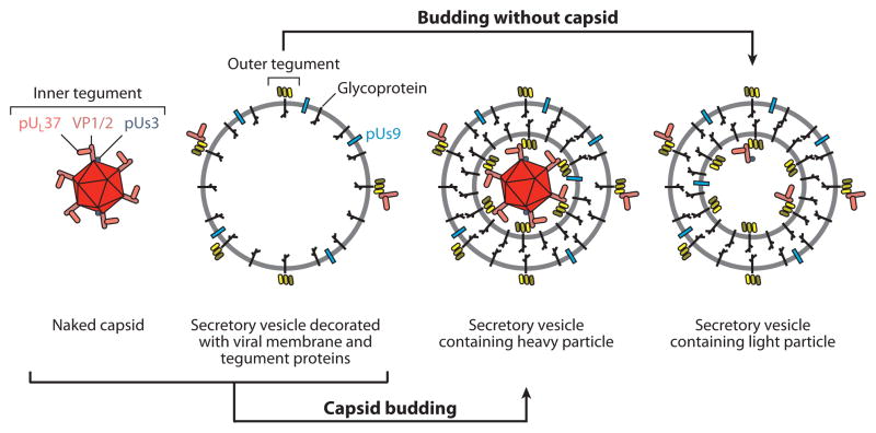
Four types of cytoplasmic viral particles present during the egress stage of infection. Unenveloped (naked) capsids newly egressed from the nucleus acquire inner tegument proteins beginning with VP1/2, which binds capsids directly. The composition of these particles and the retrograde transport complex that is released into the cytosol following entry are similar, if not equivalent (see Figure 3). Outer tegument proteins associate primarily with viral membrane proteins resident in the biosynthetic pathway (secretory vesicle), which may also acquire some inner tegument proteins in the absence of capsids. Membrane proteins consist of glycoproteins and nonglycosylated membrane-associated proteins that include pUs9. Infectious virions (heavy particles) form when capsids bud into membranes decorated with viral membrane and outer tegument proteins and subsequently exocytose from the cell. Light particles are noninfectious secreted viral particles that consist of a viral envelope and tegument proteins but lack a capsid. The three membrane-associated intracellular particles expose membrane protein tails and tegument proteins to the cytosolic surface. Among the exposed proteins, pUs9 is enriched in the vesicle membrane surface and may direct trafficking of particles to the distal axon.
References
-
- Abaitua F, Souto RN, Browne H, Daikoku T, O’Hare P. Characterization of the herpes simplex virus (HSV)-1 tegument protein VP1-2 during infection with the HSV temperature-sensitive mutant tsB7. J Gen Virol. 2009;90:2353–63. - PubMed
-
- Addison C, Rixon FJ, Palfreyman JW, O’Hara M, Preston VG. Characterisation of a herpes simplex virus type 1 mutant which has a temperature-sensitive defect in penetration of cells and assembly of capsids. Virology. 1984;138:246–59. - PubMed
Publication types
MeSH terms
Grants and funding
LinkOut - more resources
Full Text Sources
Other Literature Sources
