

18FDG PET-CT imaging detects arterial inflammation and early atherosclerosis in HIV-infected adults with cardiovascular disease risk factors
- PMID: 22726233
- PMCID: PMC3469335
- DOI: 10.1186/1476-9255-9-26
18FDG PET-CT imaging detects arterial inflammation and early atherosclerosis in HIV-infected adults with cardiovascular disease risk factors
Abstract
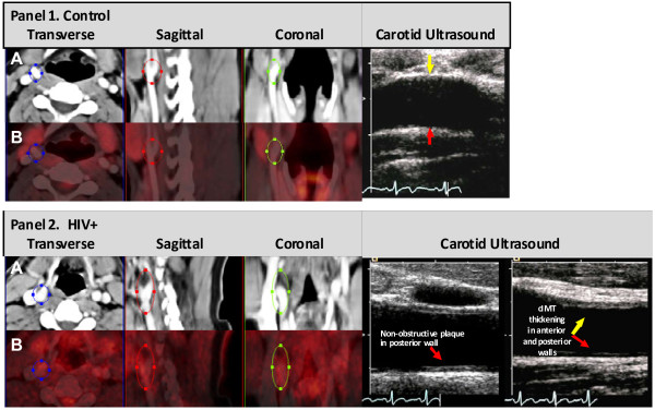
Background: Persistent vascular inflammation has been implicated as an important cause for a higher prevalence of cardiovascular disease (CVD) in HIV-infected adults. In several populations at high risk for CVD, vascular 18Fluorodeoxyglucose (18FDG) uptake quantified using 3D-positron emission-computed tomography (PET-CT) has been used as a molecular level biomarker for the presence of metabolically active proinflammatory macrophages in rupture-prone early atherosclerotic plaques. We hypothesized that 18FDG PET-CT imaging would detect arterial inflammation and early atherosclerosis in HIV-infected adults with modest CVD risk.
Methods: We studied 9 HIV-infected participants with fully suppressed HIV viremia on antiretroviral therapy (8 men, median age 52 yrs, median BMI 29 kg/m2, median CD4 count 655 cells/μL, 33% current smokers) and 5 HIV-negative participants (4 men, median age 44 yrs, median BMI 25 kg/m2, no current smokers). Mean Framingham Risk Scores were higher for HIV-infected persons (9% vs. 2%, p < 0.01). 18FDG (370 MBq) was administered intravenously. 3D-PET-CT images were obtained 3.5 hrs later. 18FDG uptake into both carotid arteries and the aorta was compared between the two groups.
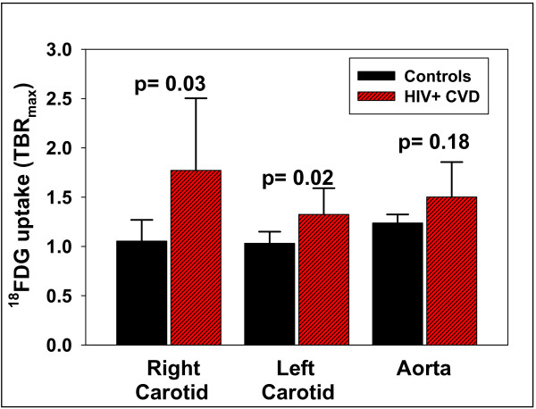
Results: Right and left carotid 18FDG uptake was greater (P < 0.03) in the HIV group (1.77 ±0.26, 1.33 ±0.09 target to background ratio (TBR)) than the control group (1.05 ± 0.10, 1.03 ± 0.05 TBR). 18FDG uptake in the aorta was greater in HIV (1.50 ±0.16 TBR) vs control group (1.24 ± 0.05 TBR), but did not reach statistical significance (P = 0.18).
Conclusions: Carotid artery 18FDG PET-CT imaging detected differences in vascular inflammation and early atherosclerosis between HIV-infected adults with CVD risk factors and healthy HIV-seronegative controls. These findings confirm the utility of this molecular level imaging approach for detecting and quantifying glucose uptake into inflammatory macrophages present in metabolically active, rupture-prone atherosclerotic plaques in HIV infected adults; a population with increased CVD risk.
Figures
References
Grants and funding
LinkOut - more resources
Full Text Sources
Medical
Research Materials
