. 2012 Sep;103(9):1640-50.
doi: 10.1111/j.1349-7006.2012.02367.x. Epub 2012 Aug 8.

Protein arginine methyltransferase 5 is a potential oncoprotein that upregulates G1 cyclins/cyclin-dependent kinases and the phosphoinositide 3-kinase/AKT signaling cascade

Affiliations

Protein arginine methyltransferase 5 is a potential oncoprotein that upregulates G1 cyclins/cyclin-dependent kinases and the phosphoinositide 3-kinase/AKT signaling cascade

Tong-You W Wei et al. Cancer Sci. 2012 Sep.

Abstract

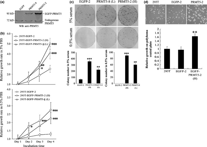
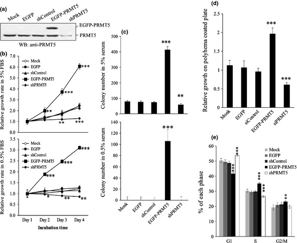
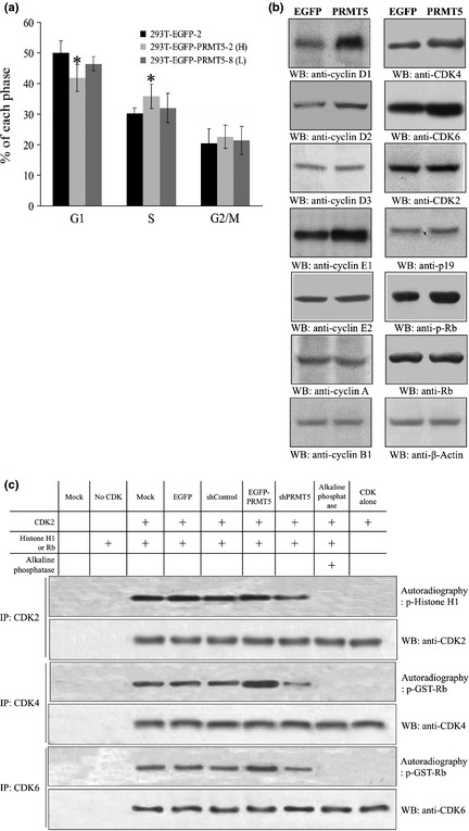
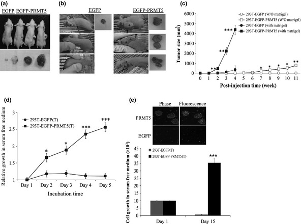
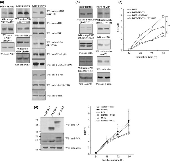
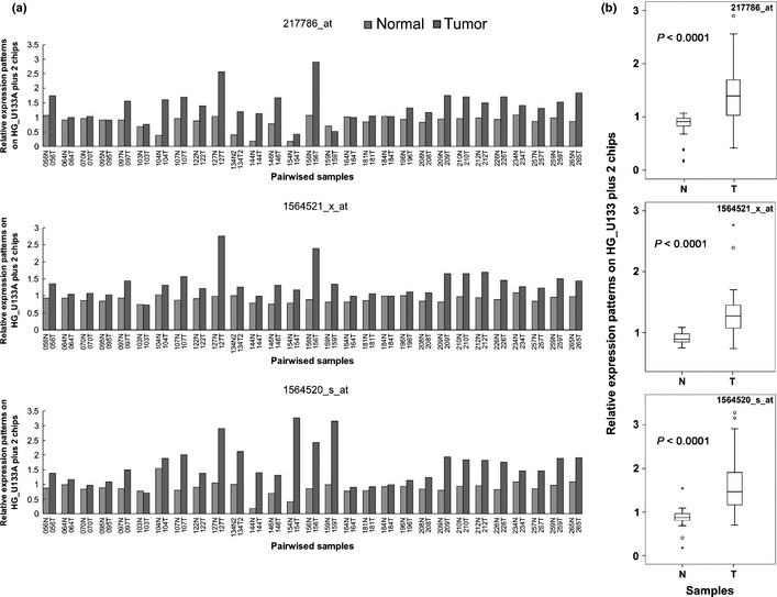
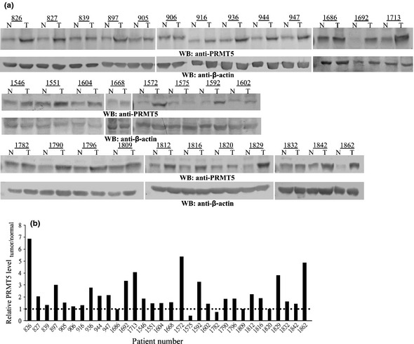
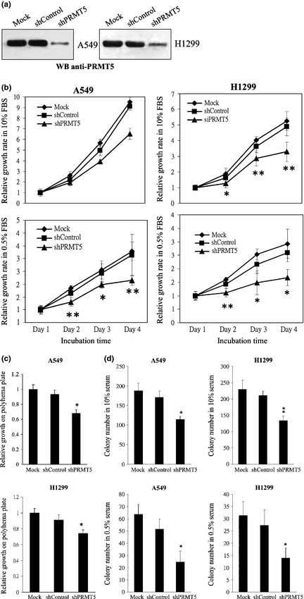
Increasing evidence suggests that PRMT5, a protein arginine methyltransferase, is involved in tumorigenesis. However, no systematic research has demonstrated the cell-transforming activity of PRMT5. We investigated the involvement of PRMT5 in tumor formation. First, we showed that PRMT5 was associated with many human cancers, through statistical analysis of microarray data in the NCBI GEO database. Overexpression of ectopic PRMT5 per se or its specific shRNA enhanced or reduced cell growth under conditions of normal or low concentrations of serum, low cell density, and poor cell attachment. A stable clone that expressed exogenous PRMT5 formed tumors in nude mice, which demonstrated that PRMT5 is a potential oncoprotein. PRMT5 accelerated cell cycle progression through G1 phase and modulated regulators of G1; for example, it upregulated cyclin-dependent kinase (CDK) 4, CDK6, and cyclins D1, D2 and E1, and inactivated retinoblastoma protein (Rb). Moreover, PRMT5 activated phosphoinositide 3-kinase (PI3K)/AKT and suppressed c-Jun N-terminal kinase (JNK)/c-Jun signaling cascades. However, only inhibition of PI3K activity, and not overexpression of JNK, blocked PRMT5-induced cell proliferation. Further analysis of PRMT5 expression in 64 samples of human lung cancer tissues by microarray and western blot analysis revealed a tight association of PRMT5 with lung cancer. Knockdown of PRMT5 retarded cell growth of lung cancer cell lines A549 and H1299. In conclusion, to the best of our knowledge, we have characterized the cell-transforming activity of PRMT5 and delineated its underlying mechanisms for the first time.


Figures

Figure 1
Figure 1
PRMT5 induces cell proliferation at low serum concentrations and under anchorage‐independent conditions. (a) Stable 293T clonal cell lines that expressed EGFP or EGFPPRMT5 at higher (clone 2, H) or lower levels of expression (clone 8, L) were subjected to western blotting with an antibody against PRMT5. (b) Clonal cell lines that expressed EGFP or PRMT5 were subjected to the MTT assay in 5% (upper) or 0.5% (lower) serum for 1–4 days. (c) Clonal cell lines that expressed EGFP or PRMT5 were subjected to a cell foci formation assay in 5% or 0.5% serum. Cell foci were fixed and stained with Giemsa solution. The number of foci were counted and plotted. (d) Clonal cell lines that expressed EGFP or PRMT5 were subjected to a polyhema‐based anchorage‐independent growth assay. The cells were allowed to grow for 2 days on polyhema plates and subsequently were trypsinized and counted with a hemocytometer. The number of cells in each group at day 2 was normalized against the number of cells that were seeded originally (i.e. 5 × 105). *, **, and *** indicate statistical significance by Student's t‐test with < 0.05, 0.01 and 0.001 respectively.
Figure 2
Figure 2
The effects of PRMT5 shRNA on cell growth in 293T cells. (a) 293T cells harboring nothing (Mock), EGFP, EGFPPRMT5, PRMT5 shRNA or scrambled shRNA were applied to Western blot adopting anti‐PRMT5 antibody. (b) 293T cells harboring nothing, EGFP, EGFPPRMT5, PRMT5 shRNA or scrambled shRNA were applied to MTT‐based cell growth assay in 5% or 0.5% serum for 1–4 days. (c) 293T cells harboring PRMT5 shRNA or scrambled control were applied to focus formation assay in 10% or 0.5% serum. (d) 293T cells harboring PRMT5 shRNA or scrambled control were subjected to polyhema‐based anchorage‐independent growth assay. (e) 293T cells harboring PRMT5 shRNA or scrambled control were analyzed by flowcytometer. The percentage cells residing in each specific phase were calculated and plotted. *, **, and *** indicate statistical significance by Student's t‐test with < 0.05, 0.01 and 0.001 respectively.
Figure 3
Figure 3
PRMT5 accelerates progression through G1 and upregulates regulators of G1 phase. (a) The EGFP and PRMT5 clonal cell lines were analyzed by flow cytometry, and the percentage of cells in each specific phase was calculated and plotted. (b) The protein level of various cell cycle regulators in the EGFP and PRMT5 clonal cell lines was analyzed by western blotting. Antibodies against cyclins A, B1, D1–D3, E1, and E2; CDK2, 4, and 6; p19; Rb protein; phospho‐Rb protein; and β‐actin were used for western blotting. (c) The kinase activities of various CDKs were assayed by performing in vitro kinase reaction where CDK2, CDK4 or CDK6 was isolated by immunoprecipitation employing specific antibodies from cells harboring nothing, EGFP empty vector, EGFPPRMT5, PRMT5 shRNA, or scrambled shRNA. Recombinant Histone H1 or Rb was used as substrate and incubated with CDKs in kinase reaction buffer in the presence of [γ‐P 32]‐ATP. *Statistical significance by Student's t‐test with < 0.05.
Figure 4
Figure 4
PRMT5 induces tumor formation in nude mice. (a–c) PRMT5 clonal cells formed tumors in nude mice. EGFP clonal cells or EGFPPRMT5 (clone 2) clonal cells were injected subcutaneously into nude mice in the absence (a) or presence (b) of Matrigel. Tumor size was measured weekly (c). (d,e) Cells from the PRMT5‐induced tumor grew in the absence of serum. PRMT5‐induced tumor cells taken from nude mice were subjected to the MTT assay (d) or counted with a hemocytometer (e) in serum‐free medium. *, **, and *** represent statistical significance by Student's t‐test with < 0.05, 0.01 and 0.001, respectively.
Figure 5
Figure 5
PRMT5 activates AKT but suppresses JNK signaling cascades. EGFP and PRMT5 clonal cells were subjected to western blotting using antibodies against (a) AKT signaling molecules, including phospho‐ and total PI3K p85, PI3K p110, phospho‐PDK1, phospho‐ and total PTEN, phospho‐ and total AKT, mTOR, phospho‐mTOR, elF4E, phospho‐IκB‐α, NF‐κB p65, phospho‐GSK‐3β, c‐Raf, phospho‐c‐Raf and β‐actin, and (b) MAPK signaling factors including phospho‐MEK1/2, phospho‐ and total ERK, phospho‐ and total p38, phospho‐ and total JNK, phospho‐ and total c‐Jun, and β‐actin. (c) An inhibitor of PI3K suppressed the growth of PRMT5 clonal cells. EGFP and PRMT5 clonal cells were subjected to the MTT‐based cell proliferation assay in the absence or presence of the PI3K inhibitor LY294002 for 1–4 days. (d) Overexpression of JNK1 or JNK2 did not alter the rate of cell proliferation induced by PRMT5. EGFP and PRMT5 clonal cells transfected with HAJNK1 or HAJNK2 were subjected to Western blot adopting anti‐HA, ‐pan JNK or ‐actin antibody, or the MTT assay for 1–4 days. *Statistical significance by Student's t‐test with < 0.05.
Figure 6
Figure 6
PRMT5 is overexpressed in patients with lung cancer. (a) The level of PRMT5 mRNA in samples from 29 patients with lung cancer was determined by microarray analysis. Overexpression of PRMT5 was observed in all three probesets (217786_at, 1564521_x_at, and 1564520_s_at). The numbers on the X axis represent the codes of the patients. (b) Box plot shows the distribution of data in grouping classification and a statistically significant difference (< 0.0001) between tumor tissues and adjacent non‐tumor tissues from the same lung cancer patients. T, tumor tissue; N, adjacent non‐tumor tissue.
Figure 7
Figure 7
Protein expression of PRMT5 in 32 samples of lung cancer tissue. (a) Biopsies from paired lung tumor (T) and adjacent normal tissues (N) were subjected to western blotting using an antibody against PRMT5 or actin. The numbers on the X axis represent the codes of the patients. Actin served as a loading control. (b) The levels of PRMT5 and actin were quantified by densitometry. The level of PRMT5 protein in each paired tissue was normalized against that of actin. The ratio of normalized PRMT5 in the tumor to that in normal tissue was calculated and plotted.
Figure 8
Figure 8
Effects of PRMT5 shRNA on the cell growth of A549 and H1299 cells. (a) A549 cells or H1299 cells harboring PRMT5 shRNA or scrambled control were applied to Western blot adopting anti‐PRMT5 antibody. (b) A549 or H1299 cells harboring PRMT5 shRNA or scrambled control were applied to MTT‐based cell growth assay in 10% or 0.5% serum for 1–4 days. (c) A549 or H1299 cells harboring PRMT5 shRNA or scrambled control were subjected to polyhema‐based anchorage‐independent growth assay. (d) A549 or H1299 cells harboring PRMT5 shRNA or scrambled control were applied to focus formation assay in 10% or 0.5% serum. * and ** represent statistical significance by Student's t‐test with < 0.05 and 0.01 respectively.

References

    1. Krapivinsky G, Pu W, Wickman K, Krapivinsky L, Clapham DE. pICln binds to a mammalian homolog of a yeast protein involved in regulation of cell morphology. J Biol Chem 1998; 273: 10811–4. - PubMed
    1. Pollack BP, Kotenko SV, He W, Izotova LS, Barnoski BL, Pestka S. The human homologue of the yeast proteins Skb1 and Hsl7p interacts with Jak kinases and contains protein methyltransferase activity. J Biol Chem 1999; 274: 31531–42. - PubMed
    1. Lee JH, Cook JR, Pollack BP, Kinzy TG, Norris D, Pestka S. Hsl7p, the yeast homologue of human JBP1, is a protein methyltransferase. Biochem Biophys Res Commun 2000; 274: 105–11. - PubMed
    1. Rho J, Choi S, Seong YR, Cho WK, Kim SH, Im DS. Prmt5, which forms distinct homo‐oligomers, is a member of the protein‐arginine methyltransferase family. J Biol Chem 2001; 276: 11393–401. - PubMed
    1. Feng Y, Wang J, Asher S et al Histone H4 acetylation differentially modulates arginine methylation by an in Cis mechanism. J Biol Chem 2011; 286: 20323–4. - PMC - PubMed

Publication types

MeSH terms

Substances