. 2012 Jun 22;19(6):731-41.
doi: 10.1016/j.chembiol.2012.04.013.

Fluxes in "free" and total zinc are essential for progression of intraerythrocytic stages of Plasmodium falciparum

Affiliations

Fluxes in "free" and total zinc are essential for progression of intraerythrocytic stages of Plasmodium falciparum

Rebecca G Marvin et al. Chem Biol. .

Abstract

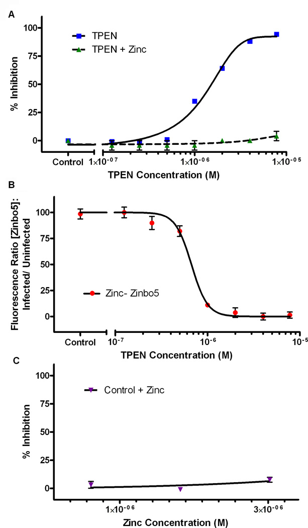
Dynamic fluxes in the concentration of ions and small molecules are fundamental features of cell signaling, differentiation, and development. Similar roles for fluxes in transition metal concentrations are less well established. Here, we show that massive zinc fluxes are essential in the infection cycle of an intracellular eukaryotic parasite. Using single-cell quantitative imaging, we show that growth of the blood-stage Plasmodium falciparum parasite requires acquisition of 30 million zinc atoms per erythrocyte before host cell rupture, corresponding to a 400% increase in total zinc concentration. Zinc accumulates in a freely available form in parasitophorous compartments outside the food vacuole, including mitochondria. Restriction of zinc availability via small molecule treatment causes a drop in mitochondrial membrane potential and severely inhibits parasite growth. Thus, extraordinary zinc acquisition and trafficking are essential for parasite development.


Figures

Figure 1
Figure 1. X-ray fluorescence two-dimensional metal maps show iron concentrated in the food vacuole and zinc concentrated nearby within separate subcellular regions
A) A typical schizont-stage parasite (top cell) and host erythrocyte (bottom cell) were analyzed for total iron and zinc. Fluorescence intensities are represented linearly on a rainbow scale with red being the maximum signal and black being the lowest signal. Phosphorus and sulfur maps reveal the location of nucleic acids and cell body, respectively. The majority of the parasite appears devoid of iron, except for the highly localized region within the parasite food vacuole. Zinc accumulation occurs within a larger region of the parasite-infected cell. Scale bar = 2 µm. B) Uninfected red blood cells (RBC), and schizont-infected red blood cells (Infected RBC) were analyzed for total iron and zinc concentrations by quantitative XFM. Infected red blood cells accumulate more Zn (p< 0.005) than erythrocytes, while Fe concentrations are not statistically different. Zinc concentrations within infected erythrocytes treated with 0.5 µM TPEN for 48 hours were not significantly different than the RBC host. Concentrations represent the means of N number of cells ± standard error. See also Table S1. C) To determine zinc/iron co-localization, signal intensities were converted to a logarithmic scale and given a single color (Fe = green, and Zn = red). The two-element overlay demonstrates that most zinc is excluded from the food vacuole, but lies within the parasite as marked by the iron depleted region. Co-localized green (Fe) and red (Zn) appear yellow in the overlay, showing a clear separation in metal localizations. Scale bar = 2 µm. D) Each developmental stage was analyzed for cellular zinc content by XFM. Erythrocytes infected with ring-stage parasites (0– 16 hrs. post merozoite invasion) had similar zinc levels as uninfected erythrocytes. The trophozoite-infected RBC (16– 28 hrs.) contained more Zn (p< 0.05) than rings, and schizont-infected RBCs (28– 48 hrs.) accumulated even more (p< 0.005).
Figure 2
Figure 2. Bioavailable zinc localizes to subcellular regions in close proximity to nuclei
An asynchronous culture of rings, early and late trophozoites, and schizonts were analyzed by epifluorescence microscopy. Fluorescence from zinc-bound Zinbo5 and DNA-bound Syto24 were given the false colors of green and red respectively, and overlaid with the DIC-transmitted light image. Fluorescence overlapping in the xy plane appears yellow when overlaid, suggesting a lack of correlation between DNA and zinc localized regions. Scale bar = 5 µm. See also Figure S1.
Figure 3
Figure 3. Late trophozoite-stage P. falciparum contain hot spots of zinc in regions that co-localize with mitochondrial probes DiOC6(3), RhodZin-3 AM, and MTG
DiOC6(3) accumulates within active mitochondria and Zinbo5 fluoresces when bound to Zn(II). RhodZin-3 AM both accumulates within mitochondria and labels zinc. MTG accumulates within mitochondria regardless of mitochondrial membrane potential. Signal overlap is visualized in yellow in overlaid images. A) Epifluorescence images of infected cells are stained for zinc and mitochondria. The yellow color in overlaid images suggests a degree of co-localization between the most intense regions of Zinbo5 and mitochondrial fluorescence. B) Confocal images of an infected cell are stained with Zinbo5 and MTG. Selected z-slices (z-thickness = 1 µm) from a z-stack dataset are displayed and demonstrate partial co-localization of fluorescence from these two probes. The entire z-stack dataset is presented as Video S1 in the Supporting Information. Scale bar = 5 µm.
Figure 4
Figure 4. RNA, DNA, and zinc levels increase as the parasite matures
A) Synchronized P. falciparum infected red blood cells were stained with Hoechst (HO) and thiazole orange (TO) to label nucleic acids within the parasite. Typical ‘Infected RBC’ and ‘RBC’ gates are indicated on a representative trophozoite-stage culture. B) In a separate sample, cells were labeled with Syto59 and TO to separate infected cells from uninfected erythrocytes. C) Syto59 and TO labeled cells were further analyzed for bioavailable zinc via Zinbo5 fluorescence. D) Zinbo5 mean fluorescence of infected cells relative to uninfected cells shows a delayed exponential increase in chelator-accessible zinc during the parasite’s final lifecycle stage, coinciding with high levels of DNA replication. Relative fluorescence represents the mean of three independent cultures (mean ± SEM). See also Figure S2.
Figure 5
Figure 5. Zinc scavenging by TPEN inhibits parasite survival
A) Synchronized, schizont-infected cells were treated for 48 hours with increasing concentrations of the zinc chelator, TPEN. Cultures were stained with Syto59 and TO and analyzed by flow cytometry to separate infected cells from normal erythrocytes. The IC50 value for TPEN inhibition is 1.38 ± 0.08 µM (IC50 ± SEM). Two molar equivalents of zinc rescued the TPEN effect. B) The TPEN treated culture was simultaneously analyzed for Zinbo5 fluorescence relative to an untreated control. The EC50 value for Zinbo5 fluorescence is 0.8 ± 0.1 µM (EC50 ± SEM). C) Control cultures were grown for 48 hours in media containing normal levels of zinc to 3× normal levels, without significant change.
Figure 6
Figure 6. Zinc insufficiency reduces the membrane potential after 48 hours
Unsynchronized parasites from each stage of the Plasmodium lifecycle were treated with 0.5 µM of the zinc chelator, TPEN. Since the accumulation of DiOC6(3) within mitochondria is dependent upon membrane potential, the intensity of the probe fluorescence was used to measure the change in membrane potential after chelator treatment. A dissipation of the DiOC6(3) probe can be seen after 2 hours of treatment, and a significant decrease in fluorescence is observed at 48 hours in trophozoite- (p < 0.005) and schizont- (p < 0.05) stage parasites. (mean ± SEM)

Comment in

References

    1. Atkinson A, Khalimonchuk O, Smith P, Sabic H, Eide D, Winge DR. Mzm1 influences a labile pool of mitochondrial zinc important for respiratory function. J Biol Chem. 2010;285:19450–19459. - PMC - PubMed
    1. Aurrecoechea C, Brestelli J, Brunk BP, Dommer J, Fischer S, Gajria B, Gao X, Gingle A, Grant G, Harb OS, et al. PlasmoDB: a functional genomic database for malaria parasites. Nucleic Acids Res. 2009;37:D539–D543. - PMC - PubMed
    1. Bannister LH, Hopkins JM, Fowler RE, Krishna S, Mitchell GH. A brief illustrated guide to the ultrastructure of Plasmodium falciparum asexual blood stages. Parasitol Today. 2000;16:427–433. - PubMed
    1. Berg JM. Zinc finger domains: hypotheses and current knowledge. Annu Rev Biophys Biophys Chem. 1990;19:405–421. - PubMed
    1. Beyersmann D, Haase H. Functions of zinc in signaling, proliferation and differentiation of mammalian cells. Biometals. 2001;14:331–341. - PubMed

Publication types

MeSH terms

LinkOut - more resources