. 2012 Jun 21;74(6):1015-22.
doi: 10.1016/j.neuron.2012.04.028.

Dual leucine zipper kinase is required for retrograde injury signaling and axonal regeneration

Affiliations

Dual leucine zipper kinase is required for retrograde injury signaling and axonal regeneration

Jung Eun Shin et al. Neuron. .

Abstract

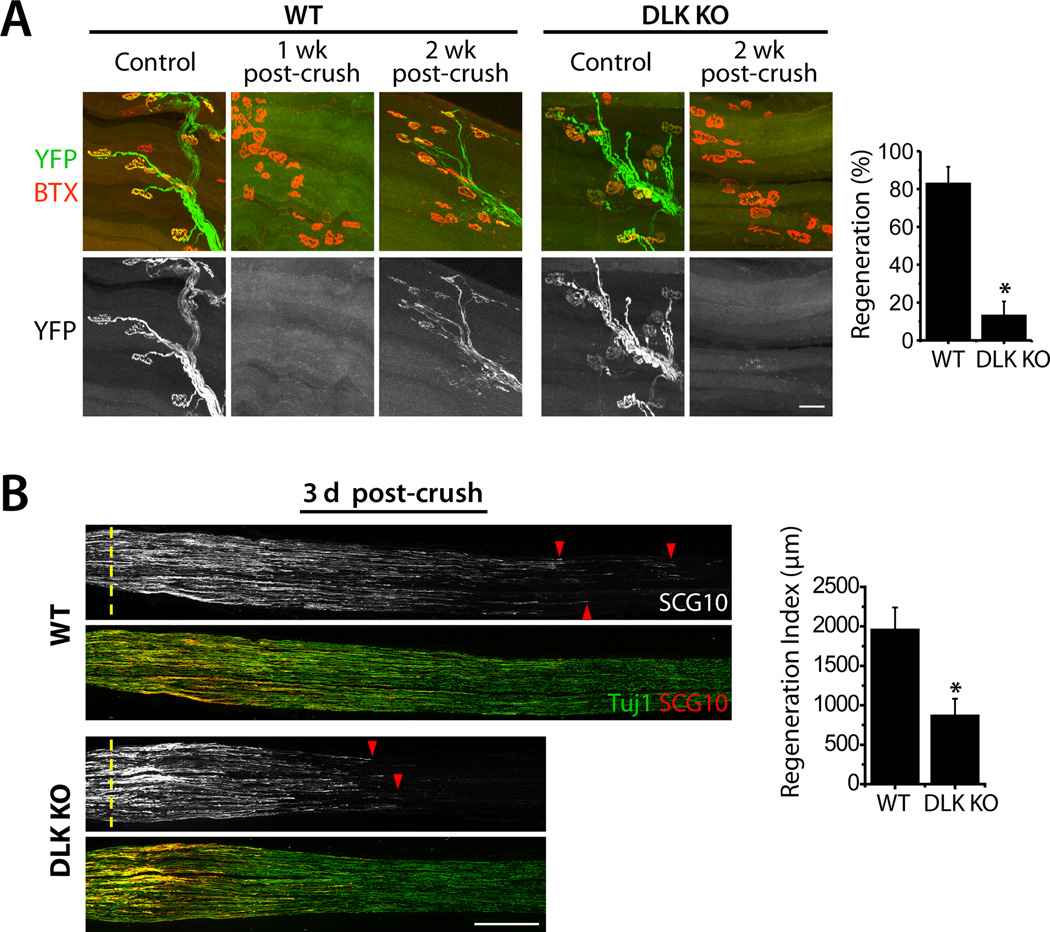
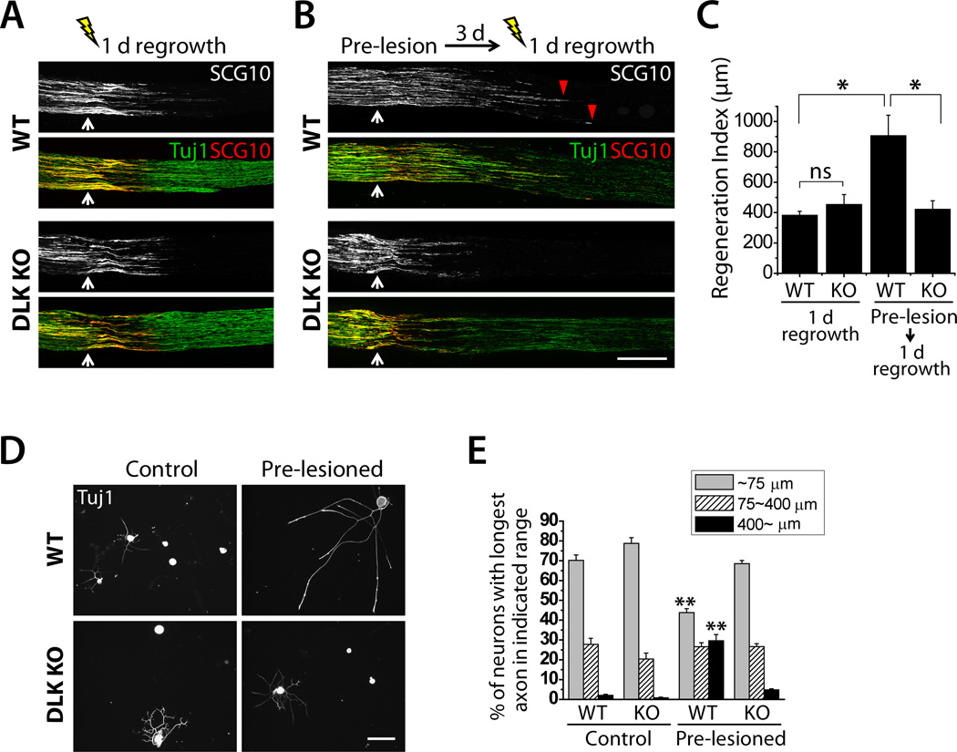
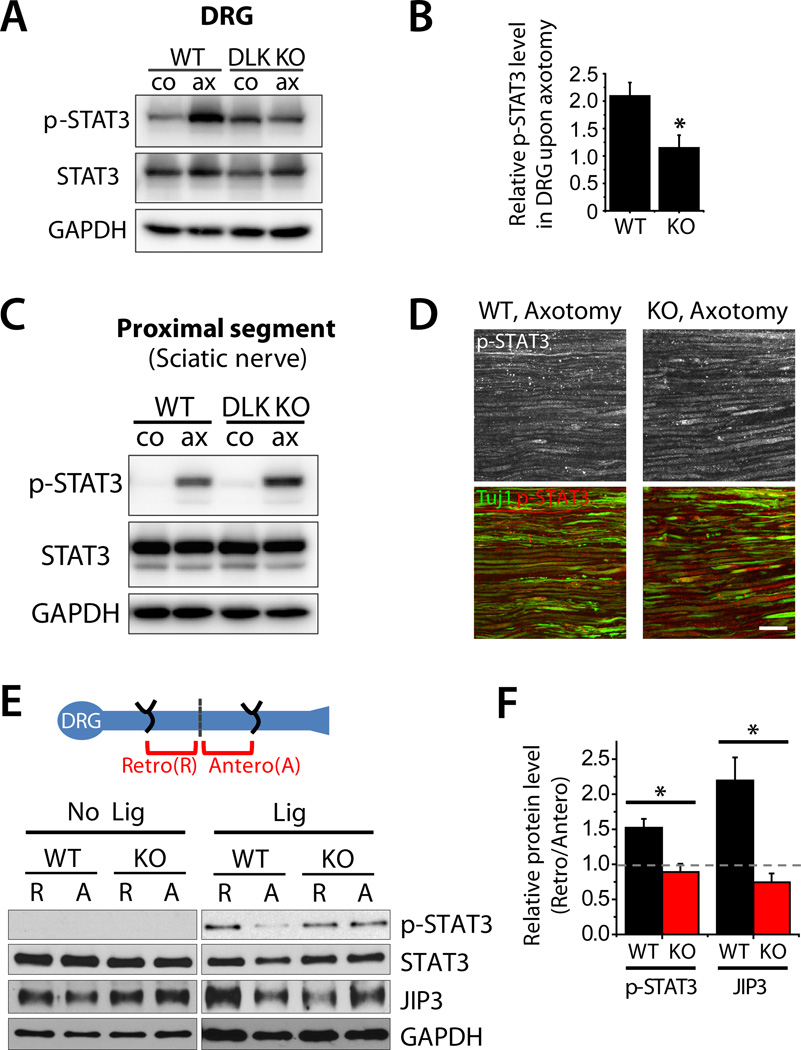
Here we demonstrate that the dual leucine zipper kinase (DLK) promotes robust regeneration of peripheral axons after nerve injury in mice. Peripheral axon regeneration is accelerated by prior injury; however, DLK KO neurons do not respond to a preconditioning lesion with enhanced regeneration in vivo or in vitro. Assays for activation of transcription factors in injury-induced proregenerative pathways reveal that loss of DLK abolishes upregulation of p-STAT3 and p-cJun in the cell body after axonal injury. DLK is not required for the phosphorylation of STAT3 at the site of nerve injury but is necessary for retrograde transport of p-STAT3 to the cell body. These data demonstrate that DLK enhances regeneration by promoting a retrograde injury signal that is required for the activation of the neuronal proregenerative program.


Conflict of interest statement

Conflict of interest: A.D., J.E.S., and Washington University may receive income based on a license by the University to Novus Biologicals.

Figures

Figure 1
Figure 1. Peripheral axon regeneration is inhibited in DLK KO mice
A, Confocal images of whole-mounted EHL muscles after crush injury of the sciatic nerve. In WT, YFP (green)-labeled axons degenerate by 1 week after injury, and robustly regenerate to the endplates labeled with α-bungarotoxin (BTX; red) by 2 weeks after injury. The axon re-targeting is significantly attenuated in HB9-Cre conditional DLK KO mice. WT (DLKF/WT; HB9-Cre; Thy-STOP-YFP15), n = 5; DLK KO (DLKF/F; HB9-Cre; Thy-STOP-YFP15), n = 4; p* < 0.001. Scale bar, 50 µm. B, Longitudinal sections of sciatic nerves from WT or Wnt1-Cre conditional DLK KO mice were immunolabeled for SCG10 and neuron-specific β3 tubulin (Tuj1) 3 days after crush injury. Axon regeneration from the crush site (dotted line) is significantly reduced in the DLK KO mice. SCG10 labels regenerating axons (red arrowheads). WT (DLKF/F), n = 4; DLK KO (DLKF/F; Wnt1-Cre), n = 3; p* < 0.05. Scale bar, 500 µm. Data are presented as means ± SEM. See also Fig. S1 and S2.
Figure 2
Figure 2. DLK is required for the accelerated axon regeneration induced by a preconditioning injury
A and B, Longitudinal sections of sciatic nerves of WT or Wnt1-Cre conditional DLK KO mice were immunolabeled for SCG10 and β3 tubulin (Tuj1) 1 day after either single crush injury (A) or double crush injury (B). Axon regeneration 1 day after crush injury is comparable between WT and DLK KO (A). Double crushed mice were given a preconditioning crush injury (Pre-lesion) for 3 days, subsequently subjected to another crush injury, and allowed to re-grow for 1 day (B). A pre-lesion significantly potentiates axon regeneration in WT (red arrowhead), but this effect is abolished in DLK KO. White arrow indicates the crush site. Scale bar, 500 µm. C, Results from (A) and (B) were quantified to generate a regeneration index. WT (DLKF/F), n = 4; DLK KO (DLKF/F; Wnt1-Cre), n = 4 for single crush, n = 5 for double crush; p* < 0.005. D, Cultured DRG neurons from WT or DLK KO were immunostained for β3 tubulin (Tuj1). A prior lesion of the sciatic nerve promotes axon regeneration in WT DRG neurons cultured for 16 hours, however this effect is abolished in DLK KO. Scale bar, 100 µm. E, Axon regrowth shown in (D) was quantified by percentage of neurons with longest axon in each range: ~75 µm, 75~400 µm, or 400~ µm. WT (DLKF/F or DLKF/WT; Wnt1-Cre); DLK KO (DLKF/F; Wnt1-Cre); n = 5; p** < 0.001 vs. WT, control and DLK KO, pre-lesioned. Data are presented as means ± SEM. See also Fig. S3.
Figure 3
Figure 3. DLK is required for retrograde transport of an injury-induced pro-regenerative signal
A, Immunoblot analysis of p-STAT3 in cell bodies following axon injury. Sciatic nerves of WT or DLK KO mice were unilaterally injured for 3 days and DRGs from either control (co) or injured (ax) side were subjected to immunoblot for p-STAT3. p-STAT3 levels are significantly elevated by axotomy (ax) in WT DRG but this increase is abolished in DLK KO DRG. Total STAT3 levels are comparable between conditions. GAPDH is shown as a loading control. B, Quantification of results shown in (A). p-STAT3 levels were quantified by normalizing the levels in the DRGs on the injured side to those in the uninjured contralateral DRGs. WT (DLKF/F); DLK KO (DLKF/F; Wnt1-Cre); n = 4; p* < 0.05. C, Sciatic nerves of WT or DLK KO mice were unilaterally injured for 1 day and the proximal nerve segments from either control (co) or injured (ax) side were subjected to immunoblot analysis of p-STAT3. p-STAT3 levels are significantly elevated in injured nerve (ax) and this increase is similar between WT and DLK KO mice, showing that DLK is not required for the local activation of STAT3. Total STAT3 levels are comparable between conditions. GAPDH is shown as a loading control. WT (DLKF/F); DLK KO (DLKF/F; Wnt1-Cre); n = 3. D, Longitudinal sections of the sciatic nerves of WT or DLK KO mice were immunostained for p-STAT3 1 day after crush injury. Confocal images visualize the proximal segments of the injured nerve and show that p-STAT3 localizes in axons. β3 tubulin (Tuj1) labels axons. WT (DLKF/F); DLK KO (DLKF/F; Wnt1-Cre). Scale bar, 50 µm. E, Schematic diagram of double ligation experiment shows that double ligation injury induces concentration of retrogradely transported cargoes (R) in the proximal segment and anterogradely transported cargoes (A) in the distal segment. In WT, p-STAT3 and JIP3 accumulate in the retrograde pool 6 hours after ligation (Lig). The accumulation of both proteins is blocked in the absence of DLK, showing that DLK is required for injury-induced retrograde transport of p-STAT3 and JIP3. Total STAT3 and JIP3 are similarly distributed in the proximal and distal segments in uninjured control nerve. GAPDH is shown as a loading control. F, Quantification of the results shown in (E). Retrograde protein accumulation is quantified as ratio of the protein levels in retrograde pool to the levels in anterograde pool. WT (DLKF/F); DLK KO (DLKF/F; Wnt1-Cre); n = 3; p* < 0.05. Data are presented as means ± SEM. See also Fig. S4.

Comment in

References

    1. Abe N, Almenar-Queralt A, Lillo C, Shen Z, Lozach J, Briggs SP, Williams DS, Goldstein LS, Cavalli V. Sunday driver interacts with two distinct classes of axonal organelles. The Journal of biological chemistry. 2009;284:34628–34639. - PMC - PubMed
    1. Abe N, Borson SH, Gambello MJ, Wang F, Cavalli V. Mammalian target of rapamycin (mTOR) activation increases axonal growth capacity of injured peripheral nerves. The Journal of biological chemistry. 2010;285:28034–28043. - PMC - PubMed
    1. Abe N, Cavalli V. Nerve injury signaling. Current opinion in neurobiology. 2008;18:276–283. - PMC - PubMed
    1. Bareyre FM, Garzorz N, Lang C, Misgeld T, Buning H, Kerschensteiner M. In vivo imaging reveals a phase-specific role of STAT3 during central and peripheral nervous system axon regeneration. Proceedings of the National Academy of Sciences of the United States of America. 2011;108:6282–6287. - PMC - PubMed
    1. Ben-Yaakov K, Dagan SY, Segal-Ruder Y, Shalem O, Vuppalanchi D, Willis DE, Yudin D, Rishal I, Rother F, Bader M, et al. Axonal transcription factors signal retrogradely in lesioned peripheral nerve. The EMBO journal. 2012 - PMC - PubMed

Publication types

Substances