. 2012 Jun 21;74(6):1023-30.
doi: 10.1016/j.neuron.2012.04.031.

Activity-dependent growth of new dendritic spines is regulated by the proteasome

Affiliations

Activity-dependent growth of new dendritic spines is regulated by the proteasome

Andrew M Hamilton et al. Neuron. .

Abstract

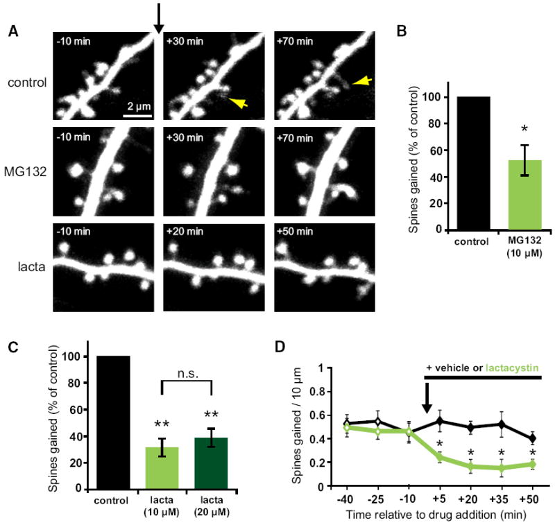
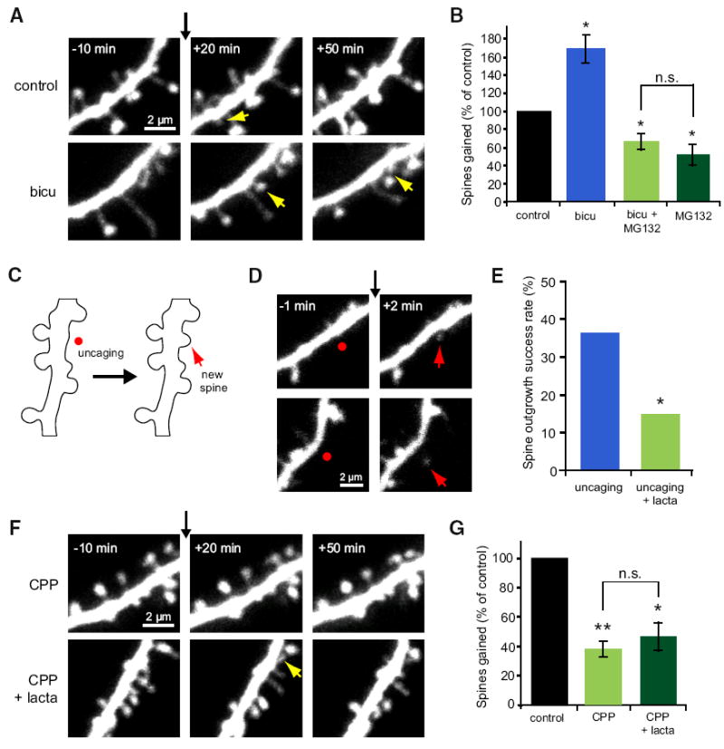
Growth of new dendritic spines contributes to experience-dependent circuit plasticity in the cerebral cortex. Yet the signaling mechanisms leading to new spine outgrowth remain poorly defined. Increasing evidence supports that the proteasome is an important mediator of activity-dependent neuronal signaling. We therefore tested the role of the proteasome in activity-dependent spinogenesis. Using pharmacological manipulations, glutamate uncaging, and two-photon imaging of GFP-transfected hippocampal pyramidal neurons, we demonstrate that acute inhibition of the proteasome blocks activity-induced spine outgrowth. Remarkably, mutation of serine 120 to alanine of the Rpt6 proteasomal subunit in individual neurons was sufficient to block activity-induced spine outgrowth. Signaling through NMDA receptors and CaMKII, but not PKA, is required to facilitate spine outgrowth. Moreover, abrogating CaMKII binding to the NMDA receptor abolished activity-induced spinogenesis. Our data support a model in which neural activity facilitates spine outgrowth via an NMDA receptor- and CaMKII-dependent increase in local proteasomal degradation.


Figures

Figure 1
Figure 1. Acute Inhibition of the Protea-some Rapidly Decreases New Spine Growth
(A) Images of dendrites from EGFP-expressing neurons at 7–12 DIV before and after the addition of vehicle, MG132, or lactacystin at t = 0 (black arrow). Yellow arrows indicate new spines. (B) MG132 (10 μM) decreased the rate of spine outgrowth (green bar; 34 spines, 5 cells) as compared to vehicle control (black bar; 68 spines, 6 cells; p < 0.05). (C) Lactacystin (10 μM) decreased the rate of spine outgrowth (light green bar; 33 spines, 6 cells) as compared to vehicle control (black bar; 131 spines, 8 cells; p < 0.01). Doubling the concentration of lactacystin (20 μM) inhibited spine outgrowth to a comparable extent (dark green bar; 25 spines, 6 cells; p < 0.001). (D) New spine growth was significantly reduced within 5 min after addition of lactacystin (10 μM) and remained significantly reduced for all subsequent time points (p < 0.05). Error bars represent SEM.
Figure 2
Figure 2. Synaptic Activity Promotes New Spine Growth in a Proteasome- and NMDA Receptor-Dependent Manner
(A) Images of dendrites from EGFP-expressing neurons at 5–10 DIV before and after the addition of vehicle or bicuculline (30 μM) at t = 0 (black arrow). Yellow arrows indicate new spines. (B) Elevated neural activity in response to bicuculline increased the rate of new spine addition (blue bar; 116 spines, 6 cells) as compared to vehicle control (black bar; 68 spines, 6 cells; p < 0.01). Inhibiting the proteasome with MG132 blocked the bicu-culline-induced increase in spine outgrowth and decreased baseline new spine growth (light green bar; 46 spines, 7 cells; p < 0.05) to a level similar to that in the presence of MG132 alone (dark green bar; data from Figure 1; p = 0.4). (C) Schematic of uncaging-induced spine outgrowth. A new spine (red arrow) was induced by focal photolysis of MNI-caged glutamate (red dot) adjacent to a section of dendrite devoid of spines. (D) Images of dendrites from EGFP-expressing neurons at 7–8 DIV before and after focal photolysis of MNI-glutamate (red dots) at t = 0 (black arrow). Red arrows indicate new spines. (E) Uncaging-induced spine outgrowth (blue bar; 16 successes out of 44 trials on 15 cells) was significantly reduced in the presence of lactacystin (10 μM; green bar; 6 successes out of 40 trials on 12 cells; p < 0.05). (F) Images of dendrites from EGFP-expressing neurons at 7–11 DIV before and after the addition of CPP (30 μM) or CPP + lactacystin (10 μM) at t = 0 (black arrow). Yellow arrows indicate new spines. (G) The rate of new spine addition decreased in the presence of CPP (light green bar; 39 spines, 8 cells) as compared to vehicle control (black bar; 105 spines, 8 cells; p < 0.001). No further decrease in spine outgrowth was observed with simultaneous blockade of the proteasome with lactacystin (dark green bar; 45 spines, 7 cells; p = 0.4). Error bars represent SEM.
Figure 3
Figure 3. Mutation of Serine 120 to Alanine of the Rpt6 Proteasomal Subunit Blocks Activity-Induced New Spine Growth
(A) Images of dendrites from hippocampal neurons transfected with EGFP, EGFP + Rpt6-WT, or EGFP + Rpt6-S120A at 9–10 DIV before and after the addition of vehicle or bicuculline at t = 0 (black arrow). Yellow arrows indicate new spines. (B) Transfection with Rpt6-WT (gray bar; 121 spines, 7 cells; p < 0.05) did not interfere with the bicuculline-induced increase in spine outgrowth (blue bar; 149 spines, 8 cells; p < 0.05) relative to vehicle-treated controls (black bar; 73 spines, 7 cells). In contrast, Rpt6-S120A transfection reduced spine outgrowth in bicuculline-treated cells (green bar; 56 spines, 7 cells; p < 0.001) relative to vehicle-treated controls. (C) Transfection with Rpt6-S120A decreased the rate of spine outgrowth (gray bar; 32 spines, 6 cells) as compared to cells transfected with EGFP alone (black bar; 74 spines, 6 cells; p < 0.01). Treatment of Rpt6-S120A transfected neurons with lactacystin (light green bar; 33 spines, 6 cells, p = 0.48) or with CPP (dark green bar; 44 spines, 7 cells, p = 0.24) did not further reduce rates of spine outgrowth over that observed for untreated S120A transfected cells. Error bars represent SEM.
Figure 4
Figure 4. CaMKII Activity and Its Interaction with the NMDA Receptor Subunit GluN2B Are Necessary for Activity-Dependent Spine Outgrowth
(A) Images of dendrites from EGFP-expressing hippocampal neurons at 7–10 DIV before and after the addition of myristoylated PKI 14–22 (20 μM), Rp-cAMPS (5 μM), or KN-93 (30 μM) at t = 0 (black arrow). Yellow arrows indicate new spines. (B) Inhibition of PKA with PKI 14–22 (dark gray bar; 58 spines, 7 cells; p = 0.9) or Rp-cAMPS (light gray bar; 66 spines, 7 cells; p = 0.4) did not alter rates of spine outgrowth relative to vehicle-treated controls (black bar; 54 spines, 7 cells). In contrast, inhibition of CaMKs with KN-93 decreased spine outgrowth (light green bar; 40 spines, 7 cells; p < 0.001) but did not further decrease spine outgrowth in cells transfected with Rpt6-S120A (dark green bar; 34 spines, 7 cells; p = 0.6). (C) Images of dendrites from EGFP-expressing WT or GluN2B L1298A/R1300Q knockin (GluN2B KI) mouse neurons at 8–11 DIV, before and after treatment with vehicle, bicuculline (30 μM), or lactacystin (10 μM) at t = 0 (black arrow). Yellow arrows indicate new spines. (D) In neurons from WT mice, treatment with bicuculline increased spine outgrowth (solid blue bar; 76 spines, 5 cells; p = 0.01) relative to vehicle-treated controls (solid black bar; 40 spines, 4 cells), while lactacystin decreased spine outgrowth (solid green bar; 16 spines, 5 cells) relative to controls (solid black bar; 54 spines, 5 cells). In contrast, in neurons from GluN2B KI mice, neither bicuculline (open blue bar; 41 spines, 5 cells; p = 0.6) nor lactacystin (open green bar; 42 spines, 5 cells; p = 0.8) altered spine outgrowth relative to vehicle-treated controls (open black bar; 45 spines, 5 cells). Error bars represent SEM.

References

    1. Acconcia F, Sigismund S, Polo S. Ubiquitin in trafficking: the network at work. Exp Cell Res. 2009;315:1610–1618. - PubMed
    1. Bhatt DH, Zhang S, Gan WB. Dendritic spine dynamics. Annu Rev Physiol. 2009;71:261–282. - PubMed
    1. Bingol B, Schuman EM. Activity-dependent dynamics and sequestration of proteasomes in dendritic spines. Nature. 2006;441:1144–1148. - PubMed
    1. Bingol B, Sheng M. Deconstruction for reconstruction: the role of proteolysis in neural plasticity and disease. Neuron. 2011;69:22–32. - PubMed
    1. Bingol B, Wang CF, Arnott D, Cheng D, Peng J, Sheng M. Autophosphorylated CaMKIIalpha acts as a scaffold to recruit proteasomes to dendritic spines. Cell. 2010;140:567–578. - PubMed

Publication types

MeSH terms

Substances

LinkOut - more resources