. 2012 Jun 21;74(6):1045-58.
doi: 10.1016/j.neuron.2012.04.025.

Dynamic FoxG1 expression coordinates the integration of multipolar pyramidal neuron precursors into the cortical plate

Affiliations

Dynamic FoxG1 expression coordinates the integration of multipolar pyramidal neuron precursors into the cortical plate

Goichi Miyoshi et al. Neuron. .

Abstract

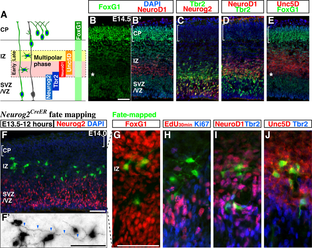
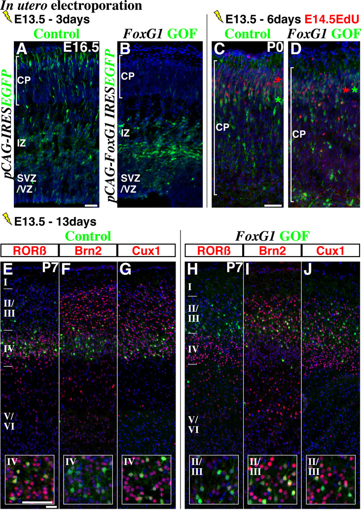
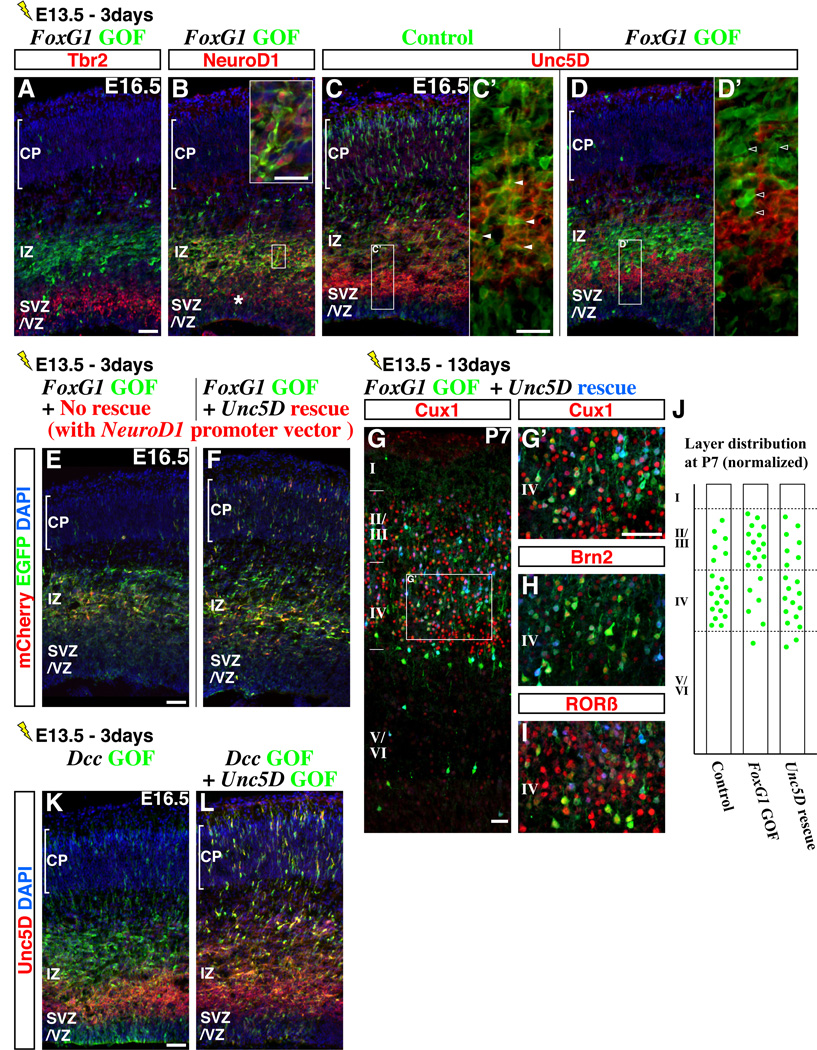
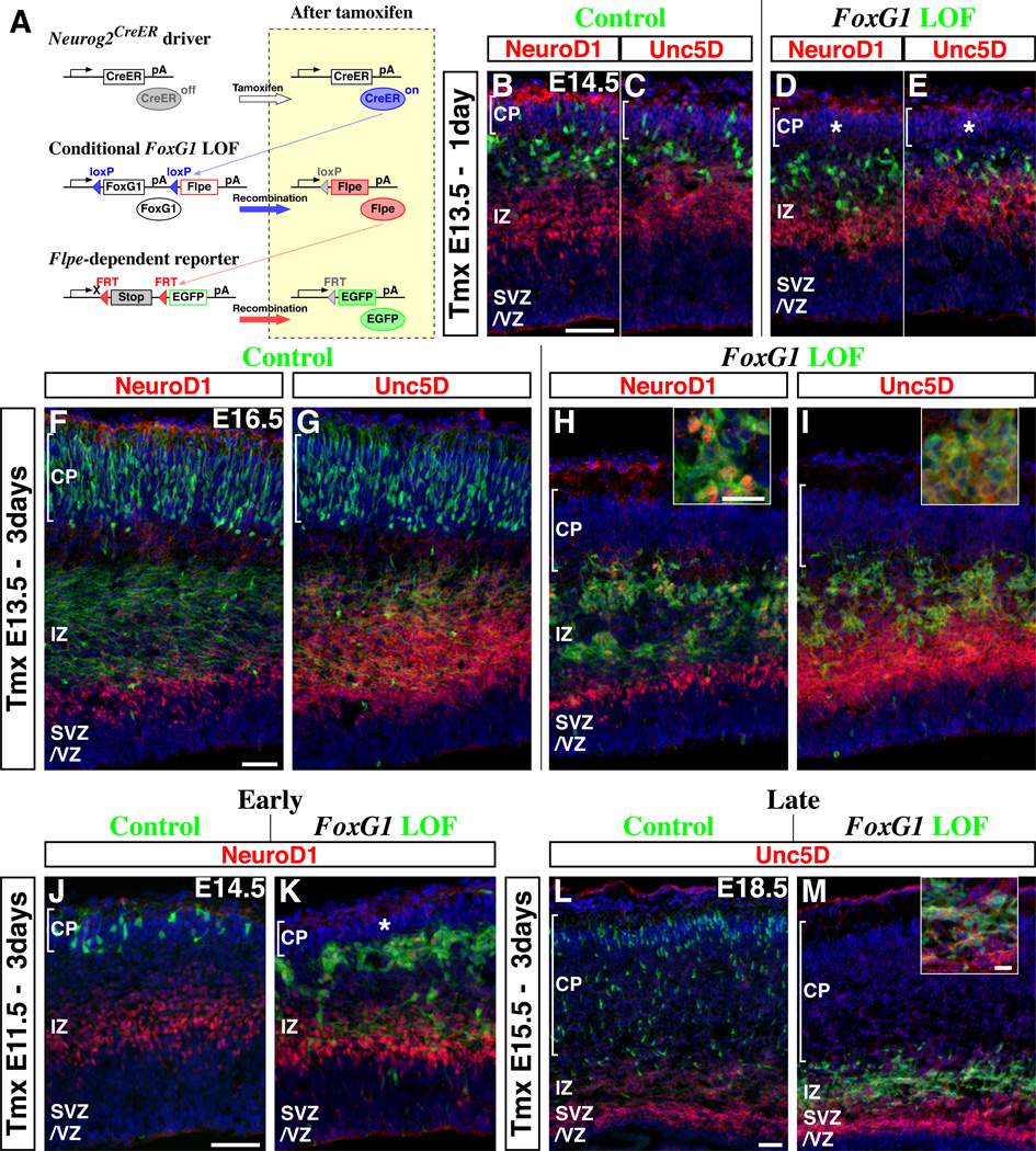
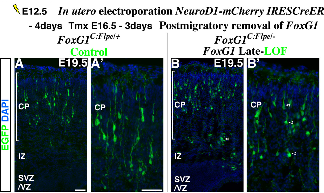
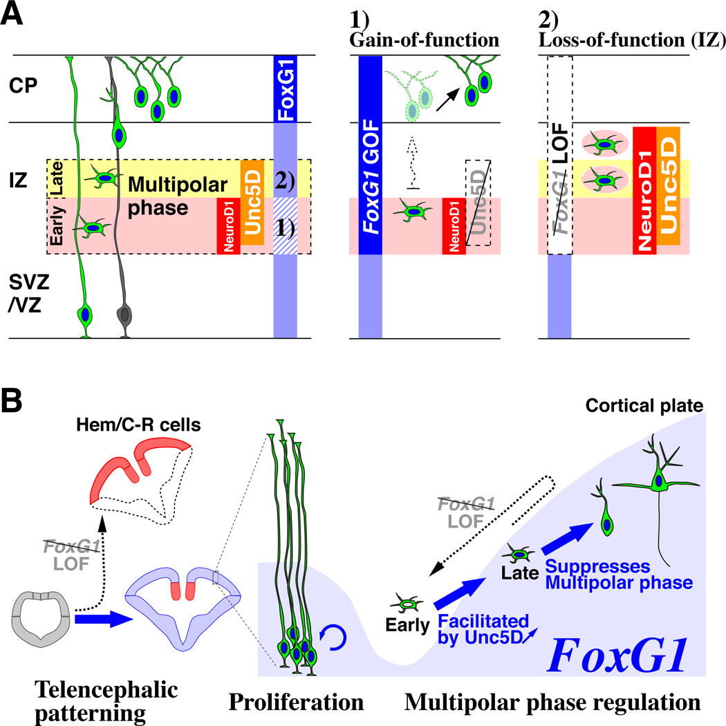
Pyramidal cells of the cerebral cortex are born in the ventricular zone and migrate through the intermediate zone to enter into the cortical plate. In the intermediate zone, these migrating precursors move tangentially and initiate the extension of their axons by transiently adopting a characteristic multipolar morphology. We observe that expression of the forkhead transcription factor FoxG1 is dynamically regulated during this transitional period. By utilizing conditional genetic strategies, we show that the downregulation of FoxG1 at the beginning of the multipolar cell phase induces Unc5D expression, the timing of which ultimately determines the laminar identity of pyramidal neurons. In addition, we demonstrate that the re-expression of FoxG1 is required for cells to transit out of the multipolar cell phase and to enter into the cortical plate. Thus, the dynamic expression of FoxG1 during migration within the intermediate zone is essential for the proper assembly of the cerebral cortex.


Figures

Figure 1
Figure 1. FoxG1 expression is dynamically regulated during the multipolar phase of pyramidal neuron development
(A) A schematic drawing comparing the expression of genes in pyramidal neuron precursors as they proceed from the proliferative zone to the cortical plate. The area surrounded by dotted lines indicates the region in which pyramidal neuron precursors adopt a multipolar identity. CP: cortical plate, IZ: intermediate zone, SVZ/VZ: subventricular and ventricular zones (B, B’) FoxG1 protein is expressed at different levels in E14.5 cortical cells at distinct stages of their maturation (See also Supplemental Figures S1A to S1C). FoxG1 expression is down-regulated (asterisk) in cells concomitant with their initiation of NeuroD1 expression. We observe similar results from the use of two different polyclonal antibodies for FoxG1. (C) A section adjacent to (B) indicating the region where the expression of Neurog2 and Tbr2 overlap. (D) Comparison of Tbr2 and NeuroD1 expression. (E) Down-regulation of FoxG1 (indicated by asterisk) occurs in the lower IZ (early multipolar phase) slightly below (i.e., before) initiation of Unc5D expression. Notably, in the uppermost IZ (late multipolar phase) Unc5D-expressing cells re-express FoxG1 below (i.e., prior) to where Unc5D-expression is extinguished. (F–J) Multipolar cells are labeled with EGFP by an acute (12 hour) fate mapping of the Neurog2-expressing population. This is achieved by combining the Neurog2-CreER driver and the R26R-CAG-loxPstop-EGFP reporter lines (See also Supplemental Figures S1D to S1G) and initiated through tamoxifen administration at E13.5. Note that EGFP-expressing cells have already shut off Neurog2 by this time. The majority of EGFP-labeled cells are found within the intermediate zone and possess multipolar morphology. (F’) In order to better visualize the cell morphology during the multipolar phase, a picture with EGFP expression is shown with reverse contrast to highlight their morphology. Tangentially oriented process resembling axon (arrowheads) are found in the multipolar cells. (G) Dependent on the position within the intermediate zone, multipolar cells express distinct levels of FoxG1 protein. (H) Multipolar cells are neither labeled by acute pulse-chase analysis using EdU (DNA analog) nor by antibodies against the Ki67 antigen suggesting that they are postmitotic. (I, J) Many Neurog2 fate-mapped cells with multipolar morphology expressed NeuroD1 and Unc5D but only low levels of Tbr2. Scale bars: 50µm
Figure 2
Figure 2. Failure to down-regulate FoxG1 delays the migration of pyramidal neuron precursors at the multipolar phase and alters their laminar fate
In utero electroporation of either (A) control (pCAG-IRES EGFP) or (B) FoxG1 gain-of-function (GOF) (pCAG-FoxG1-IRES EGFP) vectors under the regulation of a ubiquitous CAG promoter were carried out at E13.5 and brains were analyzed at E16.5. Three days after manipulation, while most control cells have entered the cortical plate (A), the majority of FoxG1 gain-of-function cells (B) were found in the lower part of the intermediate zone and displayed multipolar morphologies. (C, D) 6 days later, many FoxG1 gain-of-function cells delayed in migration by this manipulation now enter the cortical plate (bracket indicates cortical plate). Control cells (C) were generally found in lower positions compared to cells born one day after the electroporation (single pulse of EdU at E14.5), while FoxG1 gain-of-function cells (D) were intermingled with them. (E–G) At P7, the majority of EGFP-labeled control cells were found in layer IV and expressed molecular profiles consistent with cells within this layer (i.e., RORβ-on, Brn2-low, Cux1-on, insets). (H–J) The majority of FoxG1 gain-of-function cells were located in layers II/III and possessed molecular features consistent with this location (insets, RORβ-off, Brn2-high and Cux1-on). Note that in case of FoxG1 gain-of-function, a few cells were also found within the white matter and lacked expression of the layer specific markers we have examined. See also Supplemental Figures S2 and S3. Scale bars: 50µm
Figure 3
Figure 3. FoxG1 down-regulation promotes an early to late multipolar phase transition through induction of Unc5D
Migrating pyramidal neuron precursors that failed to down-regulate FoxG1 were delayed within the lower part of the intermediate zone (Figure 2). These cells shut off Tbr2 (A) but ectopically maintained NeuroD1 expression (B) (in a region above the normal expression domain, asterisk), suggesting that they become arrested at the early multipolar phase. (C, C’) Control cells within the intermediate zone expressed Unc5D (arrowheads in C’). (D, D’) FoxG1 gain-of-function cells, although they possess multipolar morphology, failed to express Unc5D protein (open arrowheads). (E) As we have previously shown, three days after electroporation at E13.5, FoxG1 gain-of-function cells (with pCAG-FoxG1-IRES EGFP) remained within the lower part of the intermediate zone. Note that mCherry was expressed in NeuroD1-expressing cells by co-introducing a pNeuroD1-IRES mCherry control vector. (F) When Unc5D expression was restored in NeuroD1-expressing FoxG1 gain-of-function cells (by using a pNeuroD1-Unc5D-IRES mCherry vector), a subset of these pyramidal neuron precursors migrated normally into the cortical plate after three days (green and red cells) and after 13 days at P7 (G, G’, H, I), we observed cells located in both layers II/III and layer IV (see summary in J). The partial restoration in the laminar location of this population at P7 was consistent with the degree of rescue in migration we observed after three days (F, compare with Figure 2A). In FoxG1 gain-of-function cells, both rescued (in layer IV) and non-rescued (in layers II/III) populations express molecular signatures appropriate to their laminar locations. Specifically, Unc5D-rescued cells in layer IV showed molecular expression profiles consistent with them being layer IV cells, i.e., Cux1-on (G’), Brn2-low (H) and RORβ-on (I). (J) Schematized layer distribution of Control (pCAG-IRES EGFP) (Figures 2E to 2G), FoxG1 gain-of-function (pCAG-FoxG1-IRES EGFP) (Figures 2H to 2J) and Unc5D-rescued FoxG1 gain-of-function (pCAG-FoxG1-IRES EGFP + pNeuroD1-Unc5D-IRES mCherry) experiments. Note that the numbers of EGFP-labeled cells (including the low-expressors) within the cortical plate is normalized (20 cells) and represented in this scheme. (K) Similar to FoxG1 gain-of-function, Dcc over-expression (pCAG-Dcc-IRES EGFP) delays cell migration at the intermediate zone. (L) Unc5D over-expression (pCAG-Unc5D-IRES EGFP) rescues the impaired migration phenotype observed by Dcc over-expression. Thus, a precise balance between Dcc versus Unc5D expression is important for cells to migrate through the intermediate zone (See also Supplemental Figures S4). This balance appears to be critically controlled by transient FoxG1 down-regulation as FoxG1 gain-of-function affects Unc5D (D, D’) but not Dcc expression (data not shown). Scale bars: 50µm, except for C’ and D’: 20µm
Figure 4
Figure 4. Up-regulation of FoxG1 is required for pyramidal neuron precursors to transit out of the multipolar cell phase and to enter into the cortical plate
(A) Schematic drawings of our genetic strategy to mosiacally remove FoxG1 at the multipolar cell phase and to selectively follow the fate of recombined cells. (Top) Neurog2-CreER driver was used to target multipolar cells at specific time points. (Middle) A conditional FoxG1 loss-of-function (LOF) allele that expresses Flpe after Cre-mediated recombination (Supplemental Figure S5). (Bottom) A Flpe-dependent EGFP reporter line was utilized to visualize the cells in which the FoxG1 conditional loss-of-function allele has been recombined. (Right) Subsequent to tamoxifen administration, the CreER expressed in Neurog2-positive cells becomes active (top) and recombines the FoxG1 conditional allele resulting in the removal of FoxG1 coding region and initiation of Flpe expression from the FoxG1 locus (middle), Flpe in turn removes a stop cassette in the reporter, allowing us to permanently trace the fate of manipulated cells with EGFP (Bottom). (B–I) Comparison of control (FoxG1 heterozygous) versus FoxG1 loss-of-function cells at various time points after tamoxifen administration at E13.5. (B, C) One day after tamoxifen administration, some control EGFP-labeled cells are already found inside the cortical plate (See also Supplemental Figure S1E). (D, E) By contrast, FoxG1-null cells remain excluded from the cortical plate (asterisk) and generally maintained multipolar morphologies. Note that neither control nor FoxG1-null cells located right below the cortical plate express NeuroD1 (B, D) or Unc5D (C, E). (F, G) Three days after tamoxifen administration, the majority of control cells are found within the cortical plate. Note that in these cortices, EGFP-labeled axonal fibers derived from the cortical plate cells are readily evident within the intermediate zone. (H, I) FoxG1-null cells in littermate embryos were only found below the cortical plate and maintained multipolar morphologies. Note also that the majority of FoxG1-null cells now express NeuroD1 (H, inset) and Unc5D (I, inset) suggesting that these cells have reverted back to the early multipolar phase. In addition, we observed aggregation of FoxG1-null cells at this time point (H, I). (J–M) Similar FoxG1 loss-of-function experiments were carried out at earlier (E11.5: J, K) or later (E15.5: L, M) time points. At both early and late time points, FoxG1-null cells maintained multipolar morphologies, did not enter the cortical plate (asterisk in K), re-initiated genes specifically expressed during the early multipolar phase (NeuroD1 in K and Unc5D in M, inset) and formed aggregates. Hence, FoxG1 up-regulation during the late multipolar cell phase seems universally required for pyramidal cells throughout development. See also Supplemental Figures S6A and S6B for an analysis after 7 days of manipulation. Scale bars: 50µm, except for insets in H, I and M: 20µm
Figure 5
Figure 5. The role of FoxG1 in the multipolar cells is distinct from its function in postmigratory neurons within the cortical plate
In order to remove FoxG1 in postmigratory pyramidal neuron precursors inside the cortical plate, we have introduced a pNeuroD1-mCherry-IRES CreER vector into the ventricular zone by in utero electroporation at E12.5. This manipulation allows us to simultaneously visualize the morphologies of postmitotic cells through their expression of mCherry, as well as enabling us to recombine the conditional FoxG1 loss-of-function allele at a desired time point. As the FoxG1 loss-of-function allele expresses Flpe after gene removal (A scheme in Figure 4A), FoxG1 mutant cells can be visualized with EGFP directed from the Flpe-activated reporter line (R26R-CAG-FRTstop-EGFP) (Miyoshi et al., 2010). At E16.5, a time by which the majority of cells electroporated at E12.5 have completed migration and settled inside the cortical plate (data not shown), tamoxifen administration was carried out to remove FoxG1 in these cells. These embryos were analyzed at E19.5. (A, high magnification in A’) The control experiment was carried out in the FoxG1-C:Flpe/+ background. Pyramidal neurons labeled by EGFP are heterozygous for FoxG1. (B, B’) The loss-of-function experiment was carried out in the FoxG1-C:Flpe/− background. Note that control and FoxG1-null cells have pyramidal morphologies and occupy similar positions within the cortex. In addition, removal of FoxG1 gene function in postmigratory cells did not result in up-regulation of NeuroD1 and Unc5D (data not shown). Although the dendritic branching of mutant pyramidal neurons appears somewhat retarded compared to the control population (A, A’), they are not different from the internal control cells (Supplemental Figure S6D’, red without green cells, arrowheads). Most importantly, these cells do not morphologically or molecularly resemble the multipolar population. Note that for the purpose of presentation, cells labeled red by mCherry are not visualized in these panels. For the mCherry expression and also for the detailed comparison of wild type, heterozygous and null cells for FoxG1 in both FoxG1C:Flpe/+ and FoxG1C:Flpe/− backgrounds, see Supplemental Figures S6C and S6D. Scale bars: 50µm
Figure 6
Figure 6. Dynamic FoxG1 expression during the postmitotic multipolar cell phase coordinates pyramidal neuron development
(A, left panel) FoxG1 expression (blue vertical bar) is dynamically regulated throughout pyramidal neuron development. Especially within the intermediate zone (IZ), FoxG1 is transiently down-regulated at the beginning of the multipolar cell phase (Pink) and subsequently re-initiated during the late multipolar phase (Yellow). (A, middle panel) FoxG1 down-regulation is necessary for multipolar cells to initiate Unc5D expression and to rapidly proceed from the early to late multipolar phase. Mis-regulation of FoxG1 at this early phase delays entry into the cortical plate and redirects the laminar fate of postmitotic multipolar cells. (A, right panel) Multipolar cells need to re-initiate FoxG1 expression during the late phase in order to enter the cortical plate; otherwise, they remain in the intermediate zone and regress to the early multipolar phase. (B) Model diagram summarizing the roles of FoxG1 during pyramidal neuron development. FoxG1 expression and function (arrows) are indicated in blue. At the early stage when the telencephalon is emerging from the anterior regions of the neural tube, FoxG1 is required for the patterning by suppressing cortical hem (Hem) / Cajal-Retzius (C-R) cell fate (Hanashima et al., 2004; Muzio and Mallamaci, 2005; Shen et al., 2006b). After this period, FoxG1 is further required for the proliferation of neocortical progenitors (Hanashima et al., 2002; Martynoga et al., 2005). In this study, we have demonstrated the requirement for FoxG1 during the postmitotic period, when pyramidal neurons transit through their multipolar phase of development. At the beginning of the multipolar phase (Early), transient down-regulation of FoxG1 allows cells to initiate Unc5D expression, which is crucial for the rapid transition from the early to late phase. Failure to undergo this transition delays entry of pyramidal neuron precursors into the cortical plate, resulting in a switch in their laminar identity (See middle panel of A). Subsequent to this, FoxG1 expression is re-initiated during the late multipolar phase (Late) and is required for pyramidal neuron precursors to enter into the cortical plate. As indicated by the dashed arrow, failure to re-express FoxG1 at this late phase results in a regression of pyramidal neuron precursors into the early multipolar phase and permanently prevents them from entering the cortical plate (See right panel of A). Hence, as illustrated here, FoxG1 has iterative roles during pyramidal neuron development in patterning, proliferation and postmitotic regulation of the multipolar cell phase.

References

    1. Ackerman SL, Kozak LP, Przyborski SA, Rund LA, Boyer BB, Knowles BB. The mouse rostral cerebellar malformation gene encodes an UNC-5-like protein. Nature. 1997;386:838–842. - PubMed
    1. Alcantara S, Ruiz M, De Castro F, Soriano E, Sotelo C. Netrin 1 acts as an attractive or as a repulsive cue for distinct migrating neurons during the development of the cerebellar system. Development. 2000;127:1359–1372. - PubMed
    1. Alfano C, Viola L, Heng JI, Pirozzi M, Clarkson M, Flore G, De Maio A, Schedl A, Guillemot F, Studer M. COUP-TFI promotes radial migration and proper morphology of callosal projection neurons by repressing Rnd2 expression. Development. 2011;138:4685–4697. - PubMed
    1. Angevine JBJ, Sidman RL. Autoradiographic Study of Cell Migration during Histogenesis of Cerebral Cortex in the Mouse. Nature. 1961;192:766–768. - PubMed
    1. Ariani F, Hayek G, Rondinella D, Artuso R, Mencarelli MA, Spanhol-Rosseto A, Pollazzon M, Buoni S, Spiga O, Ricciardi S, et al. FOXG1 is responsible for the congenital variant of Rett syndrome. Am J Hum Genet. 2008;83:89–93. - PMC - PubMed

Publication types

MeSH terms

Substances