. 2012 Jun 21;74(6):1114-24.
doi: 10.1016/j.neuron.2012.04.036.

A real-world size organization of object responses in occipitotemporal cortex

Affiliations

A real-world size organization of object responses in occipitotemporal cortex

Talia Konkle et al. Neuron. .

Abstract

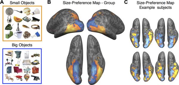
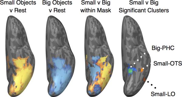
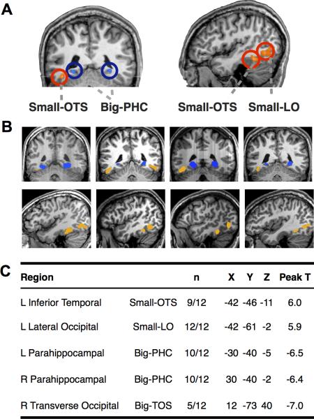
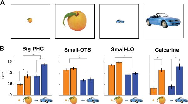
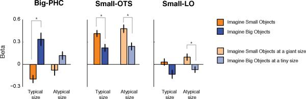
While there are selective regions of occipitotemporal cortex that respond to faces, letters, and bodies, the large-scale neural organization of most object categories remains unknown. Here, we find that object representations can be differentiated along the ventral temporal cortex by their real-world size. In a functional neuroimaging experiment, observers were shown pictures of big and small real-world objects (e.g., table, bathtub; paperclip, cup), presented at the same retinal size. We observed a consistent medial-to-lateral organization of big and small object preferences in the ventral temporal cortex, mirrored along the lateral surface. Regions in the lateral-occipital, inferotemporal, and parahippocampal cortices showed strong peaks of differential real-world size selectivity and maintained these preferences over changes in retinal size and in mental imagery. These data demonstrate that the real-world size of objects can provide insight into the spatial topography of object representation.


Figures

Figure 1
Figure 1
(A) Example objects of small and big real-world sizes. Note that all images were presented at the same retinal size. The stimulus set contained 200 small and 200 big objects. (B) Size-preference maps in the group data. Voxels with a greater response to small objects than big objects are shown in orange; Voxels with a greater response to big objects than small object are shown in blue. The data are plotted on the inflated brain of one participant. There is a medial-to-lateral arrangement of big-to-small object preferences along the ventral surface. (C) Size-preference maps for 4 example subjects.
Figure 2
Figure 2
Responses in left ventral temporal cortex of an example subject, reflecting i) small objects > rest, T>2.0, (ii) big objects > rest, T>2.0, (iii) the size-preference map masked by these small or big object-responsive voxels, and (iv) the regions of significant differential selectivity for small vs. big objects resulting from a whole-brain contrast, FDR<0.05.
Figure 3
Figure 3
(A) Whole-brain contrasts of small vs. big objects. Results of a random-effects analysis, small>big contrast, (n=12, P<0.002, cluster threshold=10), plotted on the brain of one sample subject (sagittal section, x=−42, coronal section, y=−42). The bilateral region with preference for big objects on the ventral surface is shown (Big-PHC). Two small-preference regions were found, one ventral/anterior (Small-OTS) and one lateral/posterior (Small-LO). (B) These regions of interest are shown for 4 participants. (c) Table indicating the regions identified from the group random effects analysis. Each region's anatomical location and label are indicated, followed by the number of subjects (n) who showed this region of interest in a single-subject analysis. For each region, the Talairach coordinates of the voxel with the peak t-value (small>big contrast) are reported.
Figure 4
Figure 4
Retinal Size Manipulation Results. A) Objects of small and big real-world sizes were presented at small and big retinal sizes on the screen. B) Activations in independently-localized Big-PHC, Small-OTS, and Small-LO, and anatomically-defined early visual cortex regions (Calcarine) were measured with ROI GLMs and the beta weights for the four conditions are plotted for the left hemisphere ROIs. Error bars reflect ± 1 S.E.M. The Big-PHC region showed effects of both the real-world and the retinal size, while the small regions showed only preference for the real-world of objects with no modulation to retinal size. The early visual control region showed modulation by retinal size, with no effect of real-world size.
Figure 5
Figure 5
Mental Imagery Results. Activations in independently-localized Big-PHC, Small-OTS, and Small-LO regions in left hemisphere are shown. Orange bars show data for imagined objects with a small real-world size (e.g. strawberries) and blue bars show data for imagined objects with a big real-world size (e.g. pianos). Bars with saturated colors reflect conditions where observers imagined typically-sized objects. Bars with unsaturated colors reflect conditions where the objects were imagined at atypical sizes.
Figure 6
Figure 6
Relationship of these regions to eccentricity bands and to other well-characterized regions. Left: Group size-preference maps, with the big and small regions of peak selectivity shown from a single representative subject. Right: Functionally-localized regions from the same single subject shown. Inner, middle, and outer eccentricity rings are shown in light, medium, and dark blue respectively. LOC and pFS (objects>scrambled) are shown in yellow. FFA (faces>objects) is shown in pink; this participant did not have an OFA region. PPA and TOS (scenes>objects) are shown in green. The Small-OTS and Small-LO regions are shown in orange and the Big-PHC and Big-TOS regions are shown in blue, also indicated with white arrows.

References

    1. Aguirre GK, Zarahn E, D'Esposito M. An area within human ventral cortex sensitive to “building” stimuli: evidence and implications. Neuron. 1998;21:373–383. - PubMed
    1. Arcaro MJ, McMains SA, Singer BD, Kastner S. Retinotopic organization of human ventral visual cortex. J. Neurosci. 2009;29:10638–10652. - PMC - PubMed
    1. Attneave F. Some informational aspects of visual perception. Psychol. Rev. 1954;61:183–193. - PubMed
    1. Bar M. Visual objects in context. Nat. Rev. Neurosci. 2004;5:617–629. - PubMed
    1. Beauchamp MS, Lee KE, Haxby JV, Martin A. Parallel visual motion processing streams for manipulable objects and human movements. Neuron. 2002;34:149–159. - PubMed

Publication types

LinkOut - more resources