Review
. 2012:109:75-112.
doi: 10.1016/B978-0-12-397863-9.00003-1.

Immunoproteasomes: structure, function, and antigen presentation

Affiliations
Review

Immunoproteasomes: structure, function, and antigen presentation

Deborah A Ferrington et al. Prog Mol Biol Transl Sci. 2012.

Abstract

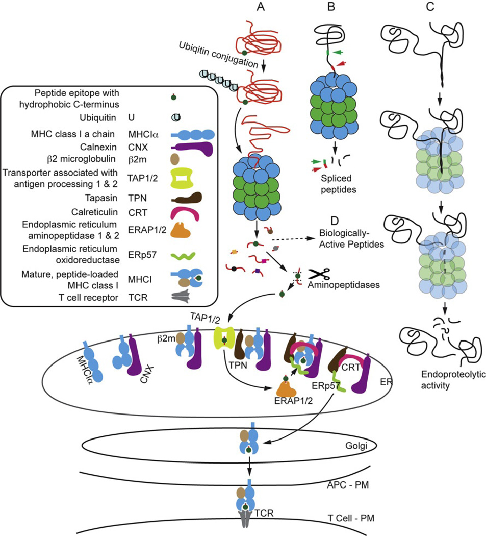
Immunoproteasomes contain replacements for the three catalytic subunits of standard proteasomes. In most cells, oxidative stress and proinflammatory cytokines are stimuli that lead to elevated production of immunoproteasomes. Immune system cells, especially antigen-presenting cells, express a higher basal level of immunoproteasomes. A well-described function of immunoproteasomes is to generate peptides with a hydrophobic C terminus that can be processed to fit in the groove of MHC class I molecules. This display of peptides on the cell surface allows surveillance by CD8 T cells of the adaptive immune system for pathogen-infected cells. Functions of immunoproteasomes, other than generating peptides for antigen presentation, are emerging from studies in immunoproteasome-deficient mice, and are complemented by recently described diseases linked to mutations or single-nucleotide polymorphisms in immunoproteasome subunits. Thus, this growing body of literature suggests a more pleiotropic role in cell function for the immunoproteasome.


Figures

Fig. 1
Fig. 1
Sequence homology for proteasome catalytic subunits. Percentage of the amino acid sequence that is identical (boxed in gray) or similar (boxed in white) for proteasome catalytic subunits. (A) Comparison of the primary sequence for catalytic subunits from mouse is shown. (B) Comparison of the primary sequence for each catalytic subunit comparing mouse versus human is shown. Percentages were obtained using the ClustalW algorithm from the UniProt Consortium.
Fig. 2
Fig. 2
Sequence alignment of standard and immunoproteasome subunits. (A) Sequence from humans includes the last three residues of the propeptide, followed by residues 1–57 of the mature protein. The dash represents the site of autocatalytic cleavage, which is performed by the active-site Thr1. The conserved Gly adjacent to Thr1 is essential for efficient autocatalytic processing of the propeptide. Conserved residues essential for catalytic activity in the mature protein include Thr1, Asp17, and Lys33 (dark gray, white letters). The S1 binding pocket that imparts specificity of cleavage includes residues 20, 31, 35, 45, 49, and 53 (light gray). Note that in LMP 7, the murine sequence substitutes a Met in place of Val31 in humans. (B) Sequence of the β2 and MECL subunits highlighting four amino acids that contribute to the S1 binding pocket of the adjacent β1 or LMP 2 subunit. Residues 114, 116, 118, and 120 are highlighted in gray. Stars indicate nonconser-vative substitutions between the β2 and MECL subunits. The sequence of the β5 and LMP 7 subunits highlights two amino acid substitutions at positions 115 and 116, which are critical for substrate binding.
Fig. 3
Fig. 3
I-proteasome-dependent pathways of protein degradation. I-proteasome-dependent proteolysis in an antigen-presenting cell (APC) generates peptides and polypeptides for MHC class I-restricted antigen presentation, and for functions other than antigen presentation. (A) The antigen-processing pathway for degradation of a cytosolic protein leading to loading of peptides into MHC class I and antigen presentation to the T cell receptor (TCR). (B) Degradation of a protein by i-proteasome resulting in the generation of a spliced peptide candidate for antigen presentation. (C) I-proteasome-mediated endoproteolytic activity generating large, potentially biologically active polypeptides from a precursor protein. Small peptides may also be produced and would be available for antigen presentation. (D) Some of the small peptides produced by i-proteasome activity may have biological activity. PM, plasma membrane; ER, endoplasmic reticulum.
Fig. 4
Fig. 4
Proteasome-dependent activation of NF-κB. Receptor-mediated pathways of NF-κB activation include the classical pathway, which involves the binding of a ligand (i.e., cytokines, virus, or bacteria) to a receptor (i.e., IFN-γ or toll-like receptors), and subsequent activation of intracellular kinases that phosphorylate the inhibitory protein, IκB. Phosphorylation is the signal for targeted ubiquitination of IκB, followed by degradation by the proteasome. The release of the inhibitory protein from the transcription factor dimer exposes the nuclear localization signal, which facilitates the movement of the NF-κB dimer into the nucleus. The alternative pathway uses signals from ligand-bound TNF receptors, which trigger the phosphorylation of p100 or p105 by intracellular kinases and the subsequent endoproteolytic degradation of p100 or p105 to form the active transcription factor p52 or p50. Translocation and localization of heterodimers (i.e., p50/p65 or p52/ relB) to the NF-κB binding sites initiates transcription of the NF-κB-responsive gene. In contrast, binding of the p50 or p52 homodimers inhibits transcription of NF-κB responsive genes.

References

    1. Ciehanover A, Hod Y, Hershko A. A heat-stable polypeptide component of an ATP-dependent proteolytic system from reticulocytes. Biochem Biophys Res Commun. 1978;81:1100–1105. - PubMed
    1. Etlinger JD, Goldberg AL. A soluble ATP-dependent proteolytic system responsible for the degradation of abnormal proteins in reticulocytes. Proc Natl Acad Sci USA. 1977;74:54–58. - PMC - PubMed
    1. Brown MG, Driscoll J, Monaco JJ. Structural and serological similarity of MHC-linked LMP and proteasome (multicatalytic proteinase) complexes. Nature. 1991;353:355–357. - PubMed
    1. Glynne R, Powis SH, Beck S, Kelly A, Kerr LA, Trowsdale J. A proteasome-related gene between the two ABC transporter loci in the class II region of the human MHC. Nature. 1991;353:357–360. - PubMed
    1. Kelly A, Powis SH, Glynne R, Radley E, Beck S, Trowsdale J. Second proteasome-related gene in the human MHC class II region. Nature. 1991;353:667–668. - PubMed

MeSH terms

Substances

LinkOut - more resources