. 2012 Aug 9;45(12):2157-63.
doi: 10.1016/j.jbiomech.2012.05.037. Epub 2012 Jun 21.

Three-dimensional modular control of human walking

Affiliations

Three-dimensional modular control of human walking

Jessica L Allen et al. J Biomech. .

Abstract

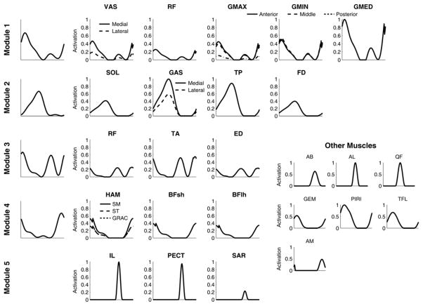
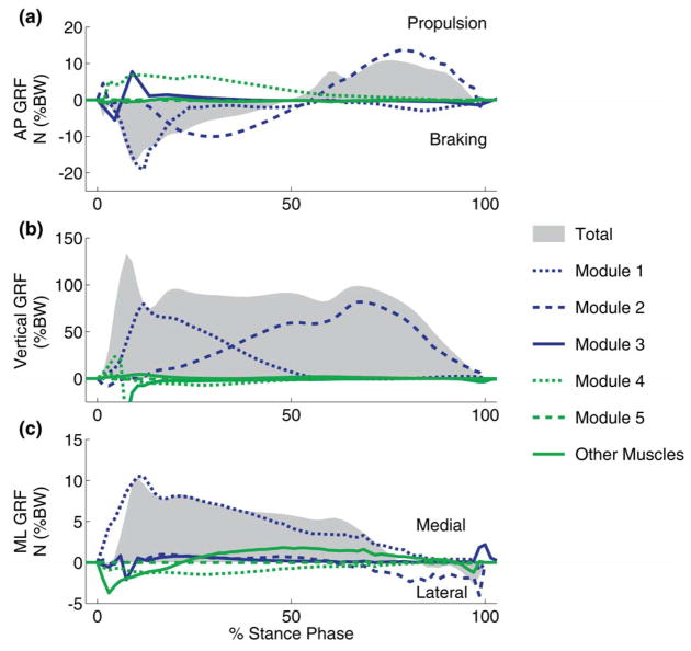
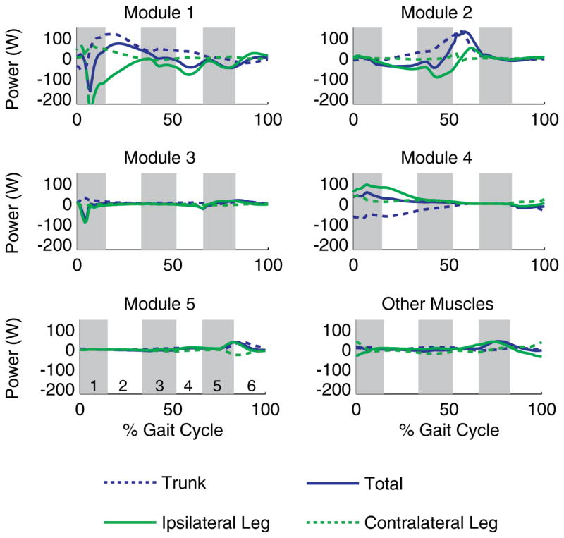
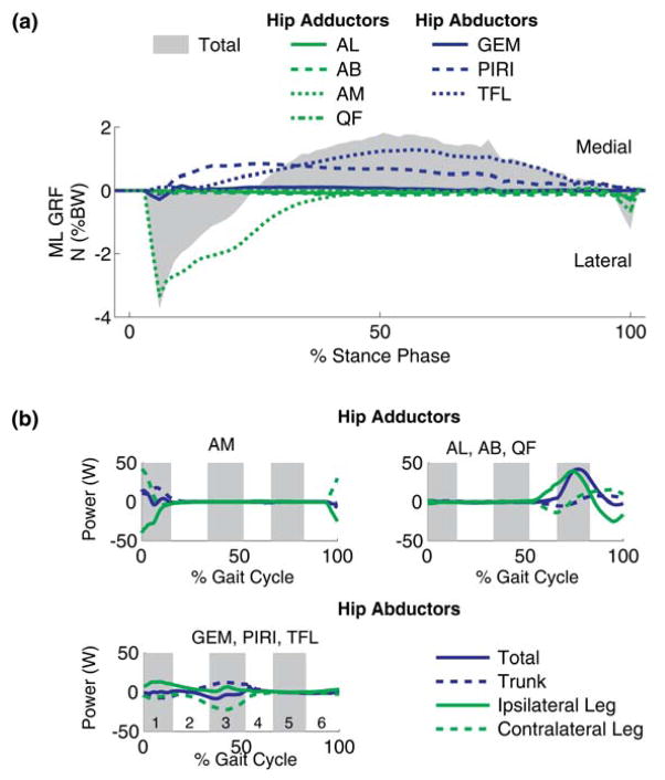
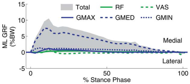
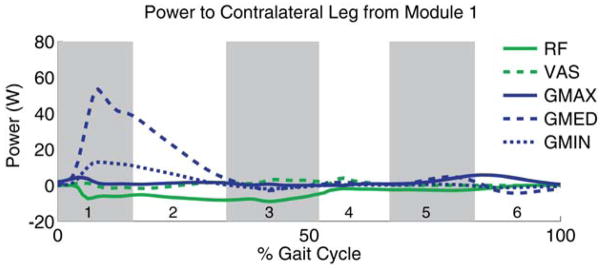
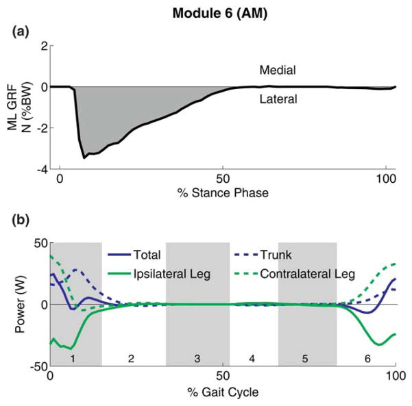
Recent studies have suggested that complex muscle activity during walking may be controlled using a reduced neural control strategy organized around the co-excitation of multiple muscles, or modules. Previous computer simulation studies have shown that five modules satisfy the sagittal-plane biomechanical sub-tasks of 2D walking. The present study shows that a sixth module, which contributes primarily to mediolateral balance control and contralateral leg swing, is needed to satisfy the additional non-sagittal plane demands of 3D walking. Body support was provided by Module 1 (hip and knee extensors, hip abductors) in early stance and Module 2 (plantarflexors) in late stance. In early stance, forward propulsion was provided by Module 4 (hamstrings), but net braking occurred due to Modules 1 and 2. Forward propulsion was provided by Module 2 in late stance. Module 1 accelerated the body medially throughout stance, dominating the lateral acceleration in early stance provided by Modules 4 and 6 (adductor magnus) and in late stance by Module 2, except near toe-off. Modules 3 (ankle dorsiflexors, rectus femoris) and 5 (hip flexors and adductors except adductor magnus) accelerated the ipsilateral leg forward in early swing whereas Module 4 decelerated the ipsilateral leg prior to heel-strike. Finally, Modules 1, 4 and 6 accelerated the contralateral leg forward prior to and during contralateral swing. Since the modules were based on experimentally measured muscle activity, these results provide further evidence that a simple neural control strategy involving muscle activation modules organized around task-specific biomechanical functions may be used to control complex human movements.


Conflict of interest statement

Conflict of interest statement

There is no conflict of interest regarding the publication of this manuscript.

Figures

Figure 1
Figure 1
Experimentally derived module patterns (left column, Modules 1–4) and the corresponding muscles excited by each module (rows). Module 1 included VAS (3-component vastus), RF (rectus femoris), GMAX (3-component gluteus maximus), GMIN (3-component gluteus minimus), and GMED (3 –component gluteus medius). Module 2 included SOL (soleus), GAS (medial and lateral gastrocnemius), TP (tibialis posterior), and FD (flexor digitorum longus). Module 3 included RF, TA (tibialis anterior), and ED (extensor digitorum longus). Module 4 included HAM (medial hamstrings = semimembranosus (SM) and semitendinosus (ST), gracilis (GRAC)), lateral hamstrings = biceps femoris long head (BFlh), and BFsh (biceps femoris short head)). Finally, Module 5 (Neptune et al. 2009) included IL (iliacus, psoas), PECT (pectinius) and SAR (sartorius). No experimental data were available for Module 5, thus a bimodal pattern was used. All muscles within a module received the same excitation timing and pattern, although the magnitude was allowed to vary. AM (3-component adductor magnus), AL (adductor longus), AB (adductor brevis), QF (quadratus femoris), GEM (gemellus), PIRI (piriformus) and TFL (tensor fascia lata), for which no experimental data were available, were not included in Modules 1–5 at first to allow them to have distinct anatomical and modular function. Thus, individual bimodal patterns were also used for these muscles.
Figure 2
Figure 2
Tracking results for the simulation controlled by five modules (plus excitation of remaining muscles). The simulated joint angles and ground reaction forces (blue lines) agree well with the experimental data (grey bars). The grey bars represent experimental means ± 2 SD.
Figure 3
Figure 3
Module contributions to the (a) anterior-posterior (AP), (b) vertical and (c) mediolateral (ML) ground reaction forces. Total is the sum of all muscles.
Figure 4
Figure 4
Mechanical power delivered to the trunk, ipsilateral and contralateral leg by each module. Total represents the sum of the mechanical power delivered to all segments. Positive and negative power values indicate a module acts to accelerate or decelerate the segments, respectively. The alternating shaded regions represent different phases of the gait cycle: 1 –1st double support/contralateral pre-swing, 2 – first half of ipsilateral single support/contralateral swing, 3 – second half of ipsilateral single support/contralateral swing, 4 – ipsilateral pre-swing/contralateral 1st double support, 5 – first half of ipsilateral swing/contralateral single support, and 6 – second half of ipsilateral swing/contralateral single support.
Figure 5
Figure 5
Contributions from individual muscles not controlled by a module to (a) ML GRF and (b) power transfer among segments. The alternating shaded regions represent different phases of the gait cycle (see Fig. 3 caption).
Figure 6
Figure 6
ML GRF contributions from individual muscles in Module 1.
Figure 7
Figure 7
Power delivered to the contralateral leg by Modules 1. The power transferred to the contralateral leg by Module 1 is primarily due to GMED and GMIN. The alternating shaded regions represent different phases of the gait cycle (see Fig. 3 caption).
Figure 8
Figure 8
Module 6 (AM) (a) generated a medial GRF during the beginning of stance and (b) decelerated the ipsilateral leg (negative power) during late swing and early stance while generating energy to the contralateral leg and trunk (positive power). The alternating shaded regions represent different phases of the gait cycle (see Fig. 3 caption).

Similar articles

Cited by

References

    1. Anderson FC. Doctoral Dissertation. The University of Texas; Austin, Austin, TX: 1999. A dynamic optimization solution for a complete cycle of normal gait.
    1. Arnold AS, Thelen DG, Schwartz MH, Anderson FC, Delp SL. Muscular coordination of knee motion during the terminal-swing phase of normal gait. J Biomech. 2007;40 (15):3314–24. - PMC - PubMed
    1. Cappellini G, Ivanenko YP, Poppele RE, Lacquaniti F. Motor Patterns in Human Walking and Running. J Neurophysiol. 2006;95 (6):3426–3437. - PubMed
    1. Clark DJ, Ting LH, Zajac FE, Neptune RR, Kautz SA. Merging of Healthy Motor Modules Predicts Reduced Locomotor Performance and Muscle Coordination Complexity Post-Stroke. J Neurophysiol. 2010;103 (2):844–857. - PMC - PubMed
    1. d’Avella A, Saltiel P, Bizzi E. Combinations of muscle synergies in the construction of a natural motor behavior. Nat Neurosci. 2003;6 (3):300–8. - PubMed

Publication types