. 2012 Aug 10;47(3):422-33.
doi: 10.1016/j.molcel.2012.05.014. Epub 2012 Jun 21.

The Akt-SRPK-SR axis constitutes a major pathway in transducing EGF signaling to regulate alternative splicing in the nucleus

Affiliations

The Akt-SRPK-SR axis constitutes a major pathway in transducing EGF signaling to regulate alternative splicing in the nucleus

Zhihong Zhou et al. Mol Cell. .

Erratum in

Abstract

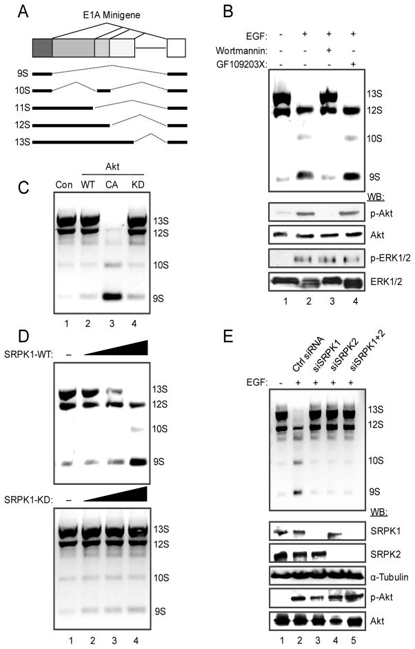
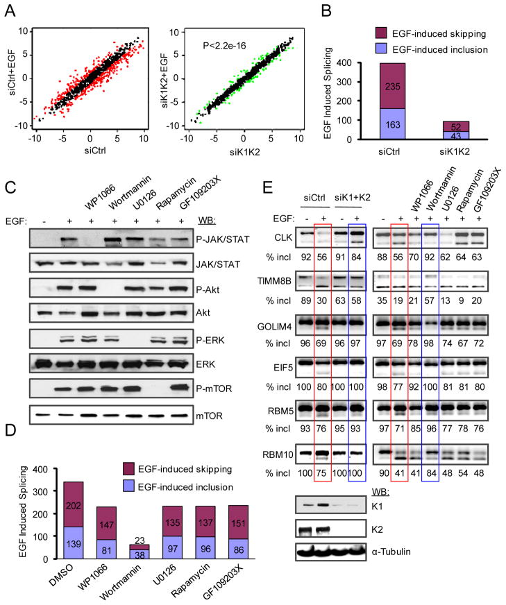
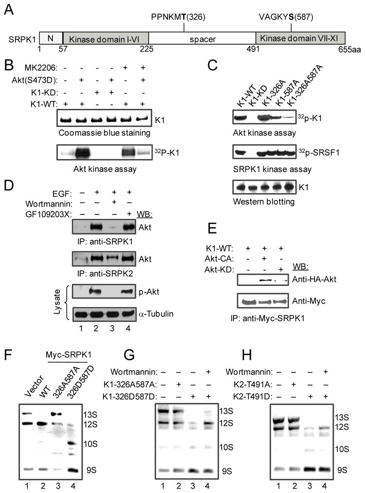
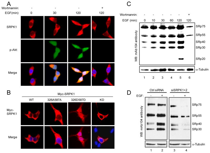
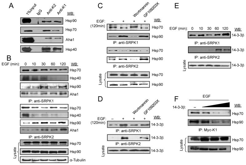
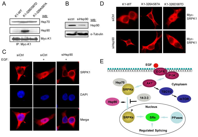
Pre-mRNA splicing is regulated by developmental and environmental cues, but little is known about how specific signals are transduced in mammalian cells to regulate this critical gene expression step. Here, we report massive reprogramming of alternative splicing in response to EGF signaling. By blocking individual branches in EGF signaling, we found that Akt activation plays a major role, while other branches, such as the JAK/STAT and ERK pathways, make minor contributions to EGF-induced splicing. Activated Akt next branches to SR protein-specific kinases, rather than mTOR, by inducing SRPK autophosphorylation that switches the splicing kinases from Hsp70- to Hsp90-containing complexes. This leads to enhanced SRPK nuclear translocation and SR protein phosphorylation. These findings reveal a major signal transduction pathway for regulated splicing and place SRPKs in a central position in the pathway, consistent with their reputed roles in a large number of human cancers.


Figures

Figure 1
Figure 1
EGF regulates E1A splicing via activated Akt and SRPKs. (A) Structure of the E1A minigene. The common three isoforms (13S, 12S, and 9S) are products of three alternative 5′ splice sites in competition for the same downstream 3′ splice site. Additional splicing events produce the 11S and 10S isoforms, which are much less abundant compared to the three common isoforms. (B) E1A splicing in response to EGF treatment. EGF signaling induced the switch from the proximal 5′ splice sites (for 13S and to some extent 12S) to the distal 5′ splice site (for 9S). The switch could be blocked by the PI3K inhibitor Wortmannin, but not by the PKC inhibitor GF109203X. The effects of EGF and Wortmannin on Akt activation were determined by Western blotting on the lower panels. The ERK/MAPK pathway was unaffected in HEK293T cells by either inhibitor. (C) Induced E1A splicing switch by a constitutively activated (CA) Akt, but not by a kinase dead (KD) Akt. (D) Overexpression of SRPK1, but not the kinase dead mutant, induced a similar switch in E1A splicing to favor the production of the 9S isoform. (E) Requirement for SRPKs in EGF-induced splicing. The effect of EGF on E1A splicing was blocked by siRNA-mediated knockdown of either SRPK1 or SRPK2 or both (upper panel). The knockdown was efficient and specific as determined by Western blotting (lower panel). Control siRNA had no effect and Akt was still active after SRPK knockdown in EGF-treated cells.
Figure 2
Figure 2
SRPKs are the major branch in the EGF pathway for global regulation of alternative splicing. (A) EGF induced widespread splicing changes from a global scale, which was largely diminished in SRPK1/2 knockdown cells (p<2.2e-16 according to KS-test). Red dots in the left panel represent splicing ratio changes ≥ 2. Although some changes were detectable in response to double knockdown of SRPK1 and SRPK2 (green dots in the right panel), the magnitude of ratio changes in those cases was much lower. (B) EGF-induced alterative splicing before and after knocking down SRPK1/2. (C) EGF activated multiple signaling branches, including the JAK/STAT, PI3K/Akt, ERK/MAPK and mTOR pathways, each of which could be blocked by a specific inhibitor. (D) Wortmannin effectively blocked EGF-induced splicing, while inhibition of all other pathways had much less effects (see the degree of individual responses in Table S1). (E) A selective panel of splicing events induced by EGF was examined by RT-PCR under different treatment conditions. SRPK knockdown and Wortmannin treatment showed a similar effect in each case, while other inhibitors had minor, if any, effects. The efficiency of SRPK knockdown was shown at bottom.
Figure 3
Figure 3
Activated Akt triggers SRPK1 autophosphorylation. (A) Schematic presentation of SRPK1 domains with two candidate Akt-mediated phosphorylation sites highlighted in bold, which were deduced from in vivo and in vitro phosphorylation experiments analyzed by mass spectrometry (Fig. S3A). (B) In vitro kinase assay. Purified activated Akt phosphorylated WT SRPK1, which could be blocked by Akt inhibitor MK2206 (1 μM). Activated Akt was unable to phosphorylate the kinase dead SRPK1 mutant while MK2206 alone could induce SRPK1 autophosphorylation. (C) Akt-mediated phosphorylation of SRPK1 at T326 and S587. Double Alanine mutations on these two sites abolished Akt-mediated phosphorylation, but had no effect on the kinase activity of SRPK1 on the SR protein SRSF1. (D) Interaction of both SRPK1 and SRPK2 with Akt only after Akt activation by EGF. (E) Co-IP experiments further showed the interaction of SRPK1 only with activated Akt. (F) Akt-mediated phosphorylation in SRPK1 is necessary and sufficient to induce splicing switch on the E1A reporter. The 326A587A mutant appears to act in a dominant negative fashion whereas the phospho-mimicking 326D587D mutant was more potent than WT SRPK1 in inducing the splicing switch. (G and H) The phospho-mimicking SRPK mutants bypassed the requirement for Akt signaling in regulated splicing. In both cases, the phospho-mimicking mutants (K1-326D587D or K2-T491D) could induce E1A splicing in the presence of Wortmannin.
Figure 4
Figure 4
Induction of SRPK nuclear translocation and dynamic regulation of SR protein phosphorylation in response to EGF signaling. (A) EGF induced SRPK1 translocation to the nucleus and Wortmannin blocked the translocation. (B) Phospho-mimicking SRPK1 entered the nucleus in the absence of EGF signaling. (C) EGF induced SR protein phosphorylation whereas blockage of EGF signaling by Wortmannin triggered extensive dephosphorylation of SR proteins. (D) SRPKs are responsible for EGF-induced SR protein phosphorylation. In the absence of EGF treatment, SRPK knockdown had a detectable but modest effect on the steady state of SR protein phosphorylation (compare lane 1 with lane 3). EGF induced dramatic reduction of SR protein phosphorylation in SRPK knockdown cells (compare lane 2 with lane 4). These data suggest increased competition by SR protein phosphatases when SR protein kinases were reduced by RNAi in EGF-treated cells.
Figure 5
Figure 5
EGF signaling modulates the interaction of SRPKs with heat shock and 14-3-3 proteins. (A) Co-immunoprecipitation of SRPK1 and SRPK2 with Hsp70 and Hsp90 along with their co-chaperones in the presence of serum. (B) EGF-induced release of Hsp70 and its co-chaperone Hsp40 from SRPKs while enhanced the association of the splicing kinases with Hsp90 and its co-chaperone Aha1. (C) The differential interaction of SRPKs with heat shock proteins required the activation of the PI3K pathway. (D-F) EGF induced the interaction of SRPKs with 14-3-3β. Both SRPKs showed similar association in proliferating HEK293T cells (D). Interestingly, SRPK2 seems to be more inducible than SRPK1 in association with 14-3-3β in response to EGF signaling (E). Increased 14-3-3β was able to progressively suppress the interaction of SRPKs with both Hsp70 and Hsp90 in EGF-treated cells (F).
Figure 6
Figure 6
A critical role of Hsp90 in facilitating SRPK nuclear translocation in response to EGF signaling. (A) The phospho-mimicking mutant K1-326D587D gained elevated association with Hsp90 compared to WT SRPK1 and the double Alanine mutant K1-326A587A. (B) RNAi-mediated knockdown of Hsp90. (C) Hsp90 knockdown blocked EGF-induced SRPK1 nuclear translocation. (D) The phospho-mimicking SRPK1 mutant entered the nucleus in the absence of EGF signaling (upper panel), but in an Hsp90-dependent fashion (lower panel). (E) Summary of EGF signaling through the Akt-SRPK-SR axis to regulate alternative splicing in the nucleus. Hsp70 is responsible for inhibiting nuclear import of the splicing kinases, Hsp90 knocks off Hsp70 to facilitate nuclear import, and 14-3-3 functions to prevent excessive accumulation of the splicing kinases in the nucleus.

References

    1. Amin EM, Oltean S, Hua J, Gammons MV, Hamdollah-Zadeh M, Welsh GI, Cheung MK, Ni L, Kase S, Rennel ES, Symonds KE, Nowak DG, Royer-Pokora B, Saleem MA, Hagiwara M, Schumacher VA, Harper SJ, Hinton DR, Bates DO, Ladomery MR. WT1 mutants reveal SRPK1 to be a downstream angiogenesis target by altering VEGF splicing. Cancer Cell. 2011;20:768–780. - PMC - PubMed
    1. Aubol BE, Chakrabarti S, Ngo J, Shaffer J, Nolen B, Fu XD, Ghosh G, Adams JA. Processive phosphorylation of alternative splicing factor/splicing factor 2. Proc Natl Acad Sci USA. 2003;100:12601–12606. - PMC - PubMed
    1. Blaustein M, Pelisch F, Tanos T, Muñoz MJ, Wengier D, Quadrana L, Sanford JR, Muschietti JP, Kornblihtt AR, Cáceres JF, Coso OA, Srebrow A. Concerted regulation of nuclear and cytoplasmic activities of SR proteins by Akt. Nat Struct Mol Biol. 2005;12:1037–1044. - PubMed
    1. Carracedo A, Pandolfi PP. The PTEN-PI3K pathway: of feedbacks and cross-talks. Oncogene. 2008;27:5527–5541. - PubMed
    1. Cho S, Hoang A, Sinha R, Zhong XY, Fu XD, Krainer AR, Ghosh G. Interaction between the RNA binding domains of Ser-Arg splicing factor 1 and U1-70K snRNP protein determines early spliceosome assembley. Proc Natl Acad Sci USA. 2011;108:8233–8238. - PMC - PubMed

Publication types

MeSH terms

Substances

Associated data