. 2013 Jan;34(1):200-10.
doi: 10.1016/j.neurobiolaging.2012.05.014. Epub 2012 Jun 22.

Morphological and molecular changes in aging rat prelimbic prefrontal cortical synapses

Affiliations

Morphological and molecular changes in aging rat prelimbic prefrontal cortical synapses

Erik B Bloss et al. Neurobiol Aging. 2013 Jan.

Abstract

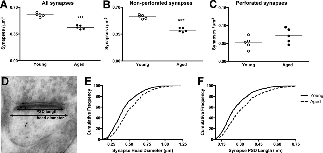
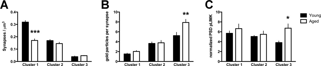
Age-related impairments of executive functions appear to be related to reductions of the number and plasticity of dendritic spine synapses in the prefrontal cortex (PFC). Experimental evidence suggests that synaptic plasticity is mediated by the spine actin cytoskeleton, and a major pathway regulating actin-based plasticity is controlled by phosphorylated LIM kinase (pLIMK). We asked whether aging resulted in altered synaptic density, morphology, and pLIMK expression in the rat prelimbic region of the PFC. Using unbiased electron microscopy, we found an approximate 50% decrease in the density of small synapses with aging, while the density of large synapses remained unchanged. Postembedding immunogold revealed that pLIMK localized predominantly to the postsynaptic density where it was increased in aging synapses by approximately 50%. Furthermore, the age-related increase in pLIMK occurred selectively within the largest subset of prelimbic PFC synapses. Because pLIMK is known to inhibit actin filament plasticity, these data support the hypothesis that age-related increases in pLIMK may explain the stability of large synapses at the expense of their plasticity.


Figures

Figure 1
Figure 1. EM sampling, disector analysis, and pLIMK immunogold
A EM blocks encompassed rat PL PFC (black box), and serial sections were collected from layer I (white dashed lines, approximately 100 µm from layer II). B Example of axospinous synapses identified by the disector analysis (Note: black dots denote synapses contained in both planes; asterisks denote synapses contained only in 1 plane). C Representative serial images of a PL layer I synapse with pLIMK immunogold labeling. Note the prominent localization of pLIMK to the PSD (arrowheads). “ax” denotes an axonal bouton, “sp” denotes a dendritic spine. Scale bars: B 500 nm; C 200 nm.
Figure 2
Figure 2. Age-related alterations of synapse density and morphology in PFC
A Axospinous synapse density decreased with age by approximately 30%. B,C Similar changes were seen when the analysis was restricted to non-perforated synapses, while the density of perforated synapses was unchanged between the ages. D–F Measurements of synaptic head diameter and PSD length revealed age-related shifts towards larger synapses. ***, p < 0.0001. Graphs: circles, individual animals; line, group mean.
Figure 3
Figure 3. pLIMK is increased in the PSD of aging non-perforated synapses
A pLIMK gold particles were assigned to synaptic bins; see Materials and Methods for specific bin details. B Across all synapses, the number of pLIMK gold particles was higher in the PSD bin of aged compared to young rats. C,D Similar age effects on PSD pLIMK were seen in non-perforated synapses, but not in perforated synapses. ***, p < 0.001. Graphs: circles, individual animals; line, group mean.
Figure 4
Figure 4. Higher number and density of pLIMK in the PSD of a subset of non-perforated synapses in the aged PFC
A Cluster analysis was used to mathematically define classes of synapses based on PSD size; aging resulted in a ~50% decrease in synapse density in cluster 1 (smallest synapses). See Materials and Methods and Table 1 for details of the PSD cluster analysis. B,C The number (B) and density (C) of pLIMK immunogold particles within the PSD bin are plotted by age and synapse size (cluster). Exclusively in cluster 3 (largest) synapses, a significant age effect was observed in the number (B) and density (C) of PSD pLIMK. *, p < 0.05; **, p < 0.01; ***, p < 0.001. A significant cluster effect on normalized PSD pLIMK was present in the young group such that pLIMK density was significantly lower in the cluster 3 (largest) synapses compared to cluster 1 (smallest) synapses; no such cluster effect was present in aged animals (data not shown); see Results for details. Graphs represent mean ± SEM.

References

    1. Adams MM, Shah RA, Janssen WG, Morrison JH. Different modes of hippocampal plasticity in response to estrogen in young and aged female rats. Proc Natl Acad Sci U S A. 2001;98:8071–8076. - PMC - PubMed
    1. Altun M, Bergman E, Edstrom E, Johnson H, Ulfhake B. Behavioral impairments of the aging rat. Physiol Behav. 2007;92:911–923. - PubMed
    1. Arber S, Barbayannis FA, Hanser H, Schneider C, Stanyon CA, Bernard O, Caroni P. Regulation of actin dynamics through phosphorylation of cofilin by LIM-kinase. Nature. 1998;393:805–809. - PubMed
    1. Arnsten AF, Paspalas CD, Gamo NJ, Yang Y, Wang M. Dynamic Network Connectivity: A new form of neuroplasticity. Trends Cogn Sci. 2010;14:365–375. - PMC - PubMed
    1. Barense MD, Fox MT, Baxter MG. Aged rats are impaired on an attentional set-shifting task sensitive to medial frontal cortex damage in young rats. Learn Mem. 2002;9:191–201. - PMC - PubMed

Publication types