. 2012 Aug;181(2):452-62.
doi: 10.1016/j.ajpath.2012.04.010. Epub 2012 Jun 22.

NK cells modulate the inflammatory response to corneal epithelial abrasion and thereby support wound healing

Affiliations

NK cells modulate the inflammatory response to corneal epithelial abrasion and thereby support wound healing

Qiong Liu et al. Am J Pathol. 2012 Aug.

Abstract

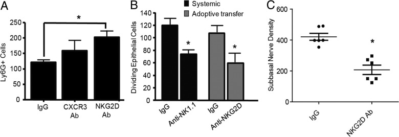
Natural killer (NK) cells are lymphocytes of the innate immune system that have crucial cytotoxic and regulatory roles in adaptive immunity and inflammation. Herein, we consider a role for these cells in corneal wound healing. After a 2-mm central epithelial abrasion of the mouse cornea, a subset of classic NK cells migrated into the limbus and corneal stroma, peaking at 24 hours with an eightfold increase over baseline. Depletion of γδ T cells significantly reduced NK cell accumulation (>70%; P < 0.01); however, in neutrophil-depleted animals, NK cell influx was normal. Isolated spleen NK cells migrated to the wounded cornea, and this migration was reduced by greater than 60% (P < 0.01) by ex vivo antibody blocking of NK cell CXCR3 or CCR2. Antibody-induced depletion of NK cells significantly altered the inflammatory reaction to corneal wounding, as evidenced by a 114% increase (P < 0.01) in neutrophil influx at a time when acute inflammation is normally waning. Functional blocking of NKG2D, an activating receptor for NK cell cytotoxicity and cytokine secretion, did not inhibit NK cell immigration, but significantly increased neutrophil influx. Consistent with excessive neutrophil accumulation, NK depletion and blocking of NKG2D also inhibited corneal nerve regeneration and epithelial healing (P < 0.01). Findings of this study suggest that NK cells are actively involved in corneal healing by limiting the innate acute inflammatory reaction to corneal wounding.


Figures

Figure 1
Figure 1
NKp46+ cells in the mouse cornea. A: At 24 hours after central corneal epithelial abrasion, corneal whole mounts were prepared for microscopic analysis using a DeltaVision microscope (Applied Precision). A montage of images (30-μm deep Z-stack, 0.3-μm increments) showing limbal vessels stained with anti-CD31-APC and cells positive for anti-NKp46-FITC. The NKp46+ cells were negative for anti-CD3-PE (B), positive for anti-EOMES-PE (C), and negative for anti-IL-22-PE (D, arrow). The NKp46+ cells were negative for anti-RORγt-APC (E, arrows), although cells stained with GL3 (D and E) (ie, γδ T cells) in the same tissue were positive for IL-22, RORγt, and CD94 (F). In normal corneas, the NKp46+ cells were occasionally positive for anti-CD94.
Figure 2
Figure 2
Factors that affect NK cell accumulation in corneas after central corneal epithelial abrasion. A: Time course of NKp46+ cell accumulation, plotting total cells counted in the stroma in nine 150 × 150-μm microscopic fields across the cornea. B: Distribution of NKp46+ cells in the corneal stroma at 24 hours after injury. C, center; L, limbus; PC, paracenter; PL, paralimbus; WM, wound margin. C: TCRδ−/− mice had significantly fewer NKp46+ cells at 24 hours after injury than did control wild-type mice. Treatment of wild-type mice with antibody GL3 before epithelial abrasion also inhibited accumulation of these cells. Treatment of wild-type mice with GL3 at 14 hours after injury was less inhibitory. D: TCRδ−/− and Icam-1−/− mice failed to accumulate NKp46+ cells within the time frame of influx of these cells in wild-type mice. E: NKp46+ cell influx was counted at 24 hours after epithelial abrasion. Depletion of neutrophils with anti-Ly6G failed to prevent NKp46+ cell influx, and systemic treatment of TCRδ−/− mice with rIL-17A failed to increase NKp46+ cells, although this cytokine has previously been reported to significantly increase neutrophils. NKp46+ cell influx was significantly reduced in mice deficient in CD11b (Mac-1−/−), CD11a (CD11a−/−), and ICAM-1 (ICAM-1−/−). F: Anti-NK1.1 was used to treat mice before epithelial abrasion. At 24 hours after abrasion, corneas were labeled with antibody GL3 to detect γδ T cells. GL3+ cells were not statistically different from those in control IgG-treated mice. G: The number of adoptively transferred NKp46+PKH67+ cells from LFA-1−/− (Cd11a−/−) mice that migrated into the corneal stroma at 30 hours after epithelial abrasion was significantly decreased when compared with adoptively transferred wild-type NK cells. H: Distribution in the stroma at 30 hours after epithelial abrasion from limbus to the corneal center in wild-type mice of adoptively transferred NKp46+PKH67+ spleen NK cells from wild-type and Cd11a−/−mice. *P < 0.01 versus wild- type control mice; P < 0.01 versus LFA-1−/− mice; n = 4 to 6 per condition; mean ± SD.
Figure 3
Figure 3
Migration of NK cells into corneal stroma was affected by chemokines. A: Protein antibody array, culture supernatant from six pooled corneas collected 18 hours after central epithelial abrasion and cultured with serum-free medium for 12 hours. B: Topical administration of anti-IP10 every 4 hours after corneal abrasion. NKp46+ cells, GL3+ cells (γδ T cells), and Ly6G+ cells (neutrophils) were counted in the corneal limbus at 24 hours after corneal injury. C: ELISA results showing IP-10 (CXCL10, pg/mL) and MCP-3 (CCL7, pg/mL) in lysates of pooled corneas at 18 hours with and without (Control) central epithelial abrasion (mean ± SD; n = 3; *P < 0.01). ELISA results showing MCP-1 (CCL2, ng/6 corneas) from corneal lysates at different times with and without central epithelial abrasion. D: Adoptive transfer of spleen NK cells with ex vivo blocking using anti-CXCR3, anti-CCR2, or anti-NKG2D. NKp46+ cell counts in the cornea at 30 hours after corneal injury (mean ± SD; *P < 0.01 versus wild-type IgG-treated control mice, n = 4 to 6 per condition). Distribution of NKp46+ cells within the corneal stroma is plotted in the accompanying graphs (mean ± SD; *P < 0.01 versus cells treated ex vivo with anti-NKG2D; n = 4 to 6 per condition).
Figure 4
Figure 4
Effects of NK cell depletion. A: Neutrophil counts in the region of the original wound margin at 24 or 30 hours after central corneal epithelial abrasion in mice either depleted of NK cells via systemic injection of anti-NK1.1 before injury or in mice with engineered deletion of CD11b (mean ± SD; *P < 0.01 versus 30 hours in control mice). B: Epithelial wound closure over time after NK depletion (mean ± SD; P < 0.01 for the 18- and 24-hour measurements, n = 4). C: Epithelial cell division counted in the basal layer of the epithelium at 24 hours after abrasion, comparing IgG-treated control mice with those treated with anti-NK1.1 at 24 hours before injury to deplete NK cells. Inset: Representative image of the ocular surface at 24 hours after abrasion in an NK cell-depleted mouse showing fluorescein staining of the open epithelial wound. D: Corneal subbasal nerve density at 96 hours after epithelial abrasion comparing IgG-treated control mice with those depleted of NK cells with anti-NK1.1 (mean ± SD; *P < 0.versus IgG-treated control mice; n = 4 to 6 per condition). Inset shows typical subbasal nerve density in the IgG-treated control mice.
Figure 5
Figure 5
Effects of adoptive transfer spleen NK cells after blocking NKG2D. A: Neutrophil counts in the region of the original wound margin at 24 hours after central corneal epithelial abrasion in mice after adoptive transfer of IgG and anti-NKG2D-blocked spleen NK cells. B: Epithelial cell divisions counted in the basal layer of the epithelium at 24 hours after abrasion after adoptive transfer of IgG and anti-NKG2D-blocked spleen NK cells compared with systemic anti-NK1.1 (ie, NK depletion). C: Data from 96 hours subbasal nerve healing after adoptive transfer of IgG and anti-NKG2D-blocked spleen NK cells. Subbasal nerve density in regions of the original wound margin is plotted for each cornea. Data plotted as mean ± SD; *P < 0.01 versus IgG-treated control mice; n = 4 to 6 per condition.

Similar articles

Cited by

References

    1. Toulon A., Breton L., Taylor K.R., Tenenhaus M., Bhavsar D., Lanigan C., Rudolph R., Jameson J., Havran W.L. A role for human skin-resident T cells in wound healing. J Exp Med. 2009;206:743–750. - PMC - PubMed
    1. Havran W.L., Jameson J.M. Epidermal T cells and wound healing. J Immunol. 2010;184:5423–5428. - PMC - PubMed
    1. Yamagami S., Hamrah P., Miyamoto K., Miyazaki D., Dekaris I., Dawson T., Lu B., Gerard C., Dana M.R. CCR5 chemokine receptor mediates recruitment of MHC class II-positive Langerhans cells in the mouse corneal epithelium. Invest Ophthalmol Vis Sci. 2005;46:1201–1207. - PubMed
    1. Hamrah P., Huq S.O., Liu Y., Zhang Q., Dana M.R. Corneal immunity is mediated by heterogeneous population of antigen-presenting cells. J Leukoc Biol. 2003;74:172–178. - PubMed
    1. Knickelbein J.E., Watkins S.C., McMenamin P.G., Hendricks R.L. Stratification of antigen-presenting cells within the normal cornea. Ophthalmol Eye Dis. 2009;1:45–54. - PMC - PubMed

Publication types

MeSH terms