Review
. 2012 Oct;10(10):1066-78.
doi: 10.1016/j.cgh.2012.06.003. Epub 2012 Jun 19.

Diagnosis and management of eosinophilic esophagitis

Affiliations
Review

Diagnosis and management of eosinophilic esophagitis

Evan S Dellon. Clin Gastroenterol Hepatol. 2012 Oct.

Abstract

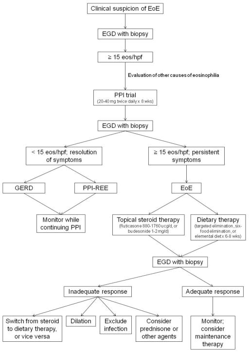
Eosinophilic esophagitis (EoE) is a chronic inflammatory condition characterized by esophageal dysfunction and eosinophilic infiltrate in the esophageal epithelium in the absence of other potential causes of eosinophilia. EoE is increasing in incidence and prevalence, and is a major cause of gastrointestinal morbidity among children and adults. EoE is thought to be immune mediated, with food or environmental antigens stimulating a T-helper (Th)-2 inflammatory response. An increased understanding of the pathogenesis of EoE has led to the evolution of diagnostic and treatment paradigms. We review the latest approach to diagnosis of EoE and present consensus diagnostic guidelines. We also discuss the clinical, endoscopic, and histologic features of EoE and challenges to diagnosis. Finally, we present the 3 major treatment options for EoE: pharmacologic therapy, dietary modification, and endoscopic dilation.


Conflict of interest statement

Disclosures: No conflicts of interested pertaining to this manuscript. Dr. Dellon has received research support from AstraZeneca, Meritage Pharma, Olympus, NIH, ACG, and AGA. Dr. Dellon has been a consultant for Oncoscope.

Figures

Figure 1
Figure 1
Endoscopic findings of EoE include fixed esophageal rings (A), narrow caliber esophagus (B; also note the pale/congested mucosa with decreased vascularity), longitudinal furrows running parallel to the axis of the esophagus (C), white plaques or exudates (D), and crêpe-paper mucosa which tears passage of the endoscope alone (E). Often, a combination of these findings can be seen at the same time (F).
Figure 1
Figure 1
Endoscopic findings of EoE include fixed esophageal rings (A), narrow caliber esophagus (B; also note the pale/congested mucosa with decreased vascularity), longitudinal furrows running parallel to the axis of the esophagus (C), white plaques or exudates (D), and crêpe-paper mucosa which tears passage of the endoscope alone (E). Often, a combination of these findings can be seen at the same time (F).
Figure 2
Figure 2
Histologic findings of EoE include a brisk infiltrate of eosinophils in the esophageal epithelium, eosinophilic microabscesses (clusters of 4 or more eosinophils), spongiosis, and basal zone hyperplasia (A). In addition to an eosinophilic infiltrate, spongiosis, and basal zone hyperplasia, this sample shows eosinophil degranulation with extracellular eosinophil granules dispersed throughout the epithelium (B). If an esophageal biopsy contains tissue deeper than epithelium, fibrosis of the lamina propria can often be seen (C).
Figure 2
Figure 2
Histologic findings of EoE include a brisk infiltrate of eosinophils in the esophageal epithelium, eosinophilic microabscesses (clusters of 4 or more eosinophils), spongiosis, and basal zone hyperplasia (A). In addition to an eosinophilic infiltrate, spongiosis, and basal zone hyperplasia, this sample shows eosinophil degranulation with extracellular eosinophil granules dispersed throughout the epithelium (B). If an esophageal biopsy contains tissue deeper than epithelium, fibrosis of the lamina propria can often be seen (C).
Figure 3
Figure 3
Endoscopic dilation for treatment of EoE. A through-the-scope balloon dilator has been inflated to a diameter of 12mm in a patient with EoE and a narrow caliber esophagus (A). When the balloon is deflated, a longitudinal rent is noted, indicating good dilation effect (B).
Figure 3
Figure 3
Endoscopic dilation for treatment of EoE. A through-the-scope balloon dilator has been inflated to a diameter of 12mm in a patient with EoE and a narrow caliber esophagus (A). When the balloon is deflated, a longitudinal rent is noted, indicating good dilation effect (B).
Figure 4
Figure 4
An approach to diagnosis and management of EoE.

Similar articles

Cited by

References

    1. Liacouras CA, Furuta GT, Hirano I, Atkins D, Attwood SE, Bonis PA, Burks AW, Chehade M, Collins MH, Dellon ES, Dohil R, Falk GW, Gonsalves N, Gupta SK, Katzka DA, Lucendo AJ, Markowitz JE, Noel RJ, Odze RD, Putnam PE, Richter JE, Romero Y, Ruchelli E, Sampson HA, Schoepfer A, Shaheen NJ, Sicherer SH, Spechler S, Spergel JM, Straumann A, Wershil BK, Rothenberg ME, Aceves SS. Eosinophilic esophagitis: Updated consensus recommendations for children and adults. J Allergy Clin Immunol. 2011;128:3–20.e6. - PubMed
    1. Landres RT, Kuster GG, Strum WB. Eosinophilic esophagitis in a patient with vigorous achalasia. Gastroenterology. 1978;74:1298–1301. - PubMed
    1. Picus D, Frank PH. Eosinophilic esophagitis. AJR Am J Roentgenol. 1981;136:1001–3. - PubMed
    1. Lee RG. Marked eosinophilia in esophageal mucosal biopsies. Am J Surg Pathol. 1985;9:475–9. - PubMed
    1. Attwood SE, Smyrk TC, Demeester TR, Jones JB. Esophageal eosinophilia with dysphagia. A distinct clinicopathologic syndrome. Dig Dis Sci. 1993;38:109–16. - PubMed

Publication types

Substances