. 2012 Sep;303(5):L449-59.
doi: 10.1152/ajplung.00301.2011. Epub 2012 Jun 22.

TNFR1-dependent pulmonary apoptosis during ischemic acute kidney injury

Affiliations

TNFR1-dependent pulmonary apoptosis during ischemic acute kidney injury

Laura E White et al. Am J Physiol Lung Cell Mol Physiol. 2012 Sep.

Abstract

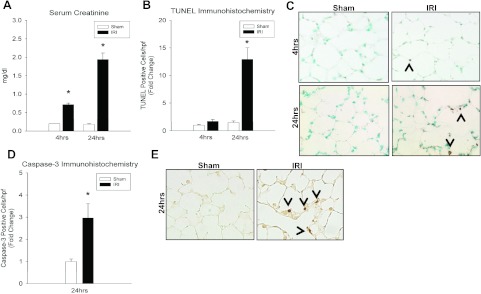
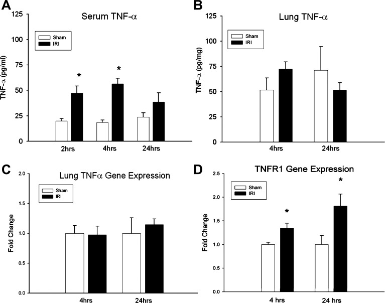
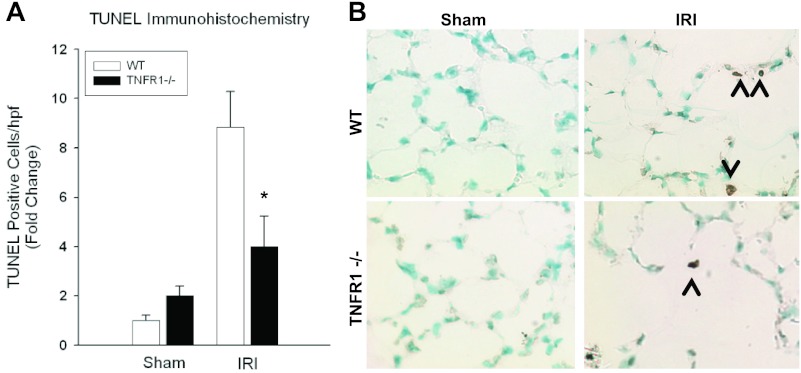
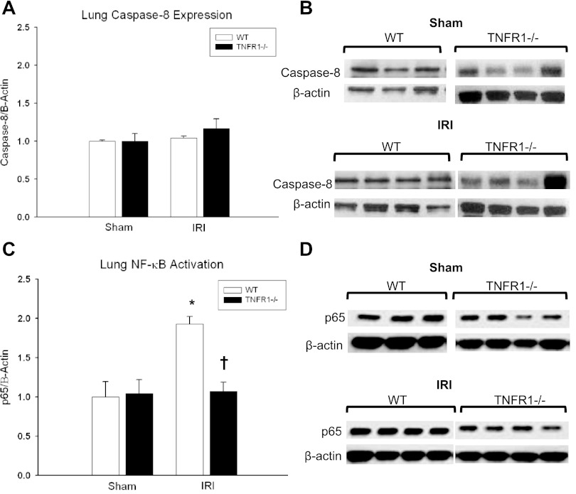
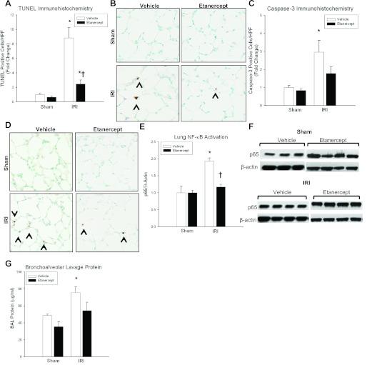
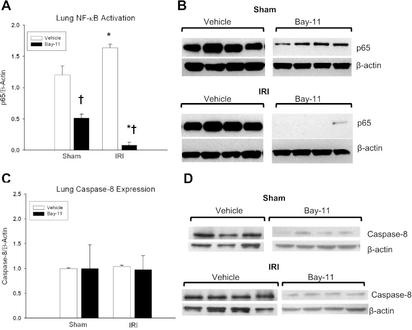
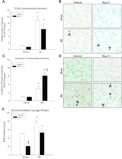
Despite advancements in renal replacement therapy, the mortality rate for acute kidney injury (AKI) remains unacceptably high, likely due to remote organ injury. Kidney ischemia-reperfusion injury (IRI) activates cellular and soluble mediators that incite a distinct pulmonary proinflammatory and proapoptotic response. Tumor necrosis factor receptor 1 (TNFR1) has been identified as a prominent death receptor activated in the lungs during ischemic AKI. We hypothesized that circulating TNF-α released from the postischemic kidney induces TNFR1-mediated pulmonary apoptosis, and we aimed to elucidate molecular pathways to programmed cell death. Using an established murine model of kidney IRI, we characterized the time course for increased circulatory and pulmonary TNF-α levels and measured concurrent upregulation of pulmonary TNFR1 expression. We then identified TNFR1-dependent pulmonary apoptosis after ischemic AKI using TNFR1-/- mice. Subsequent TNF-α signaling disruption with Etanercept implicated circulatory TNF-α as a key soluble mediator of pulmonary apoptosis and lung microvascular barrier dysfunction during ischemic AKI. We further elucidated pathways of TNFR1-mediated apoptosis with NF-κB (Complex I) and caspase-8 (Complex II) expression and discovered that TNFR1 proapoptotic signaling induces NF-κB activation. Additionally, inhibition of NF-κB (Complex I) resulted in a proapoptotic phenotype, lung barrier leak, and altered cellular flice inhibitory protein signaling independent of caspase-8 (Complex II) activation. Ischemic AKI activates soluble TNF-α and induces TNFR1-dependent pulmonary apoptosis through augmentation of the prosurvival and proapoptotic TNFR1 signaling pathway. Kidney-lung crosstalk after ischemic AKI represents a complex pathological process, yet focusing on specific biological pathways may yield potential future therapeutic targets.


Figures

Fig. 1.
Fig. 1.
Effect of kidney ischemia-reperfusion injury (IRI) on renal function and lung apoptosis. A: serum creatinine (mg/dl) was increased in wild-type (WT) mice at both 4 h (0.2 ± 0 vs. 0.72 ± 0.1*, P = 10 × 10−7) and 24 h (0.18 ± 0.02 vs. 1.94 ± 0.17*, P = 0.007) after kidney IRI vs. sham. B: kidney IRI produced more TUNEL-positive cells at 24 h (fold change, FC = 1.46 ± 0.31 vs. 12.9 ± 2.1*; absolute value, AV = 0.19 ± 0.04 vs. 1.68 ± 0.27*; P = 0.008) but not at 4 h (FC = 1.0 ± 0.15 vs. 1.69 ± 0.36; AV = 0.13 ± 0.02 vs. 0.22 ± 0.05; P = 0.13) compared with sham controls. C: representative lung micrographs of TUNEL staining for WT mice at 4 and 24 h after IRI vs. sham demonstrating increased apoptosis in IRI-treated mice at 24 h. D: WT mice demonstrated significantly increased cleaved caspase-3-positive cells at 24 h (FC = 1 ± 0.11 vs. 2.96 ± 0.64*; AV = 0.55 ± 0.06 vs. 1.63 ± 0.35*; P = 0.02) after kidney IRI compared with sham. E: representative lung micrographs of caspase-3 immunohistochemistry demonstrating increased caspase-3-positive cells at 24 h after kidney IRI. n ≥ 5/group, *P < 0.05 for IRI vs. sham.
Fig. 2.
Fig. 2.
Effect of ischemic acute kidney injury (AKI) on TNF-α levels and pulmonary TNF receptor (TNFR) 1 expression. A: serum TNF-α levels measured by ELISA (pg/mg) increased after kidney IRI at 2 h (20.1 ± 2.3 vs. 47.5 ± 6.9*, P = 0.04) and at 4 h (18.5 ± 2.5 vs. 56.4 ± 5.4*, P = 9.16 × 10⁻⁵), but not at 24 h (23.7 ± 4.3 vs. 38.6 ± 9.0, P = 0.16) compared with sham. B: lung tissue TNF-α levels did not increase after kidney IRI at either 4 h (51.6 ± 12.0 vs. 72.4 ± 7.2, P = 0.16) or 24 h (71.1 ± 23.5 vs. 51.6 ± 7.3, P = 0.47) compared with sham controls. C: RT-PCR confirmed no increase in lung TNF-α expression at either 4 (1 ± 0.1 vs. 0.98 ± 0.1, P = 0.5) or 24 (1 ± 0.3 vs. 1.1 ± 0.09, P = 0.6) h of ischemic AKI. n ≥ 5/group, D: relative fold change for WT lung TNFR1 gene expression by quantitative RT-PCR demonstrated increased expression at 4 h (FC = 1 ± 0.05 vs. 1.34 ± 0.11*; AV = 0.48 ± 0.02 vs. 0.65 ± 0.05*; P = 0.03) and 24 h (FC = 1 ± 0.19 vs. 1.81 ± 0.25*; AV = 0.41 ± 0.8 vs. 0.75 ± 0.1*; P = 0.03) compared with sham. *P < 0.05 vs. sham.
Fig. 3.
Fig. 3.
Effect of TNFR1−/− on pulmonary apoptosis after kidney IRI. A: kidney IRI produced fewer lung TUNEL-positive cells at 24 h for TNFR1−/− mice compared with WT mice (FC = 4 ± 1.23 vs. 8.84 ± 1.44*; AV = 0.76 ± 0.23 vs. 1.68 ± 0.27*; P = 0.047), but no difference was seen during sham laparotomy (FC = 2 ± 0.39 vs. 1 ± 0.2, AV = 0.38 ± 0.07 vs. 0.19 ± 0.04; P = 0.056). B: representative lung micrographs of TUNEL staining demonstrating decreased lung apoptosis after IRI in TNFR1−/− mice at 24 h.
Fig. 4.
Fig. 4.
Lung TNFR1 signaling after ischemic AKI. A and B: no difference in caspase-8 expression (Complex II) was demonstrated after kidney IRI in WT vs. TNFR1−/− animals (FC = 1.04 ± 0.06 vs. 1.2 ± 0.32, P = 0.37). C and D: lung NF-κB p65 (Complex I) expression was decreased in TNFR1−/− animals compared with WT (FC = 1.9 ± 0.1 vs. 1.07 ± 0.11†, P = 0.047) after ischemic AKI. Compared with sham, WT animals had an increased in lung NF-κB expression after ischemic AKI (FC = 1 ± 0.2 vs. 1.9 ± 0.1*; AV = 0.26 ± 0.05 vs. 0.51 ± 0.03*; P = 0.005). *P < 0.05 for IRI vs. sham, †P < 0.05 for WT vs. TNFR1−/−.
Fig. 5.
Fig. 5.
Effect of TNF-α inhibition after kidney IRI. A: kidney IRI produced more lung TUNEL-positive cells at 24 h for vehicle-treated compared with Etanercept-treated groups (FC = 8.84 ± 1.44 vs. 2.47 ± 0.53†, AV = 1.68 ± 0.27 vs. 0.47 ± 0.1†, P = 0.009); however, there was no difference in TUNEL positivity after sham laparotomy (FC = 1 ± 0.21 vs. 0.63 ± 0.16, AV = 0.19 ± 0.04 vs. 0.12 ± 0.03, P = 0.205). B: representative lung micrographs of TUNEL staining for vehicle and Etanercept groups at 24 h. C: kidney IRI produced no difference in caspase-3-positive cells between vehicle-treated and Etanercept-treated mice (FC = 2.96 ± 0.64 vs. 1.78 ± 0.37, AV = 1.63 ± 0.35 vs. 0.98 ± 0.2, P = 0.142). There was also no difference between vehicle-treated and Etanercept-treated mice after sham laparotomy (FC = 1 ± 0.11 vs. 0.84 ± 0.1, AV = 0.55 ± 0.06 vs. 0 ± 0.05, P = 0.341). D: representative lung micrographs of caspase-3 immunohistochemistry for vehicle and Etanercept groups at 24 h. E and F: NF-κB p65 expression was decreased in Etanercept-treated mice compared with vehicle-treated mice following kidney IRI (FC = 1.9 ± 0.1 vs. 1.17 ± 0.08†, AV = 0.56 ± 0.04 vs. 0.51 ± 0.03†, P = 0.008), but there was no difference after sham laparotomy (FC = 1 ± 0.07 vs. 1 ± 0.2, AV = 0.48 ± 0.03 vs. 0.26 ± 0.05, P = 0.49). G: bronchoalveolar lavage (BAL) protein was decreased in Etanercept-treated mice during kidney IRI (75.8 ± 6.6 vs. 54.6 ± 8.4*, P = 0.01) compared with vehicle-treated groups. *P < 0.05 for IRI vs. sham; †P < 0.05 for WT vs. Etanercept.
Fig. 6.
Fig. 6.
Effect of NF-κB inhibition on TNFR1 signaling after kidney IRI. A and B: Bay-11 served as an effective NF-κB inhibitor, decreasing NF-κB expression after 24 h of kidney IRI compared with sham (AV = 1.63 ± 0.06 vs. 0.08 ± 0.05*; P = 2.75 × 10⁻⁴). NF-κB expression was also decreased in sham animals treated with Bay-11 compared with vehicle (AV = 1.2 ± 0.14 vs. 0.5 ± 0.06†, P = 9.5 × 10⁻⁵). C and D: no difference in caspase-8 expression (Complex II) was demonstrated at 24 h in vehicle (FC = 1 ± 0.03 vs. 1.04 ± 0.06; AV = 1.37 ± 0.02 vs. 1.43 ± 0.03; P = 0.21) or Bay-11 mice (FC = 1 ± 0.47 vs. 0.98 ± 0.28; AV = 0.08 ± 0.04 vs. 0.08 ± 0.02; P = 0.97) after kidney IRI compared with sham. n ≥ 4/group. *P < 0.05 for IRI vs. sham, †P < 0.05 for vehicle vs. Bay-11.
Fig. 7.
Fig. 7.
Effect of NF-κB inhibition on pulmonary apoptosis and injury. A: kidney IRI induced TUNEL positivity in lungs of both vehicle-treated and Bay 11-treated mice (FC = 8.84 ± 1.44 vs. 6.2 ± 1.7, AV = 1.68 ± 0.27 vs. 1.18 ± 0.33, P = 0.402), both of which were increased compared with sham. B: representative lung micrographs of TUNEL staining at 24 h exhibit increased apoptosis in both vehicle and Bay-11 groups. C: kidney IRI induced more caspase-3 positivity in Bay-11-treated groups compared with vehicle-treated groups (FC = 2.96 ± 0.64 vs. 6.13 ± 0.70†, AV = 1.63 ± 0.35 vs. 3.37 ± 0.38, P = 0.03), both of which were increased compared with sham. D: representative lung micrographs of cleaved caspase-3 immunohistochemistry show increased cleaved caspase-3-positive cells in both vehicle and Bay-11 groups after IRI. E: BAL protein was measured at 24 h in vehicle-treated and Bay-11-treated mice following sham or ischemic AKI. Similar to vehicle-treated mice (48.7 ± 1.7 vs. 75.8 ± 6.6*, P = 0.01), Bay-11-treated mice demonstrated increased BAL protein leak during kidney IRI compared with sham (21.1 ± 2.4 vs. 53.1 ± 8.4*, P = 0.006). Whereas there was no difference in BAL protein leak following IRI between vehicle-treated and Bay-11-treated mice (75.8 ± 6.6 vs. 53.1 ± 8.4, P = 0.09), Bay-11 mice had decreased BAL protein after sham compared with vehicle-treated groups (48.7 ± 1.7 vs. 21.1 ± 2.4†, P = 6.005 × 10⁻⁵). n ≥ 5/group, *P < 0.05 for IRI vs. sham, †P < 0.05 for vehicle vs. Bay-11.

References

    1. Cataisson C, Pearson AJ, Torgerson S, Nedospasov SA, Yuspa SH. Protein kinase C alpha-mediated chemotaxis of neutrophils requires NF-kappa B activity but is independent of TNF alpha signaling in mouse skin in vivo. J Immunol 174: 1686–1692, 2005 - PubMed
    1. Donnahoo KK, Meng X, Ayala A, Cain MP, Harken AH, Meldrum DR. Early kidney TNF-α expression mediates neutrophil infiltration and injury after renal ischemia-reperfusion. Am J Physiol Regul Integr Comp Physiol 277: R922–R929, 1999 - PubMed
    1. Feltes CM, Hassoun HT, Lie ML, Cheadle C, Rabb H. Pulmonary endothelial cell activation during experimental acute kidney injury. Shock 36: 70–76, 2011 - PMC - PubMed
    1. Geering B, Gurzeler U, Federzoni E, Kaufmann T, Simon HU. A novel TNFR1-triggered apoptosis pathway mediated by class IA PI3Ks in neutrophils. Blood 117: 5953–5962, 2011 - PubMed
    1. Golks A, Brenner D, Krammer PH, Lavrik IN. The c-FLIP-NH2 terminus (p22-FLIP) induces NF-kappaB activation. J Exp Med 203: 1295–1305, 2006 - PMC - PubMed

Publication types

MeSH terms

LinkOut - more resources