. 2012 Jul;122(7):2531-42.
doi: 10.1172/JCI58525. Epub 2012 Jun 25.

Oxidative stress fuels Trypanosoma cruzi infection in mice

Affiliations

Oxidative stress fuels Trypanosoma cruzi infection in mice

Claudia N Paiva et al. J Clin Invest. 2012 Jul.

Abstract

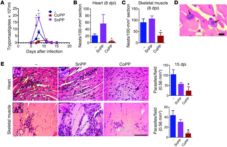
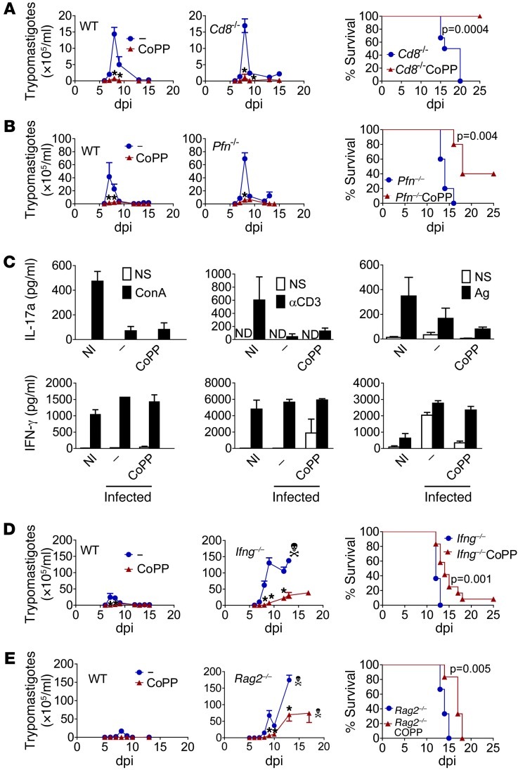
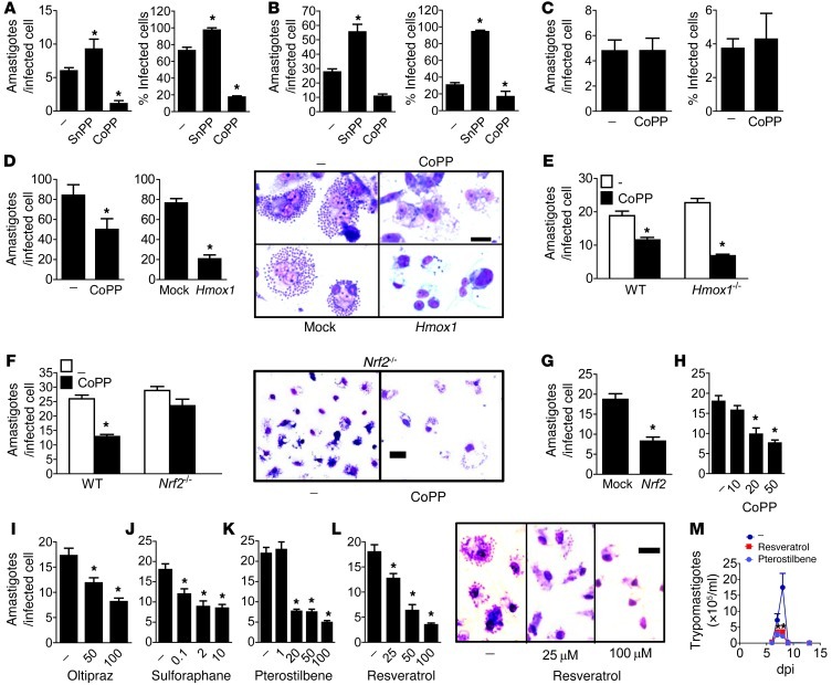
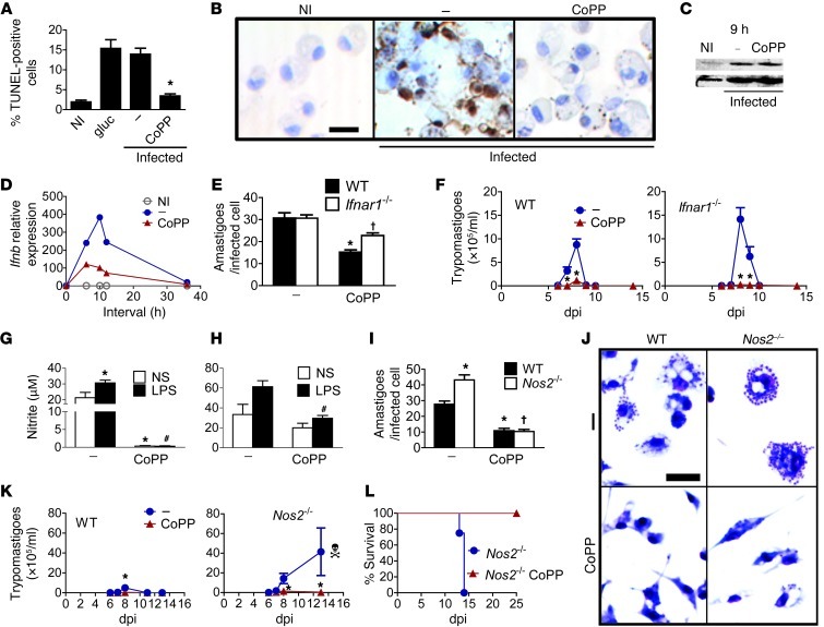
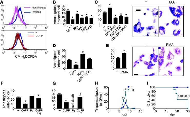
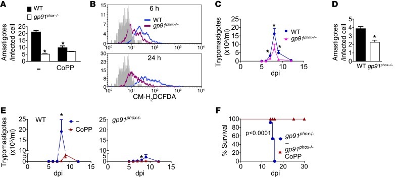
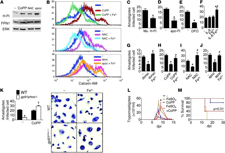
Oxidative damage contributes to microbe elimination during macrophage respiratory burst. Nuclear factor, erythroid-derived 2, like 2 (NRF2) orchestrates antioxidant defenses, including the expression of heme-oxygenase-1 (HO-1). Unexpectedly, the activation of NRF2 and HO-1 reduces infection by a number of pathogens, although the mechanism responsible for this effect is largely unknown. We studied Trypanosoma cruzi infection in mice in which NRF2/HO-1 was induced with cobalt protoporphyrin (CoPP). CoPP reduced parasitemia and tissue parasitism, while an inhibitor of HO-1 activity increased T. cruzi parasitemia in blood. CoPP-induced effects did not depend on the adaptive immunity, nor were parasites directly targeted. We also found that CoPP reduced macrophage parasitism, which depended on NRF2 expression but not on classical mechanisms such as apoptosis of infected cells, induction of type I IFN, or NO. We found that exogenous expression of NRF2 or HO-1 also reduced macrophage parasitism. Several antioxidants, including NRF2 activators, reduced macrophage parasite burden, while pro-oxidants promoted it. Reducing the intracellular labile iron pool decreased parasitism, and antioxidants increased the expression of ferritin and ferroportin in infected macrophages. Ferrous sulfate reversed the CoPP-induced decrease in macrophage parasite burden and, given in vivo, reversed their protective effects. Our results indicate that oxidative stress contributes to parasite persistence in host tissues and open a new avenue for the development of anti-T. cruzi drugs.


Figures

Figure 1
Figure 1. Treatment with CoPP reduces T. cruzi parasite burden.
(A) Mean parasitemia during acute infection (n = 7 mice per time point). The experiment was performed 5 times. Mean parasitism in (B) heart and (C) skeletal muscle at 8 dpi. (D) Infected cells infiltrating the heart from SnPP-treated mice at 8 dpi. Scale bar: 10 μm. (E) Mean parasitism in heart and skeletal muscle at 15 dpi. At least 50 fields were assessed per H&E section from each of 4 mice per group. Yellow arrows indicate parasites. Insets (×560) show parasites among inflammatory leukocytes. Experiments were performed twice, with similar results. *P < 0.05 compared with infected nontreated controls. Error bars represent SEM. Scale bars: 100 μm.
Figure 2
Figure 2. CoPP-mediated reduction of T. cruzi parasitemia does not involve adaptive immunity.
(A) Effect of CoPP or SnPP treatment on survival and mean parasitemia in wild-type control, Cd8–/– (n = 7–8), and (B) Pfn–/– mice (n = 6–9). (C) Cytokine production by splenocytes stimulated with ConA, anti-CD3 (for 24 hours), or T. cruzi antigen (Ag; for 48 hours). Splenocytes were pooled from 3–5 mice per group treated in vivo with CoPP at 9 dpi, and supernatants from 3 pools were analyzed by ELISA. (D) Effects of treatment with CoPP on survival and mean parasitemia of wild-type controls and Ifng–/– mice (n = 9–15 mice) or (E) Rag2–/– mice (n = 6). Experiments were performed twice, with similar results (AD). ND, not detected; NI, noninfected; –, infected untreated controls. *P < 0.05 compared with infected nontreated controls. Error bars represent SEM.
Figure 3
Figure 3. CoPP reduces parasitism via macrophage physiology and NRF2 target genes.
(A) Effects of in vivo treatment with CoPP or SnPP on the mean parasite burden of peritoneal macrophages at 8 dpi (n = 3 mice/group). Peritoneal cells were cultured for an additional 48 hours before cells were fixed. (B) Effects of treatment with CoPP or SnPP on the mean parasite burden of thioglycollate-elicited macrophages infected in vitro. (C) Effects of treatment with CoPP on the mean parasite burden of L929 fibroblast cells. (D) Effects of prior transfection of THP-1 cells with HO-1 or empty vector on mean parasite burden. (E) Effects of treatment with CoPP on mean parasite burden of BMMs derived from wild-type and Hmox1–/– mice or (F) Nrf2–/– mice. (G) Effects of prior transfection of THP-1 cells with Nrf2 or empty vector on mean parasite burden. (HL) Dose-dependent decreases in macrophage parasitism with the NRF2 activators. (M) Mean parasitemia during acute infection (n = 8) in mice treated with pterostilbene or resveratrol. Thioglycollate-elicited macrophages were infected with 3:1 trypomastigotes in vitro for 12 hours and treated with drugs for 48 hours. Each experiment was performed with 2 independent samples of cells, and results for 1 sample are shown. Cells were stained with Giemsa, and amastigotes were counted in each of 100 infected cells. The mean percentage of infected cells was calculated for each field, and 20 fields were assessed. All experiments were performed at least twice, with similar results. *P < 0.05 compared with infected nontreated controls. Error bars represent SEM. Scale bars: 20 μm.
Figure 4
Figure 4. Classical mechanisms of parasite clearance are not responsible for the decreased parasitism mediated by CoPP.
(A) Effects of CoPP on the mean percentage of TUNEL-positive cells (apoptotic) found in infected macrophages, as shown in B. Glucocorticoid-treated macrophages (gluc) are shown as a positive control. Amastigotes were also stained with TUNEL (33). Data represent percentages in 10–15 microscope fields (original magnification, ×40). Effects of CoPP on (C) IRF-3 phosphorylation in infected macrophages (immunoblots) and (D) the amounts of IFN-β transcripts (qRT-PCR). (E) Effects of CoPP on the mean parasite burden in infected macrophages derived from wild-type or Ifnar1–/– mice. (F) Effects of CoPP on the mean parasitemia of wild-type and Ifnar1–/– mice (n = 8 per group). (G) Effects of in vivo CoPP treatment on the mean nitrite production by LPS-stimulated peritoneal macrophages at 8 dpi. Cells were pooled from 3–5 mice/group, and results represent 3–4 pools (Griess). Effects of CoPP on infected macrophages: (H) Mean nitrite production upon LPS stimulation in vitro (results represent 3 independent samples evaluated by Griess); (I) Mean parasite burden of macrophages derived from wild-type or Nos2–/– mice (Giemsa), as shown in J. Effects of in vivo CoPP treatment on: (K) Mean parasitemia and (L) survival of wild-type and Nos2–/– mice (n = 4–6). Experiments were performed at least twice. Error bars represent SEM. *P < 0.05 compared with infected untreated wild-type; #P < 0.05 compared with LPS-stimulated infected untreated macrophages; P < 0.05 compared with infected untreated knockout mice. Scale bars: 20 μm.
Figure 5
Figure 5. Oxidative stress promotes T. cruzi parasitism.
(A) Flow cytometry quantification of ROS (CM-H2DCFDA) in thioglycollate-elicited macrophages infected in vitro and left untreated (–) or treated with CoPP for 6 hours. (B) Parasite burden (Giemsa) in thioglycollate-elicited peritoneal macrophages infected in vitro and left untreated (–) or treated for 48 hours with CoPP, bilirubin (Bb), biliverdin (Bvd), apocynin (Apoc), or NAC; (C) H2O2, CAT-PEG, SOD-PEG, or SOD-PEG plus CAT-PEG; (D) CoPP, H2O2, or CoPP plus H2O2; (E) PMA; CoPP, paraquat (Pq; 10 μM [F] or 100 μM [G]), or CoPP plus paraquat. (H) Mean parasitemia and (I) survival of mice treated with paraquat (n = 8). Experiments were performed at least twice, with similar results. Error bars represent SEM. *P < 0.05 compared with infected untreated controls. #P < 0.05 compared with CoPP-treated cells. Scale bars: 20 μm.
Figure 6
Figure 6. Effects of gp91phox on T. cruzi infection.
(A) Effects of CoPP on the parasite burden of in vitro infected macrophages derived from wild-type or gp91phox–/– mice (Giemsa). (B) Flow cytometry quantification of ROS (CM-H2DCFDA probe) in BMMs from wild-type or gp91phox–/– mice after infection. Non-labeled controls are shown in gray. (C) Mean parasitemia of wild-type and gp91phox–/– mice (n = 7). (D) Parasite burden in peritoneal macrophages taken from wild-type and gp91phox–/– mice at 8 dpi. Cells were cultured for an additional 48 hours before they were fixed and stained. The graphs show the mean ± SEM of the parasite burden in each individual mouse (n = 4 mice/group). (E) Mean parasitemia (n = 5) and (F) survival of wild-type and gp91phox–/– mice left untreated (n = 12) or treated with CoPP (n = 5). Error bars represent SEM. *P < 0.05 compared with infected untreated controls.
Figure 7
Figure 7. Cellular iron is mobilized by oxidative stress and fuels T. cruzi infection.
(A) Antioxidants increase H-ferritin (H-Ft) and FPN1 expression (immunoblot). Extracts were prepared from infected thioglycollate-elicited peritoneal macrophages treated for 12 hours with CoPP, NAC, or apocynin. (B) Antioxidants reduce the labile iron pool. The quenching of calcein fluorescence was measured by flow cytometry as an indicator of labile iron. Thioglycollate-elicited peritoneal macrophages were infected in vitro and left untreated (–) or treated for 6 hours with antioxidants (CoPP, NAC, apocynin) with or without Fe2+. (C) Parasite burden in THP-1 cells transfected with H-ferritin or mutant nonfunctional ferritin (Mu) 48 hours after infection. Parasite burden in thioglycollate-elicited peritoneal macrophages infected in vitro and left untreated (–) or treated with (D) apo-ferritin (apo-Ft); (E) DFO; Fe2SO4 alone or (F) added to IL-6, (G) dorsomorphin (dorso), (H) CoPP, (I) NAC, or (J) apocynin for 48 hours. (K) Parasite burden in thioglycollate-elicited peritoneal macrophages taken from wild-type or gp91phox–/– mice infected in vitro and treated with Fe2SO4 for 48 hours. Effects of treatment with CoPP and Fe2SO4 on (L) mean parasitemia and (M) survival of mice (n = 8–10). Error bars represent SEM. In CK, cells were stained with Giemsa, and amastigotes were counted in each of 100 infected cells and expressed as mean ± SEM. Experiments were performed at least twice. *P < 0.05 compared with infected untreated wild-type controls; #P < 0.05 compared with IL-6– (F), dorsomorphin- (G), CoPP- (H), NAC- (I), or apocynin-treated cells (J); P < 0.05 compared with infected knockout mice. Scale bars: 20 μm.

Comment in

References

    1. Gupta S, Bhatia V, Wen JJ, Wu Y, Huang MH, Garg NJ. Trypanosoma cruzi infection disturbs mitochondrial membrane potential and ROS production rate in cardiomyocytes. . Free Radic Biol Med. 2009;47(10):1414–1421. doi: 10.1016/j.freeradbiomed.2009.08.008. - DOI - PMC - PubMed
    1. Wen JJ, Garg N. Oxidative modification of mitochondrial respiratory complexes in response to the stress of Trypanosoma cruzi infection. . Free Radic Biol Med. 2004;37(12):2072–2081. doi: 10.1016/j.freeradbiomed.2004.09.011. - DOI - PubMed
    1. Wen JJ, Vyatkina G, Garg N. Oxidative damage during chagasic cardiomyopathy development: role of mitochondrial oxidant release and inefficient antioxidant defense. Free Radic Biol Med. 2004;37(11):1821–1833. doi: 10.1016/j.freeradbiomed.2004.08.018. - DOI - PubMed
    1. Wen JJ, Bhatia V, Popov VL, Garg NJ. Phenyl-alpha-tert-butyl nitrone reverses mitochondrial decay in acute Chagas’ disease. Am J Pathol. 2006;169(6):1953–1964. doi: 10.2353/ajpath.2006.060475. - DOI - PMC - PubMed
    1. McCord JM. Iron, free radicals, and oxidative injury. J Nutr. 2004;134(11):3171S–3172S. - PubMed

Publication types

MeSH terms