. 2012 Jul;122(7):2509-18.
doi: 10.1172/JCI62617. Epub 2012 Jun 25.

TBX5 drives Scn5a expression to regulate cardiac conduction system function

Affiliations

TBX5 drives Scn5a expression to regulate cardiac conduction system function

David E Arnolds et al. J Clin Invest. 2012 Jul.

Abstract

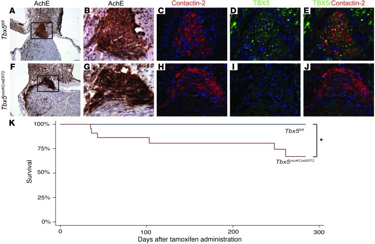
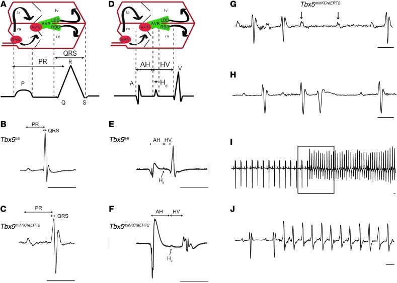
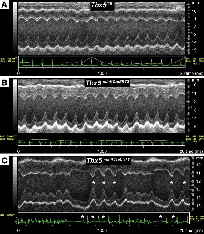
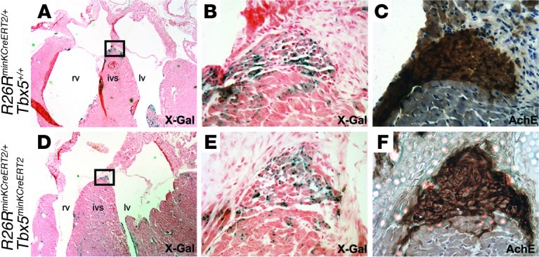
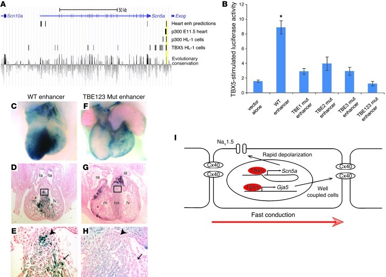
Cardiac conduction system (CCS) disease, which results in disrupted conduction and impaired cardiac rhythm, is common with significant morbidity and mortality. Current treatment options are limited, and rational efforts to develop cell-based and regenerative therapies require knowledge of the molecular networks that establish and maintain CCS function. Recent genome-wide association studies (GWAS) have identified numerous loci associated with adult human CCS function, including TBX5 and SCN5A. We hypothesized that TBX5, a critical developmental transcription factor, regulates transcriptional networks required for mature CCS function. We found that deletion of Tbx5 from the mature murine ventricular conduction system (VCS), including the AV bundle and bundle branches, resulted in severe VCS functional consequences, including loss of fast conduction, arrhythmias, and sudden death. Ventricular contractile function and the VCS fate map remained unchanged in VCS-specific Tbx5 knockouts. However, key mediators of fast conduction, including Nav1.5, which is encoded by Scn5a, and connexin 40 (Cx40), demonstrated Tbx5-dependent expression in the VCS. We identified a TBX5-responsive enhancer downstream of Scn5a sufficient to drive VCS expression in vivo, dependent on canonical T-box binding sites. Our results establish a direct molecular link between Tbx5 and Scn5a and elucidate a hierarchy between human GWAS loci that affects function of the mature VCS, establishing a paradigm for understanding the molecular pathology of CCS disease.


Figures

Figure 1
Figure 1. Removal of Tbx5 from the ventricular CCS reduces survival.
(AJ) Tbx5fl/fl and Tbx5minKCreERT2 littermates were administered tamoxifen at 6–7 weeks of age, and TBX5 expression in the VCS was evaluated by immunofluorescence at 10–11 weeks of age. (AE) Serial sections demonstrating TBX5 expression through the AV bundle (positive for acetylcholinesterase [AchE] and contactin-2)of Tbx5fl/fl mice. (FJ) In contrast, TBX5 was not detected in the AV bundle of Tbx5minKCreERT2 mice. Boxed areas in A and F are shown at higher magnification in B and G. TBX5 and contactin-2 were evaluated on serial sections (merged in E and J). Nuclei were stained with hematoxylin (A, B, F, and G) or DAPI (CE and HJ). Original magnification, ×10 (A and F), ×40 (BE and GJ). (K) Tbx5minKCreERT2 mice (n = 22) and Tbx5fl/fl littermates (n = 15) were followed longitudinally after tamoxifen administration at 6–7 weeks of age. Kaplan-Meier survival estimates demonstrated significantly decreased survival after Tbx5 removal. *P < 0.05, log-rank test.
Figure 2
Figure 2. Conduction slowing and arrhythmias after removal of Tbx5 from the ventricular CCS.
(AJ) Conduction system function in Tbx5fl/fl (B and E) and Tbx5minKCreERT2 (C and FJ) mice was evaluated by ambulatory telemetry (B, C, and GJ) and invasive EP studies (E and F). Electroanatomical correlates of ECG and EP recordings are shown in A and D, respectively. la and ra, left and right atria; AVB, AV bundle; AVN and SAN, AV and SA nodes; LBB and RBB, left and right bundle branches. PR and QRS intervals were prolonged during ambulatory telemetry analysis (representative recordings in B and C), and intracardiac recordings (representative recordings in E and F) demonstrated prolongation of AH interval, Hd, and HV interval. Mobitz type II second-degree AV block (G) occurred exclusively in Tbx5minKCreERT2 mice. PVCs (H) were more common in Tbx5minKCreERT2 mice, and ventricular tachycardia (I and J) was observed exclusively in Tbx5minKCreERT2 mice. Boxed area in I is shown at slower scale in J. Scale bars: 50 ms. Arrows in G represent nonconducted p waves. See Table 1 for quantification of ECG and EP intervals.
Figure 3
Figure 3. Normal cardiac function after loss of TBX5 in the VCS.
(A and B) Cardiac function, assessed by M-mode echocardiography, of Tbx5fl/fl controls (A) and Tbx5minKCreERT2 littermates (B) administered tamoxifen at 6–7 weeks of age and studied 4–5 weeks later in sinus rhythm. No functional difference between mutant and control mice was detected. (C) A Tbx5minKCreERT2 animal with episodic spontaneous ventricular tachycardia demonstrated rapid recovery of ventricular function during sinus beats (asterisks) that followed tachycardic episodes.
Figure 4
Figure 4. Tbx5 removal does not affect cell survival in the VCS.
X-Gal– (A, B, D, and E) and acetylcholinesterase-stained (C and F) serial sections through the AV bundle of R26RminKCreERT2/+Tbx5+/+ (AC) and R26RminKCreERT2/+Tbx5minKCreERT2 (DF) animals administered tamoxifen showed similar distribution of cells labeling the VCS fate map, which demonstrated that Tbx5 was not required for VCS cell survival. ivs, interventricular septum. Boxed regions in A and D are shown at higher magnification in B and E. Original magnification, ×2.5 (A and D), ×40 (B, C, E, and F).
Figure 5
Figure 5. Decreased Cx40 and Nav1.5 expression in the VCS after removal of TBX5.
The proximal (AJ) and distal (KT) AV bundle was identified by acetycholinesterase activity (A, B, F, G, K, L, P, and Q) and contactin-2 expression (C, D, H, I, M, N, R, and S) on serial sections from Tbx5fl/fl and Tbx5minKCreERT2 hearts. Whereas the contactin-2–positive AV bundle expressed high levels of Cx40, Nav1.5, and TBX5 in Tbx5fl/fl mice, their expression was drastically reduced in that of Tbx5minKCreERT2 mice. (C, D, H, I, M, N, R, and S) Dual-color immunofluorescence for Cx40 or Nav1.5 and contactin-2 is shown. Contactin-2 expression in E, J, O, and T is from sections adjacent to those stained for TBX5; contactin-2 and TBX5 antibodies were both raised in goat, preventing dual-color immunofluorescence on the same section. Nuclei were stained with hematoxylin (A, B, F, G, K, L, P, and Q) or DAPI (blue; CE, HJ, MO, and RT). Boxed regions in A, F, K, and P are shown at higher magnification in B, G, L, and Q. Original magnification, ×10 (A, F, K, and P); ×40 (BE, GJ, LO, and PT).
Figure 6
Figure 6. TBX5 directly regulates an enhancer downstream of Scn5a.
(A) Bioinformatic identification of a candidate enhancer downstream of Scn5a. We used previously reported data sets to identify potential TBX-responsive enhancers: bioinformatic predictions of cardiac enhancers (24); p300 ChIP-seq peaks to mark active enhancers in the E11.5 heart (23); ChIP-seq studies identifying both p300 and TBX5 binding sites in the atrial cardiomyocyte HL-1 cell line (22); and evolutionary conservation, as assessed by genomic evolutionary rate profiling score (21). A region demonstrating overlap in all 4 data sets, approximately 15 kb downstream of Scn5a, is shaded yellow. (B) The WT candidate enhancer demonstrated robust TBX5-mediated activation in dual luciferase reporter assays in HEK-293T cells. Luciferase activity was blunted by single mutation of any of 3 conserved T-box elements (TBE1 mut, TBE2 mut, or TBE3 mut) and eliminated by mutation of all 3 T-box elements (TBE123 mut). *P < 0.05 versus all other groups; n ≥ 3; mean ± SEM. (CH) VCS of transient transgenic embryos, analyzed at E13.5. Whereas the WT enhancer reproducibly drove lacZ expression from a minimal promoter (CE), mutation of the T-box elements in the enhancer resulted in blunted and regionally variable expression (FH). Shown are representative posterior (C and F) and sagittal section (D and G) views of X-Gal staining. Higher-magnification views of boxed regions in D and G demonstrated X-gal expression (E) or its absence (H) in the developing AV bundle (arrowhead) and bundle branches (arrow). Note that, in contrast to the weak, non-CCS lacZ expression in F, the more robustly stained heart in G demonstrated ectopic expression in the endocardial cushions and compact myocardium. (I) Model for the role of TBX5 in driving fast conduction in the VCS via direct regulation of Scn5a and Gja5. Original magnification, ×4 (D and G); ×40 (E and H).

References

    1. Priori SG. The fifteen years of discoveries that shaped molecular electrophysiology: time for appraisal. Circ Res. 2010;107(4):451–456. doi: 10.1161/CIRCRESAHA.110.226811. - DOI - PubMed
    1. Hatcher CJ, Basson CT. Specification of the cardiac conduction system by transcription factors. Circ Res. 2009;105(7):620–630. doi: 10.1161/CIRCRESAHA.109.204123. - DOI - PMC - PubMed
    1. Chambers JC, et al. Genetic variation in SCN10A influences cardiac conduction. Nat Genet. 2010;42(2):149–152. doi: 10.1038/ng.516. - DOI - PubMed
    1. Holm H, et al. Several common variants modulate heart rate, PR interval and QRS duration. Nat Genet. 2010;42(2):117–122. doi: 10.1038/ng.511. - DOI - PubMed
    1. Pfeufer A, et al. Genome-wide association study of PR interval. Nat Genet. 2010;42(2):153–159. doi: 10.1038/ng.517. - DOI - PMC - PubMed

Publication types

MeSH terms