. 2012 Aug;14(8):865-73.
doi: 10.1038/ncb2527. Epub 2012 Jun 24.

Tubulin nucleotide status controls Sas-4-dependent pericentriolar material recruitment

Affiliations

Tubulin nucleotide status controls Sas-4-dependent pericentriolar material recruitment

Jayachandran Gopalakrishnan et al. Nat Cell Biol. 2012 Aug.

Abstract

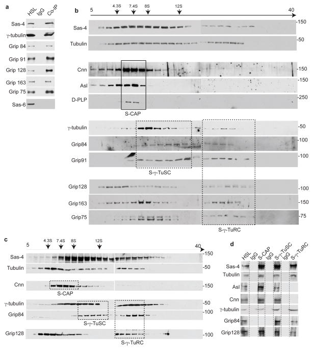
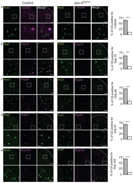
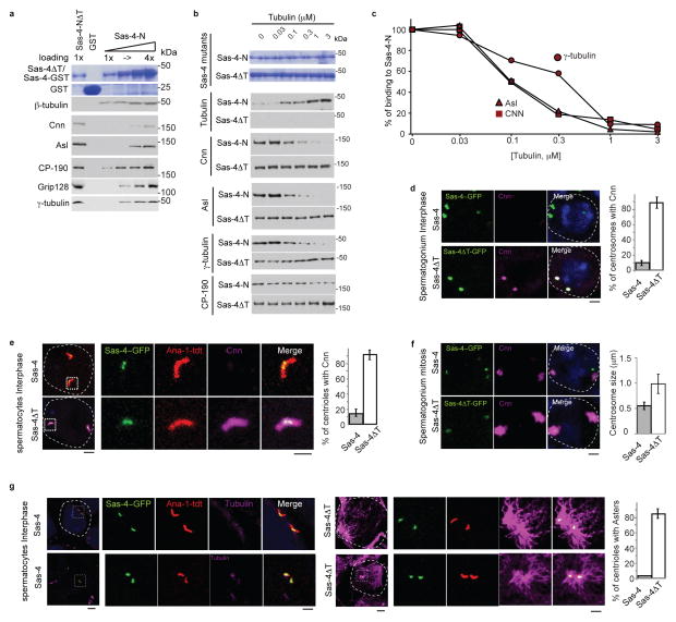
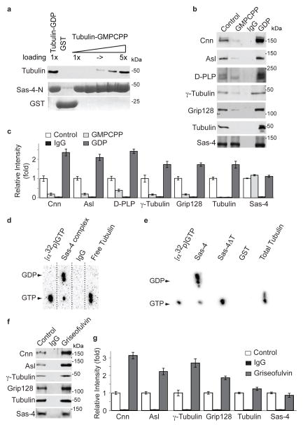
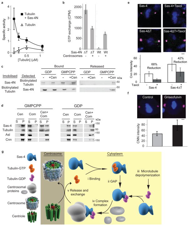
Regulated centrosome biogenesis is required for accurate cell division and for maintaining genome integrity. Centrosomes consist of a centriole pair surrounded by a protein network known as pericentriolar material (PCM). PCM assembly is a tightly regulated, critical step that determines the size and capability of centrosomes. Here, we report a role for tubulin in regulating PCM recruitment through the conserved centrosomal protein Sas-4. Tubulin directly binds to Sas-4; together they are components of cytoplasmic complexes of centrosomal proteins. A Sas-4 mutant, which cannot bind tubulin, enhances centrosomal protein complex formation and has abnormally large centrosomes with excessive activity. These results suggest that tubulin negatively regulates PCM recruitment. Whereas tubulin-GTP prevents Sas-4 from forming protein complexes, tubulin-GDP promotes it. Thus, the regulation of PCM recruitment by tubulin depends on its GTP/GDP-bound state. These results identify a role for tubulin in regulating PCM recruitment independent of its well-known role as a building block of microtubules. On the basis of its guanine-bound state, tubulin can act as a molecular switch in PCM recruitment.


Conflict of interest statement

Author Information: The authors declare that they have no competing financial interests.

Figures

Fig. 1
Fig. 1. Tubulin is present in each Sas-4 complex type
(a) Immuno-purification of Sas-4 complexes from high-speed lysate (HSL) of Drosophila embryonic extracts using anti-Sas-4 antibody revealed associations between Sas-4 with Grip proteins that are components of γ-TuRCs: Grip91, Grip84, Grip163, Grip128, and Grip75, and γ-tubulin small complexes (γ-TuSCs): Grip91 and Grip84. Embryonic extracts were used as a positive control; mouse IgG beads were used as a negative control. The use of extract depleted of centrosomes and the absence of the centriole-core protein Sas-6 indicate that the purified complexes were not centrosomes. (b) Immuno-purified Sas-4 complexes fractionate at distinct densities in a 5–40% sucrose gradient. Individual fractions are analyzed by Western blot. Tubulin co-fractionates with Sas-4 across the gradient. Co-fractionation patterns likely represent different complex types: S-CAP (Cnn, Asl, and D-PLP, proteins); dashed boxes, S-γ-TuSC, and S-γ-TuRC; dashed boxes. The fractionation pattern of S-CAP complexes in a narrow range of low-density fractions and γ-tubulin and Grip proteins fractionation at intermediate and high-densities were consistent with the previous reports, . However, the fractionation pattern of γ-TuSC and γ-TuRC proteins complexes do not exhibit clear peaks as previously demonstrated suggesting that Sas-4 interact with assembly intermediates of γ-tubulin ring proteins. (c–d) The immuno-purified Sas-4 complexes are unlikely to be part of an unstable large complex that destabilizes during immuno-purification. HSL of Drosophila embryonic extract was first fractionated in a 5–40% sucrose gradient (c) and the immuno-purifications of the distinct Sas-4 complex types were performed on distinct fractions (marked by solid boxes and named S-CAP, S-γ-TuSCs and S-γ-TuRCs) (d). Note that Sas-4 and tubulin were detected in all of the complex types. Embryonic extract was used as a positive control; mouse IgG beads were used as a negative control. In experiments (a–d), HSLs were diluted such that the tubulin concentration was below 0.2μM in order to prevent tubulin polymerization; additionally, Sas-4 complexes were purified at 4°C in the presence of nocodazole (330 nM). In experiments (b and c), inverted arrows mark the corresponding peaks of the sedimentation coefficient standards.
Fig. 2
Fig. 2. Sas-4 is essential for recruiting S-γ-tubulin complexes to centrosomes
Centriolar structures labeled by Ana-1-GFP in control testes but not in sas-4s2214 null mutant testes recruit components of S-γ-tubulin complexes as tested using antibodies specific to γ-tubulin, Grip75, Grip84, Grip91 and Grip163 (a–e). Dashed boxes mark the enlarged areas shown in the lower panels. Charts on the right show the fraction of Ana-1 positive centriolar structures (CS) that are also positive for the respective proteins tested in the control (gray filling) and in sas-4s2214 (white filling). As described previously, Ana-1-GFP-labeled centriolar structures from each testis were counted within a 20μm2 area that is ~25 μm away from the tip of a testis (dotted lines). The mean ± SEM of three independent testes are shown. (a–e) Scale bar 10 μm; and 1 μm for lower and higher magnification (inset), respectively. *** marks significant difference (P<0.001)
Fig. 3
Fig. 3. Tubulin negatively regulates PCM recruitment
(a) Comparison of the ability of Sas-4-NΔT and Sas-4-N to interact with centrosomal proteins in embryonic extracts. Increasing loading amounts of Sas-4-N (1 to 4-fold) pulls down Sas-4 interacting proteins from embryonic HSL. Sas-4-NΔT does not pull down tubulin, but pulls down approximately three times more Cnn, Asl, γ-tubulin, and Grip128 than Sas-4-N. Purified recombinant proteins are shown in Coomassie stained gels (b–c) Addition of increasing amounts of free tubulin to Sas-4-N pull down experiments from HSL proportionally inhibits Sas-4-N binding to its interacting partners (a) with an IC50 of 0.1–0.3 μM (b). There is no significant change in the ability of Sas-4-NΔT to bind Cnn and Asl in the presence of tubulin. CP-190 binding to Sas-4N did not change significantly; suggesting that tubulin specifically interferes with the binding of Cnn, Asl and γ-tubulin. The purified recombinant proteins used are shown in Coomassie stained gels. (d) In Sas-4::sas-4s2214 testes Cnn immunoreactivity (Magenta) is not detected in interphase centrosomes. In contrast, sas-4ΔT::sas-4s2214 (sas-4ΔT) interphase centrosomes contain Cnn. Dotted lines mark a cell boundary. Charts show the percentage of centrosomes positive for Cnn. The mean±SEM of six independent testes are shown. p<0.001. Scale bar, 2 μm.. (e) Unlike Sas-4::sas-4s2214, sas-4ΔT::sas-4s2214 interphase spermatocyte centrosomes contain Cnn. Centrosomes are marked by Ana-1-tdT (Red) and Sas-4-GFP (Green). Boxes mark the magnified areas. Charts show the percentage of centrosomes positive for Cnn. The mean±SEM of six independent testes are shown, p<0.001. Scale bar, 2 μm. (f) sas-4ΔT::sas-4s2214 mitotic centrosomes have increased Cnn immunoreactivity. The chart shows centrosome size for Sas-4::sas-4s2214 and sas-4ΔT::sas-4s2214, as measured by Cnn immunolabeling. The mean±SEM of six independent testes are shown. p<0.001. Scale bar, 2μm. (g) sas-4ΔT::sas-4s2214 interphase spermatocyte centrosomes emanate microtubule asters. Microtubules are stained by α-tubulin (Magenta). Boxes mark the magnified areas. The chart shows the percentage of centrosomes emanating microtubule asters in sas-4::sas-4s2214 (grey) and sas-4ΔT::sas-4s2214 (white). The mean±SEM of six independent testes are shown. p<0.001. Scale bar, 2 μm.
Fig. 4
Fig. 4. Tubulin regulates Sas-4 complex formation
(a) Sas-4-N GST (which includes the first 190 amino acids of Sas-4 and contains Sas-4’s tubulin binding site, 0.5 μM) binds over four times as much tubulin-GDP as tubulin-GMPCPP. Purified recombinant proteins (Sas-4-N and GST) are shown in Coomassie stained gels. Note that increasing loading amounts of Sas-4-N-GST (1- to 5-fold) are shown, and that Sas-4N-GST binds tubulin as much as five-fold Sas-4-N-GST. To prevent microtubule polymerization, this experiment contained nocodazole, the tubulin concentration was below the concentration necessary for microtubule polymerization, and the Sas-4-N concentration used prevents microtubule polymerization, , . (b–c) Immuno-purification of Sas-4 complexes from Drosophila embryonic HSL in the absence of additional nucleotides (control), or presence of additional GMPCPP, or GDP (2 mM). GDP enhances Sas-4’s complex association, whereas GMPCPP reduces assembly of the Sas-4 complex (b). The amount of Sas-4 was unchanged among the different experiments, however GDP enhanced the association of PCM components. Quantification of signal intensity (n=3). (c) The mean±SEM of three independent experiments are shown. (d–e) Tubulin in complex with Sas-4 contains GDP. Embryonic HSL was supplemented with [α32p]GTP. (d) Complexes purified using anti-Sas-4 antibody (Sas-4 complex) but not mock IgG (IgG) contain [α32p]GDP. In contrast, tubulin purified using an anti-tubulin antibody (tubulin) after depletion of the Sas-4 complexes, bound [α32p]GTP. (e) Pull down assay from HSL using Sas-4N-GST, but not Sas-4NΔT-GST, contained GDP. The detected GTP in the Sas-4 complex may be from γ-tubulin, which is a GTP binding protein. The relative increase in GTP detected in Sas-4NΔT may reflect the increase inγ-tubulin binding (see Fig. 1a). Standards of GDP and GTP were run in parallel and arrowheads indicate their position. (f–g) Griseofulvin enhances Sas-4 complex formation. Immuno-purification of Sas-4 complexes fro Drosophila embryonic HSL treated with 250 μM Griseofulvin. The amount of Sas-4 is unchanged among experiments. Quantification of signal intensity (n=3) (g). The mean±SEM of three independent experiments are shown.
Fig. 5
Fig. 5. GAP and guanine exchange activities in PCM recruitment
(a) Sas-4-N functions as a tubulin GAP. Specific activity of tubulin GTPase as determined by [Pi] release (μM, min-1,μM tubulin-1). Since the GTPase activity is greater at low tubulin concentrations, the observed increase in GTPase activity is unlikely to be mediated by tubulin-tubulin interactions occurring during microtubule polymerization,,. (b) Centrosomes induce guanine exchange of the tubulin-Sas-4 complex. [α32p]GTP was added to biotinylated-tubulin bound to Sas-4 (Sas-4-N) or not bound to Sas-4 (Sas-4-NΔT). Scintillation counting shows that the inclusion of centrosomes increases the GTP exchange of the Sas-4-tubulin complex but not free tubulin. (a–b) The mean±SEM of three independent experiments are shown. (c) Centrosomes disrupt the Sas-4-tubulin-GDP interaction. When GMPCPP or GDP is added to the Sas-4-N-biotinylated-tubulin-GDP complex immobilized to resin via Sas-4-N, biotinylated-tubulin remains bound to tubulin-GDP (upper row); likewise, when GMPCPP or GDP is added to the Sas-4-N-biotinylated-tubulin-GDP complex immobilized to resin via biotinylated-tubulin, Sas-4-N remains bound to tubulin-GDP (lower row). However, when centrosomes (+Cen) and GMPCPP are added together, the interaction between tubulin and Sas-4 is weakened, releasing the partner that is not immobilized to the resin; this is not observed when centrosomes and GDP are added together. (d) Centrosomes can induce Sas-4 complex disassembly allowing Sas-4 interacting proteins to remain in the centrosome while Sas-4 is released into the cytoplasm. Isolated centrosomes (Cen) were mixed with Sas-4 complexes (Com) in the presence of GMPCPP or GDP, and subjected to velocity sedimentation. Proteins bound to the centrosome are found in the pellet (P), while proteins not bound to the centrosome are found in the supernatant (S). (e) Taxol treated (1 μm) mitotic centrosomes of S2 cells have a reduced amount of Cnn (magenta). S2 cells transfected with Sas-4ΔT but not with Sas-4 are less sensitive to taxol treatment. Scale bar, 2 μm. (f) Griseofulvin treated (250 μM) mitotic centrosomes of S2 cells have an increased amount of Cnn. Scale bar 2 μm. (e–f) Signal intensity with mean±SEM of ten cells is shown. (g) Model for tubulin in regulating PCM recruitment

References

    1. Nigg EA, Raff JW. Centrioles, centrosomes, and cilia in health and disease. Cell. 2009;139:663–678. - PubMed
    1. Conduit PT, et al. Centrioles regulate centrosome size by controlling the rate of Cnn incorporation into the PCM. Curr Biol. 2010;20:2178–2186. - PubMed
    1. Kirkham M, Muller-Reichert T, Oegema K, Grill S, Hyman AA. SAS-4 is a C. elegans centriolar protein that controls centrosome size. Cell. 2003;112:575–587. - PubMed
    1. Piehl M, Tulu US, Wadsworth P, Cassimeris L. Centrosome maturation: measurement of microtubule nucleation throughout the cell cycle by using GFP-tagged EB1. Proc Natl Acad Sci U S A. 2004;101:1584–1588. - PMC - PubMed
    1. Gopalakrishnan J, et al. Sas-4 provides a scaffold for cytoplasmic complexes and tethers them in a centrosome. Nat Commun. 2011;2:359. - PMC - PubMed

Publication types

LinkOut - more resources