. 2012 Jun 24;14(7):707-16.
doi: 10.1038/ncb2523.

ER network formation requires a balance of the dynamin-like GTPase Sey1p and the Lunapark family member Lnp1p

Affiliations

ER network formation requires a balance of the dynamin-like GTPase Sey1p and the Lunapark family member Lnp1p

Shuliang Chen et al. Nat Cell Biol. .

Abstract

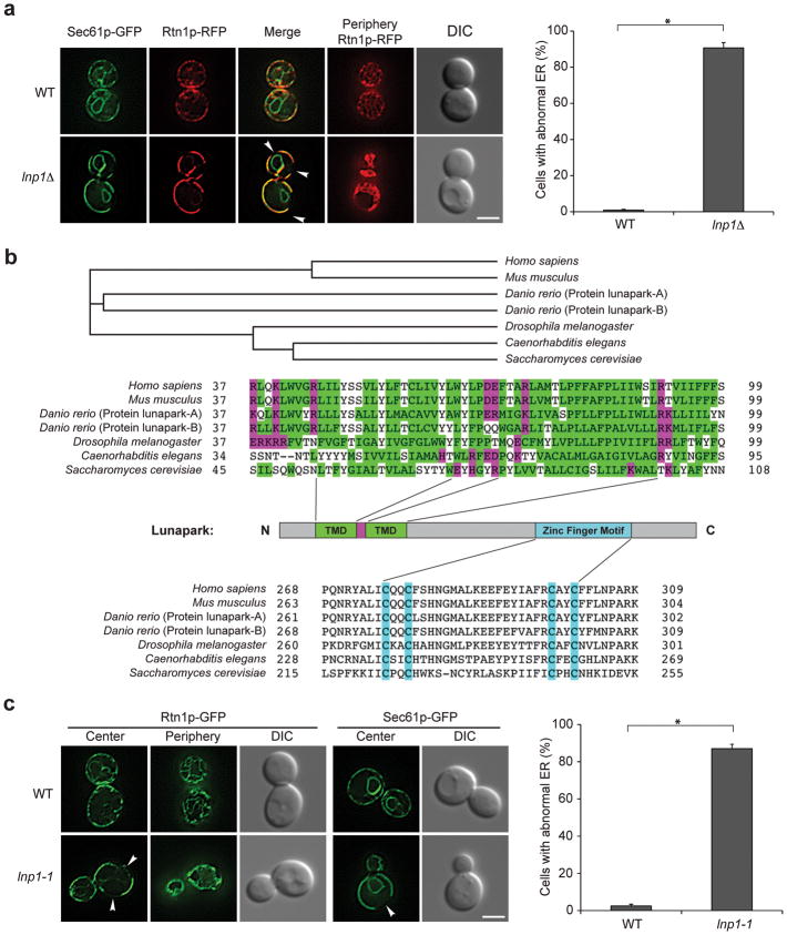
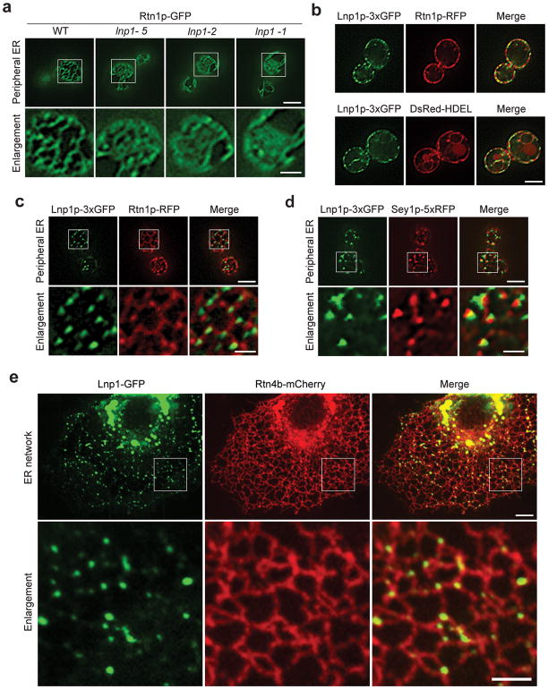
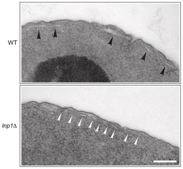
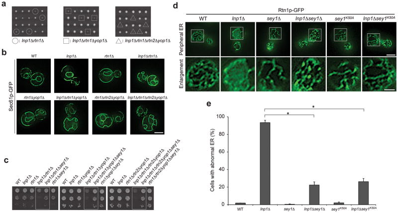
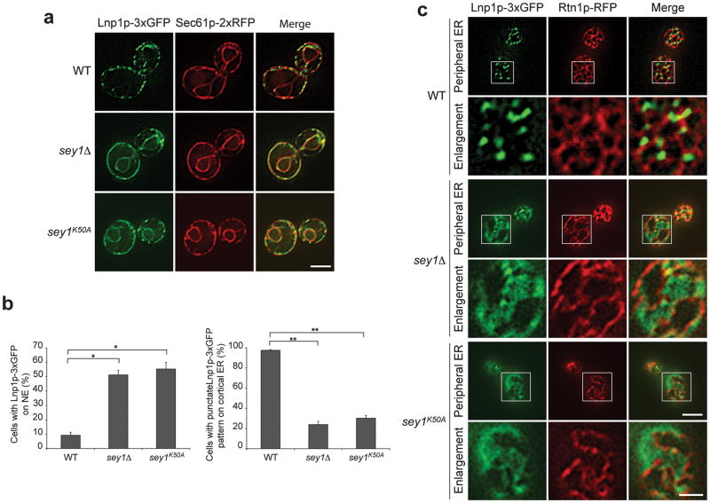
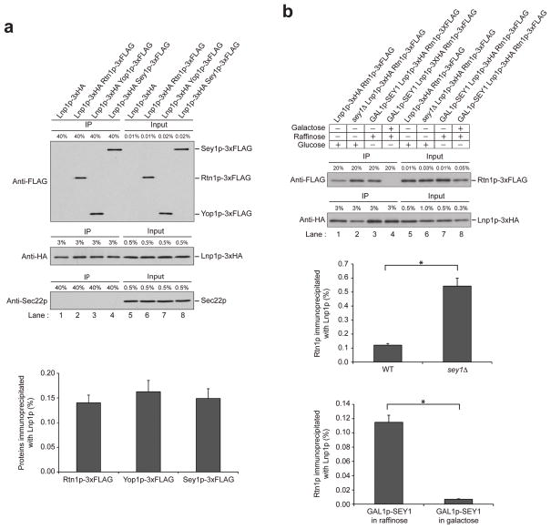
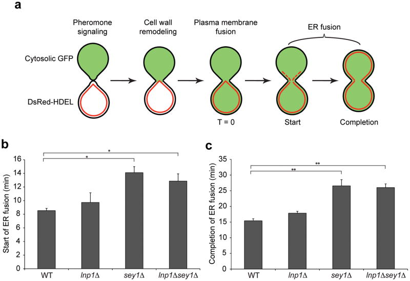
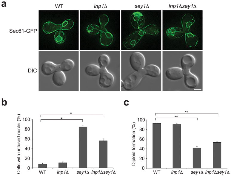
Although studies on endoplasmic reticulum (ER) structure and dynamics have focused on the ER tubule-forming proteins (reticulons and DP1/Yop1p) and the tubule fusion protein atlastin, nothing is known about the proteins and processes that act to counterbalance this machinery. Here we show that Lnp1p, a member of the conserved Lunapark family, plays a role in ER network formation. Lnp1p binds to the reticulons and Yop1p and resides at ER tubule junctions in both yeast and mammalian cells. In the yeast Saccharomyces cerevisiae, the interaction of Lnp1p with the reticulon protein, Rtn1p, and the localization of Lnp1p to ER junctions are regulated by Sey1p, the yeast orthologue of atlastin. We propose that Lnp1p and Sey1p act antagonistically to balance polygonal network formation. In support of this proposal, we show that the collapsed, densely reticulated ER network in lnp1 Δ cells is partially restored when the GTPase activity of Sey1p is abrogated.


Figures

Figure 1
Figure 1. Cortical ER morphology is abnormal in the lnp1 mutant
a, Yeast cells expressing Sec61p-GFP and Rtn1p-RFP (SFNY 2111, SFNY 2112) were grown to early log phase in YPD medium at 25°C. Subsequently, the cells were harvested and directly examined under the fluorescence microscope. The percentage of cells with abnormal ER morphology was quantified (right) from cells expressing Rtn1p-RFP. Error bars are S.E.M for three separate experiments, n=297 cells for wild type and n=258 cells for the lnp1Δ mutant. *P < 0.001 Student’s t-test. b, Alignment of the transmembrane domain and zinc finger motif of the lunapark protein from yeast to man. Hydrophobic amino acids are shown in green and charged amino acids in purple. The four cysteines that form an atypical Cys4 type zinc finger motif are highlighted in blue. The beginning and end of the transmembrane domains are marked on the Saccharomyces cerevisiae sequence. c, The zinc finger motif plays a role in ER morphology. Same as a only the following number of cells was quantitated (right), n = 334, wild type (SFNY 2088, SFNY 2094); n = 391, lnp1-1(SFNY 2116, SFNY 2120). Error bars represent S.E.M for three separate experiments *P < 0.001 Student’s t-test. The arrowheads in a and c point to areas of the cortex that lack cortical ER. The scale bar is 3 μm.
Figure 2
Figure 2. Lnp1p resides at three way junctions
a, The ER network collapses in lnp1 mutant cells. Wild type (SFNY 2088) and mutant cells expressing Rtn1p-GFP were grown to early log phase in YPD medium at 25°C and harvested. Live cells were directly examined under the fluorescence microscope and the images were deconvolved. The boxed area is enlarged in the bottom panel. For the lnp1-1 mutant (SFNY 2116), the zinc finger mutations are C223A C226A C244A C247A. For the lnp1-5 mutant (SFNY 2142), the zinc finger mutation is C223A. For the lnp1-2 mutant (SFNY 2113), the zinc finger mutations are C223A C226A. b, Lnp1p-3xGFP is present on puncta that line the tubular ER. Cells expressing Lnp1p-3xGFP and Rtn1p-RFP (SFNY 2093, top) or Lnp1p-3xGFP and DsRed-HDEL (SFNY 2100, bottom) were grown to early log phase in YPD medium at 25°C, harvested and the live cells were directly examined under the fluorescence microscope. c, Lnp1p-3xGFP localizes to three way junctions. Cells expressing Lnp1p-3xGFP and Rtn1p-RFP (SFNY 2093) were grown and examined as in a. The boxed area is enlarged in the bottom panel. d, Lnp1p-3xGFP partially overlaps or is adjacent to Sey1p-RFP at three way junctions. Yeast cells expressing Lnp1p-3xGFP and Sey1p-RFP (SFNY 2143) were grown and examined as in a. e, Human Lnp1-GFP localizes to the three way junctions of the ER marked by Rtn4b-mCherry in COS-7 cells. The boxed area is enlarged in the bottom panel. Scale bars in b and the peripheral images in a, c, and d are 3 μm. Scale bars in the enlarged images in a, c, and d are 1 μm. Scale bar in e (top) is 10 μm, and the scale bar in the enlarged images in e (bottom) is 5 μm.
Figure 3
Figure 3. The cortical ER is highly reticulated in the lnp1Δ mutant
Yeast cells were grown, harvested and thin section analysis was performed as described in the Methods. In the cross section view, the arrows point to segments of ER in wild type (black arrowheads) and lnp1Δ mutant cells (white arrowheads). Scale bar is 200 nm.
Figure 4
Figure 4. Lnp1p acts synergistically with the reticulons and Yop1p, but antagonistically with Sey1p
a, Lnp1p acts synergistically with the reticulons and Yop1p. Diploid lnp1Δ/LNP1 rtn1Δ/RTN1, lnp1Δ/LNP1 rtn1Δ/RTN1 yop1Δ/yop1Δ, and lnp1Δ/LNP1 rtn1Δ/RTN1 rtn2Δ/rtn2Δ yop1Δ/yop1Δ yeast cells were sporulated, dissected on YPD plates and the meiotic products were incubated for 3–5 days at 25°C. The lnp1Δrtn1Δ double mutant cells are marked with a white circle. The lnp1Δrtn1Δ yop1Δ triple mutant cells are marked with white squares. The lnp1Δrtn1Δrtn2Δyop1Δ quadruple mutant cells are marked with white triangles. b, Yeast cells expressing Sec61p-GFP (SFNY 2094, SFNY 2269, SFNY 2271, SFNY 2095, SFNY 2098, SFNY 2105, SFNY 2106, SFNY 2123) were grown to early log phase in YPD medium at 25°C, harvested and the livecells were directly examined under the fluorescence microscope. c, Lnp1p acts antagonistically with Sey1p.Yeast cells were grown to early stationary phase in YPD medium at 25°C, serially diluted and spotted on YPD plates. The plates were then incubated for 3–4 days at 25°C. The double, triple, quadruple, and quintuple mutant combinations examined in Fig. S3 are marked in white boxes. d, Loss of the GTPase activity of Sey1p is sufficient to suppress the ER morphology defect in the lnp1Δ mutant. Yeast cells expressing Rtn1p-GFP (SFNY 2088, SFNY 2090, SFNY 2126, SFNY 2128, SFNY 2147, SFNY 2148) were grown to early log phase in YPD medium at 25°C, harvested and the livecells were directly examined under the fluorescence microscope. Note that in our strain background sey1Δ has a more pronounced ER defect than previously reported . The loss of Yop1p does not enhance the phenotype we see. e, Quantitation of the data in d. The percentage of cells with abnormal ER morphology was determined and the number of cells quantitated is listed below. Error bars represent S.E.M. for three separate experiments, n = 277 wild type; n = 298 lnp1Δ; n= 229 sey1Δ; n = 278 lnp1Δsey1Δ; n = 172 sey1K50A; n = 333 lnp1Δsey1K50A. *P < 0.001 Student’s t-test. The scale bars in b and peripheral image in d are 3 μm. The scale bar in the enlarged images is 1 μm.
Figure 5
Figure 5. The GTPase activity of Sey1p retains Lnp1p at three way junctions
a, Yeast cells expressing Lnp1p-3xGFP and Sec61p-2xRFP (SFNY 2109, SFNY 2149, SFNY 2272) were grown to early log phase in YPD medium at 25°C, harvested and the live cells were examined directly under the fluorescence microscope. b, Left, quantitation of the nuclear envelope (NE) pattern of Lnp1p-3xGFP in wild type and sey1 mutants. The number of cells examined is listed below. Error bars represent S.E.M. for three separate experiments, n = 296 wild type; n = 288 sey1Δ; n = 281 sey1K50A. *P < 0.001 Student’s t-test. Right, the percentage of cells in which Lnp1p-3xGFP is in a punctate cortical ER pattern was quantitated. The number of cells examined is listed below. Error bars represent S.E.M. for three separate experiments, n = 348 wild type; n = 336 sey1Δ; n = 243 sey1K50A. **P < 0.0001 Student’s t-test. c, Yeast cells expressing Lnp1p-3xGFP and Rtn1p-RFP (SFNY 2093, SFNY 2133, SFNY2146) were grown to early log phase in YPD medium at 25°C, harvested and the live cells were examined as above. The scale bars in a and peripheral images in c are 3 μm. The scale bar in the enlarged images is 1 μm.
Figure 6
Figure 6. Sey1p regulates the interaction of Lnp1p with Rtn1p
a, Lnp1p interacts with Rtn1p, Yop1p and Sey1p. Yeast lysates were prepared from cells expressing either Lnp1p-3xHA and Rtn1p-3xFLAG (SFNY 2102), Lnp1p-3xHA and Yop1p-3xFLAG (SFNY 2103), or Lnp1p-3xHA and Sey1p-3xFLAG (SFNY 2104). Lysates were immunoprecipitated with anti-HA antibody and analyzed by western blot analysis using anti-FLAG, anti-HA and anti-Sec22p antibodies. Quantitation of the immunoprecipitates is shown on the bottom. Error bars represent S.D., n=3 b, Yeast lysates were prepared from cells expressing Lnp1p-3xHA and Rtn1p-3xFLAG when Sey1p was either absent or overexpressed (SFNY 2132, SFNY 2131). Overexpression of Sey1p was induced in YP medium with 0.1% galactose. Lysates were immunoprecipitated with anti-HA antibody and western blot analysis was performed using anti-FLAG antibody. The percent of the immunoprecipitate (IP) and lysate (Input) loaded on the gel is indicated at the top of each lane. Quantitation of the immunoprecipitates is shown on the bottom. Error bars represent S.E.M., n=3. *P < 0.02 Student’s t-test.
Figure 7
Figure 7. The sey1Δ mutant exhibits a delay in cortical ER fusion that is not suppressed by the loss of Lnp1p
a, Haploid MATa and MATα yeast cells expressing cytosolic GFP or DsRed-HDEL were grown to log phase in SC media. An equal number of cells of each mating type were mixed, incubated on an agarose pad of SC medium at 25°C for 40–60 min and cortical ER fusion was observed by fluorescence microscopy. Cell-cell fusion was marked by cytosolic GFP mixing (T=0). b, Quantitation of the time from T=0 to the start of ER fusion, marked by the appearance of DsRed-HDEL in the GFP expressing cell. Error bars represent S.E.M. for three separate experiments, n = 40 wild type; n = 40 lnp1Δ; n= 50 sey1Δ; n = 49 lnp1Δsey1Δ. *P < 0.02 Student’s t-test. c, Quantitation of the time it takes to complete ER fusion, from T=0 to the time it takes DsRed-HDEL to evenly spread into the GFP expressing cell. Error bars represent S.E.M. for three separate experiments, n = 40 wild type; n = 40 lnp1Δ; n= 50 sey1Δ; n = 49 lnp1Δsey1Δ. **P < 0.01 Student’s t-test.
Figure 8
Figure 8. The sey1Δ mutant exhibits a defect in nuclear fusion that is not suppressed by the loss of Lnp1p
a, Haploid MATa and MATα yeast cells expressing Sec61p-GFP were grown to early stationary phase. Equal numbers of cells of each mating type were mixed, incubated at 25°C for 2–4 hr on a YPD plate and examined for nuclear fusion under the fluorescence microscope. b, Quantitation of the data in a. The number of cells examined is listed below. Error bars represent S.E.M. for three separate experiments, n = 65 wild type; n = 55 lnp1Δ; n= 38 sey1Δ; n = 48 lnp1Δsey1Δ. *P < 0.01, Student’s t-test. c, Same as a except zygotes were picked, transferred to a YPD plate and incubated for 4 days at 25°C. Cells were considered to be diploid if they sporulated and could no longer mate with MATa and MATα tester strains. The number of cells examined is listed below. Error bars represent S.E.M., n = 85 wild type; n = 95 lnp1Δ; n= 123 sey1Δ; n = 127 lnp1Δsey1Δ. **P < 0.005 Student’s t-test. The scale bar is 3 μm.

References

    1. Du Y, Ferro-Novick S, Novick P. Dynamics and inheritance of the endoplasmic reticulum. J Cell Sci. 2004;117:2871–2878. - PubMed
    1. Voeltz GK, Prinz WA, Shibata Y, Rist JM, Rapoport TA. A class of membrane proteins shaping the tubular endoplasmic reticulum. Cell. 2006;124:573–586. - PubMed
    1. Lee C, Chen LB. Dynamic behavior of endoplasmic reticulum in living cells. Cell. 1988;54:37–46. - PubMed
    1. De Craene JO, et al. Rtn1p is involved in structuring the cortical endoplasmic reticulum. Mol Biol Cell. 2006;17:3009–3020. - PMC - PubMed
    1. Hu J, et al. Membrane proteins of the endoplasmic reticulum induce high-curvature tubules. Science. 2008;319:1247–1250. - PubMed

Publication types

MeSH terms