. 2012 Aug;154(8):1361-70; discussion 1370.
doi: 10.1007/s00701-012-1418-x. Epub 2012 Jun 23.

The relationship between Cho/NAA and glioma metabolism: implementation for margin delineation of cerebral gliomas

Affiliations

The relationship between Cho/NAA and glioma metabolism: implementation for margin delineation of cerebral gliomas

Jun Guo et al. Acta Neurochir (Wien). 2012 Aug.

Abstract

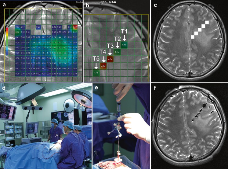
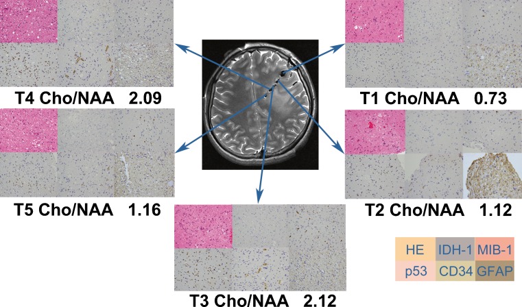
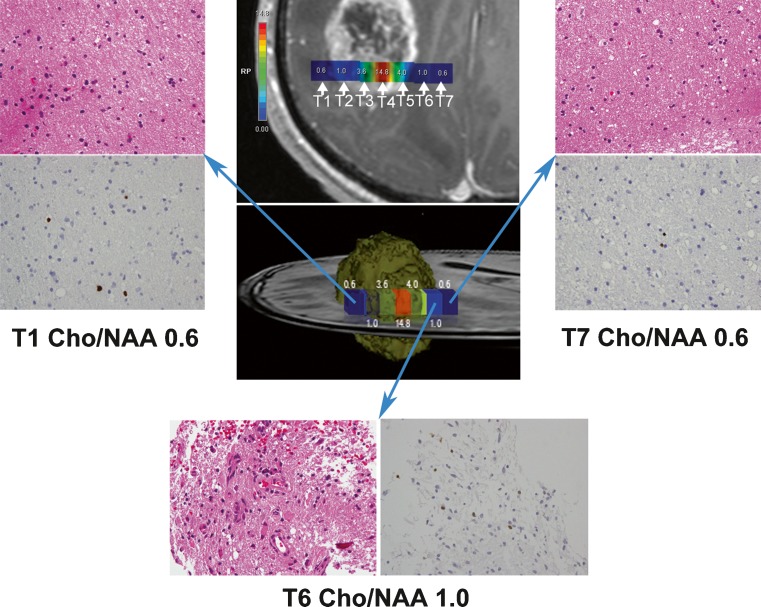
Background: The marginal delineation of gliomas cannot be defined by conventional imaging due to their infiltrative growth pattern. Here we investigate the relationship between changes in glioma metabolism by proton magnetic resonance spectroscopic imaging ((1)H-MRSI) and histopathological findings in order to determine an optimal threshold value of choline/N-acetyl-aspartate (Cho/NAA) that can be used to define the extent of glioma spread.

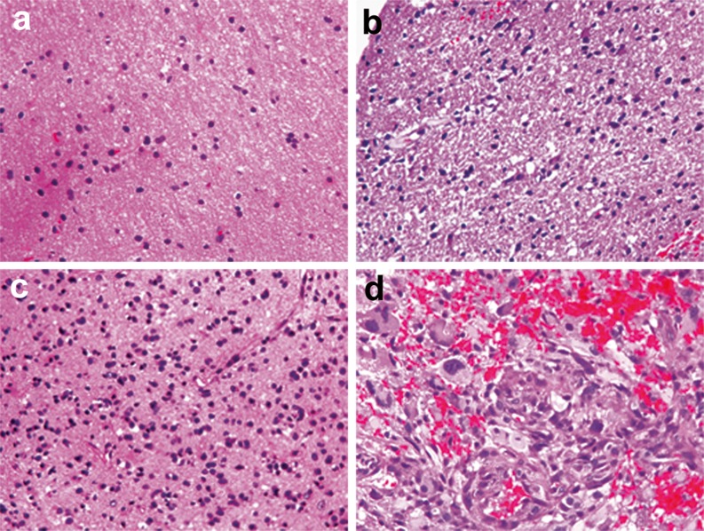
Method: Eighteen patients with different grades of glioma were examined using (1)H-MRSI. Needle biopsies were performed under the guidance of neuronavigation prior to craniotomy. Intraoperative magnetic resonance imaging (MRI) was performed to evaluate the accuracy of sampling. Haematoxylin and eosin, and immunohistochemical staining with IDH1, MIB-1, p53, CD34 and glial fibrillary acidic protein (GFAP) antibodies were performed on all samples. Logistic regression analysis was used to determine the relationship between Cho/NAA and MIB-1, p53, CD34, and the degree of tumour infiltration. The clinical threshold ratio distinguishing tumour tissue in high-grade (grades III and IV) glioma (HGG) and low-grade (grade II) glioma (LGG) was calculated.

Results: In HGG, higher Cho/NAA ratios were associated with a greater probability of higher MIB-1 counts, stronger CD34 expression, and tumour infiltration. Ratio threshold values of 0.5, 1.0, 1.5 and 2.0 appeared to predict the specimens containing the tumour with respective probabilities of 0.38, 0.60, 0.79, 0.90 in HGG and 0.16, 0.39, 0.67, 0.87 in LGG.

Conclusions: HGG and LGG exhibit different spectroscopic patterns. Using (1)H-MRSI to guide the extent of resection has the potential to improve the clinical outcome of glioma surgery.


Figures

Fig. 1
Fig. 1
a T2-weighted MR image superimposed with coloured voxels from patient 4 with grade II astrocytoma. b Five voxels (T1, T2, T3, T4, T5) with IDs 197, 182, 167, 152, 137 corresponded to Cho/NAA ratio 0.73, 1.12, 2.12, 2.09, 1.16 respectively arranged from right to left were chosen by referring to the conventional MR images and local metabolic information together. c The five voxels were labelled in the neuronavigation data sets. d Positioning and calibration of navigation probe. e Skull-mounted trajectory guide guarantees the direction of biopsy needle. f The accurate sampling for needle biopsy was confirmed by intraoperative MR scanning
Fig. 2
Fig. 2
H & E (×400) and immunohistochemical staining with IDH-1, MIB-1, p53, CD34 and GFAP antibodies (×400, respectively) were performed respectively in each biopsy sample. The maximal Cho/NAA ratio (2.12) corresponded to the maximal value of cell density, MIB-1 and p53. The minimal Cho/NAA ratio (0.73) corresponded to the minimal value of cell density, MIB-1, and p53. All specimen were strong immunopositive for GFAP and moderate immunopositive for CD34, but immunonegative for IDH-1
Fig. 3
Fig. 3
Criteria for tumour infiltration. a Without tumour infiltration (normal brain or gliosis) (×400). b Mild tumour infiltration (low cell density, mild nuclear atypia) (×400). c Moderate tumour infiltration (moderate cell density, obvious nuclear atypia, few mitoses) (×400). d Heavy tumour infiltration (high cell density, obvious nuclear atypia, abundant mitoses) (×400)
Fig. 4
Fig. 4
Contrast-enhanced T1-weighted image superimposed with coloured voxels (upper middle) from patient 5 with glioblastoma multiforme. Seven voxels (T1, T2, T3, T4, T5, T6, T7) with IDs 43, 59, 75, 91, 107, 123, 139 arranged from left to right. A 3-D ideograph of the case (centre) rendered using Photoshop. H & E stained sections (×400) and MIB-1 (×400) showed the infiltration degree of T1, T6 and T7. Both T1 (upper left) and T6 (bottom) were located in IPR but had different Cho/NAA ratios. Histopathology confirmed that specimens from T1 and T6 were infiltrated differently. T1 and T7 (upper right) were located in IPR and DPR respectively but they shared the same Cho/NAA ratio and histopathology confirmed that specimens from T1 and T7 were infiltrated similarly

References

    1. Capper D, Sahm F, Hartmann C, Meyermann R, von Deimling A, Schittenhelm J. Application of mutant IDH1 antibody to differentiate diffuse glioma from nonneoplastic central nervous system lesions and therapy-induced changes. Am J Surg Pathol. 2010;34:1199–1204. doi: 10.1097/PAS.0b013e3181e7740d. - DOI - PubMed
    1. Capper D, Weissert S, Balss J, Habel A, Meyer J, Jager D, Ackermann U, Tessmer C, Korshunov A, Zentgraf H, Hartmann C, von Deimling A. Characterization of R132H mutation-specific IDH1 antibody binding in brain tumors. Brain Pathol. 2010;20:245–254. doi: 10.1111/j.1750-3639.2009.00352.x. - DOI - PMC - PubMed
    1. Capper D, Zentgraf H, Balss J, Hartmann C, von Deimling A. Monoclonal antibody specific for IDH1 R132H mutation. Acta Neuropathol. 2009;118:599–601. doi: 10.1007/s00401-009-0595-z. - DOI - PubMed
    1. Chen J, Huang SL, Li T, Chen XL. In vivo research in astrocytoma cell proliferation with 1H-magnetic resonance spectroscopy: correlation with histopathology and immunohistochemistry. Neuroradiology. 2006;48:312–318. doi: 10.1007/s00234-006-0066-3. - DOI - PubMed
    1. Claus EB, Horlacher A, Hsu L, Schwartz RB, Dello-Iacono D, Talos F, Jolesz FA, Black PM. Survival rates in patients with low-grade glioma after intraoperative magnetic resonance image guidance. Cancer. 2005;103:1227–1233. doi: 10.1002/cncr.20867. - DOI - PubMed

Publication types