

The acute effect of running on knee articular cartilage and meniscus magnetic resonance relaxation times in young healthy adults
- PMID: 22729505
- PMCID: PMC3660554
- DOI: 10.1177/0363546512449816
The acute effect of running on knee articular cartilage and meniscus magnetic resonance relaxation times in young healthy adults
Abstract
Background: Understanding the acute response of healthy knee cartilage to running may provide valuable insight into functional properties. In recent years, quantitative magnetic resonance (MR) imaging techniques (T1(ρ) and T2 relaxation measurement) have shown tremendous potential and unique ability to noninvasively and quantitatively determine cartilage response to physiologic levels of loading occurring with physiologic levels of exercise.
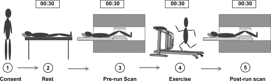
Purpose: To measure the short-term changes in MR T1(ρ) and T2 relaxation times of knee articular cartilage and meniscus in healthy individuals immediately after 30 minutes of running.
Study design: Descriptive laboratory study.
Methods: Twenty young healthy volunteers, aged 22 to 35 years, underwent 3T MR imaging of the knee before and immediately after 30 minutes of running. Quantitative assessment of the cartilage and menisci was performed using MR images with a T1(ρ) and T2 mapping technique. After adjusting for age, sex, and body mass index, repeated-measures analysis of variance was used to determine the effects of running on MR relaxation times.
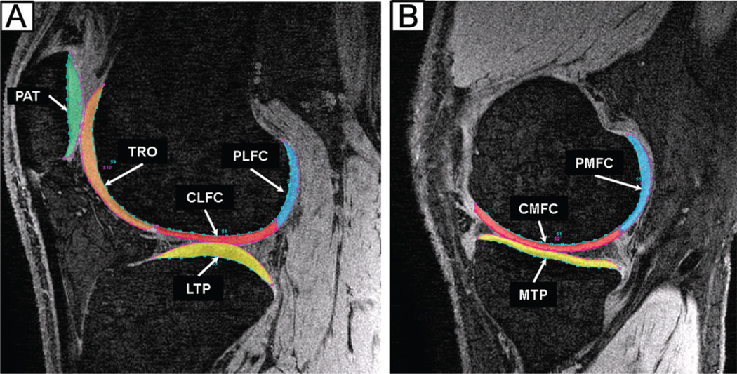
Results: The post-run T1(ρ) and T2 measurement showed significant reduction in all regions of cartilage except the lateral tibia when compared with the pre-run condition. The medial tibiofemoral (T1(ρ): 9.4%, P < .0001; T2: 5.4%, P = .0049) and patellofemoral (T1(ρ): 12.5%, P < .0001; T2: 5.7%, P = .0007) compartments experienced the greatest reduction after running. The superficial layer of the articular cartilage showed significantly higher change in relaxation times than the deep layer (T1(ρ): 9.6% vs 8.2%, P = .050; T2: 6.0% vs 3.5%, P = .069). The anterior and posterior horns of the medial meniscus (9.7%, P = .016 and 11.4%, P = .001) were the only meniscal subregions with significant changes in T1(ρ) after running.
Conclusion: Shorter T1(ρ) and T2 values after running suggest alteration in the water content and collagen fiber orientation of the articular cartilage. Greater changes in relaxation times of the medial compartment and patellofemoral joint cartilage indicate greater load sharing by these areas during running.
Figures
References
-
- Akella SV, Regatte RR, Gougoutas AJ, et al. Proteoglycan-induced changes in T1rho-relaxation of articular cartilage at 4T. Magn Reson Med. 2001;46(3):419–423. - PubMed
