

Increased tumor-infiltrating CD8(+)Foxp3(+) T lymphocytes are associated with tumor progression in human gastric cancer
- PMID: 22729557
- PMCID: PMC11029073
- DOI: 10.1007/s00262-012-1277-6
Increased tumor-infiltrating CD8(+)Foxp3(+) T lymphocytes are associated with tumor progression in human gastric cancer
Abstract
Background: CD8(+)Foxp3(+) T lymphocytes have been detected in tumors. However, the distribution, phenotypic features, and regulation of these cells in gastric cancer remain unknown.
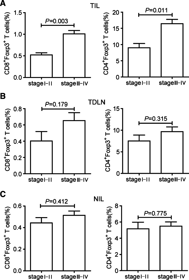
Methods: The levels of CD8(+)Foxp3(+) T lymphocytes in the peripheral blood, tumor-draining lymph nodes, non-tumor tissues, and tumor tissues of patients with gastric cancer were detected by flow cytometry. Foxp3 induction in CD8(+)Foxp3(-) T cells was investigated in vitro. The suppressive function of CD8(+)Foxp3(+) T lymphocytes was analyzed by their effect on CD4(+) T-cell proliferation and IFN-γ production. The percentages of CD8(+)Foxp3(+) T lymphocytes were evaluated for the association with tumor stage.
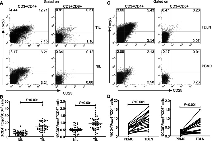
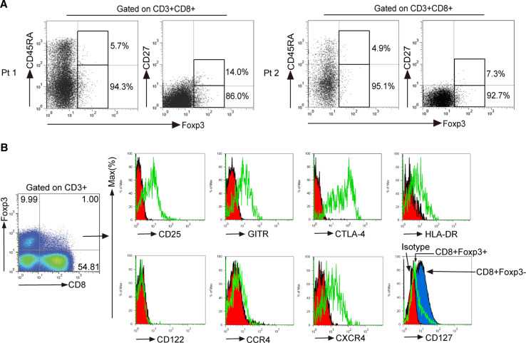
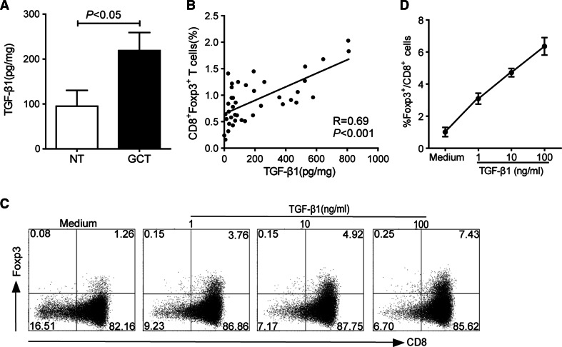
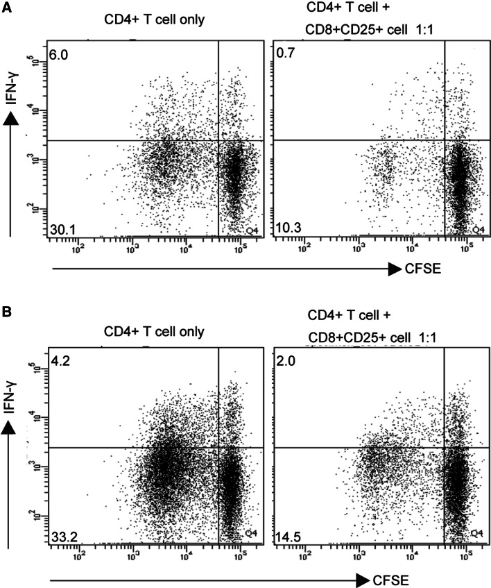
Results: The frequency of CD8(+)Foxp3(+) T lymphocytes in tumor tissues was significantly higher than that in non-tumor tissues, and similar results were also observed in tumor-draining lymph nodes compared with peripheral blood. Most intratumoral CD8(+)Foxp3(+) T lymphocytes were activated effector cells (CD45RA(-)CD27(-)). TGF-β1 levels were positively correlated with the frequency of CD8(+)Foxp3(+) T lymphocytes in tumor tissues, and in vitro TGF-β1 could induce the generation of CD8(+)Foxp3(+) T lymphocytes in a dose-dependent manner. Furthermore, intratumoral CD8(+)Foxp3(+) T lymphocytes suppressed the proliferation and IFN-γ production of CD4(+) T cells. Finally, intratumoral CD8(+)Foxp3(+) T lymphocytes were significantly increased with tumor progression in terms of tumor-node-metastasis (TNM) stage.
Conclusions: Our data have shown that increased intratumoral CD8(+)Foxp3(+) T lymphocytes are associated with tumor stage and potentially influence CD4(+) T-cell functions, which may provide insights for developing novel immunotherapy protocols against gastric cancer.
Conflict of interest statement
The authors declare that they have no conflict of interest.
Figures
Similar articles
-
Determination of a CD4+CD25-FoxP3+ T cells subset in tumor-draining lymph nodes of colorectal cancer secreting IL-2 and IFN-γ.Tumour Biol. 2016 Nov;37(11):14659-14666. doi: 10.1007/s13277-016-5345-y. Epub 2016 Sep 13. Tumour Biol. 2016. PMID: 27619682
-
B7-H4 overexpression impairs the immune response of T cells in human cervical carcinomas.Hum Immunol. 2014 Dec;75(12):1203-9. doi: 10.1016/j.humimm.2014.10.002. Epub 2014 Oct 13. Hum Immunol. 2014. PMID: 25446402
-
CD4/CD8 + T cells, DC subsets, Foxp3, and IDO expression are predictive indictors of gastric cancer prognosis.Cancer Med. 2019 Dec;8(17):7330-7344. doi: 10.1002/cam4.2596. Epub 2019 Oct 20. Cancer Med. 2019. PMID: 31631566 Free PMC article.
-
The significance of Treg cells in defective tumor immunity.Arch Immunol Ther Exp (Warsz). 2008 May-Jun;56(3):181-91. doi: 10.1007/s00005-008-0018-1. Epub 2008 May 30. Arch Immunol Ther Exp (Warsz). 2008. PMID: 18512029 Review.
-
CD4+CD8+ double-positive T cells in immune disorders and cancer: Prospects and hurdles in immunotherapy.Autoimmun Rev. 2025 Feb 28;24(3):103757. doi: 10.1016/j.autrev.2025.103757. Epub 2025 Jan 22. Autoimmun Rev. 2025. PMID: 39855286 Review.
Cited by
-
Increased tumor-infiltrating CD45RA-CCR7- regulatory T-cell subset with immunosuppressive properties foster gastric cancer progress.Cell Death Dis. 2017 Aug 17;8(8):e3002. doi: 10.1038/cddis.2017.388. Cell Death Dis. 2017. PMID: 28817117 Free PMC article.
-
The prognostic influence of tumor infiltrating Foxp3(+)CD4(+), CD4(+) and CD8(+) T cells in resected non-small cell lung cancer.J Inflamm (Lond). 2015 Nov 23;12:63. doi: 10.1186/s12950-015-0108-x. eCollection 2015. J Inflamm (Lond). 2015. PMID: 26604855 Free PMC article.
-
Regulatory T cells and the immune aging process: a mini-review.Gerontology. 2014;60(2):130-7. doi: 10.1159/000355303. Epub 2013 Nov 28. Gerontology. 2014. PMID: 24296590 Free PMC article. Review.
-
Sunitinib represses regulatory T cells to overcome immunotolerance in a murine model of hepatocellular cancer.Oncoimmunology. 2017 Sep 21;7(1):e1372079. doi: 10.1080/2162402X.2017.1372079. eCollection 2017. Oncoimmunology. 2017. PMID: 29296523 Free PMC article.
-
An immune-related prognostic signature associated with immune landscape and therapeutic responses in gastric cancer.Aging (Albany NY). 2023 Feb 22;15(4):1074-1106. doi: 10.18632/aging.204534. Epub 2023 Feb 22. Aging (Albany NY). 2023. PMID: 36812479 Free PMC article.
References
-
- Galon J, Costes A, Sanchez-Cabo F, Kirilovsky A, Mlecnik B, Lagorce-Pagès C, Tosolini M, Camus M, Berger A, Wind P, Zinzindohoué F, Bruneval P, Cugnenc PH, Trajanoski Z, Fridman WH, Pagès F. Type, density, and location of immune cells within human colorectal tumors predict clinical outcome. Science. 2006;313(5795):1960–1964. doi: 10.1126/science.1129139. - DOI - PubMed
-
- Sharma P, Shen Y, Wen S, Yamada S, Jungbluth AA, Gnjatic S, Bajorin DF, Reuter VE, Herr H, Old LJ, Sato E. CD8 tumor-infiltrating lymphocytes are predictive of survival in muscle-invasive urothelial carcinoma. Proc Natl Acad Sci USA. 2007;104(10):3967–3972. doi: 10.1073/pnas.0611618104. - DOI - PMC - PubMed
Publication types
MeSH terms
Substances
LinkOut - more resources
Full Text Sources
Other Literature Sources
Medical
Research Materials
