. 2012 Sep;194(17):4608-18.
doi: 10.1128/JB.00378-12. Epub 2012 Jun 22.

RefZ facilitates the switch from medial to polar division during spore formation in Bacillus subtilis

Affiliations

RefZ facilitates the switch from medial to polar division during spore formation in Bacillus subtilis

Jennifer K Wagner-Herman et al. J Bacteriol. 2012 Sep.

Abstract

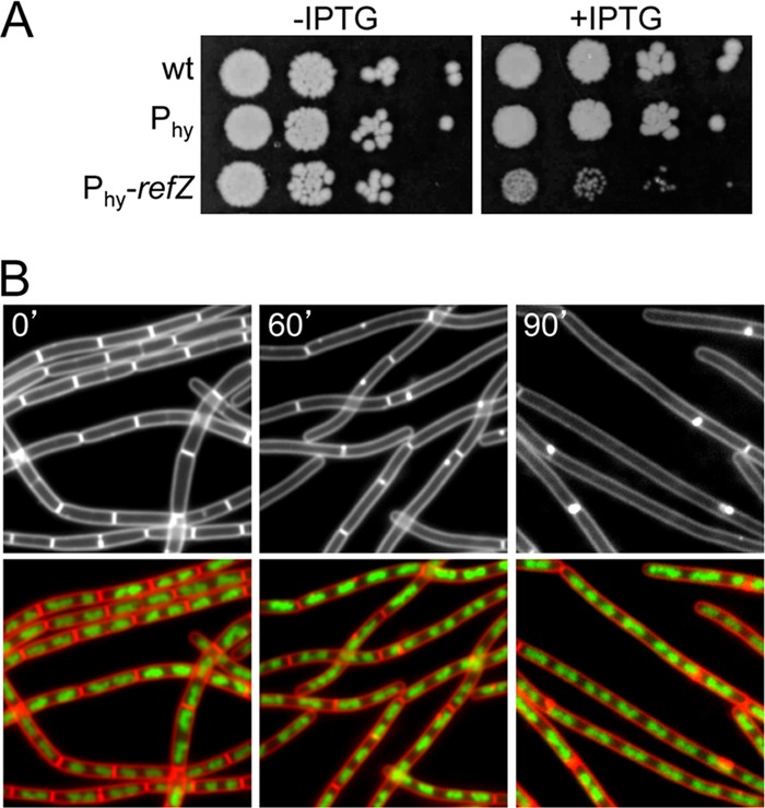
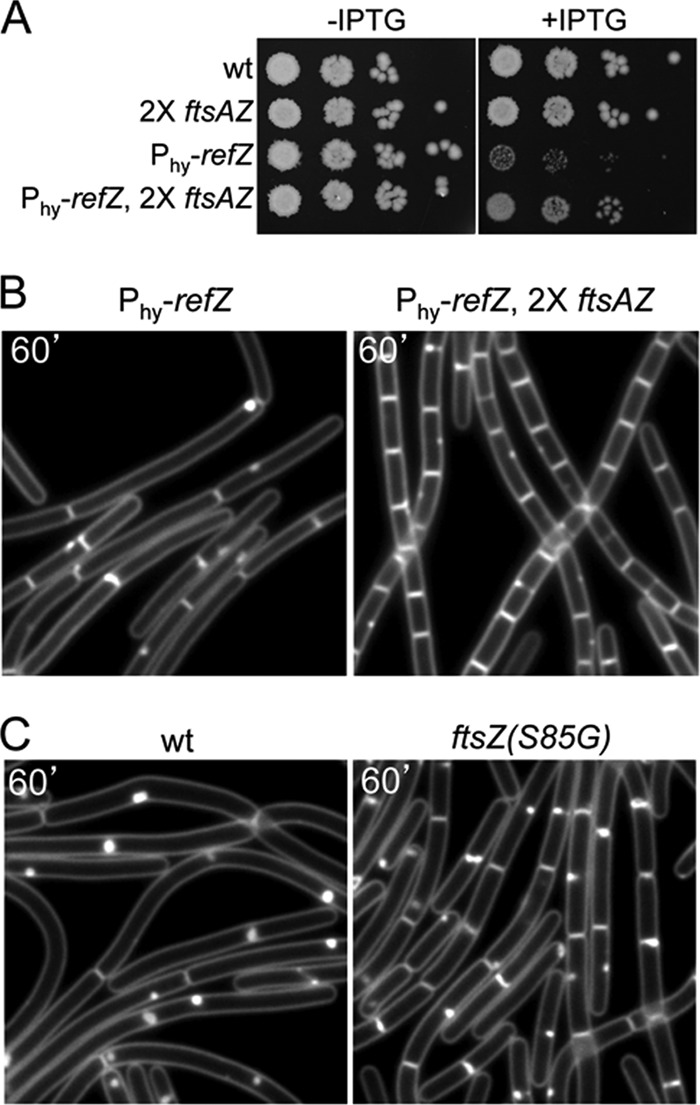
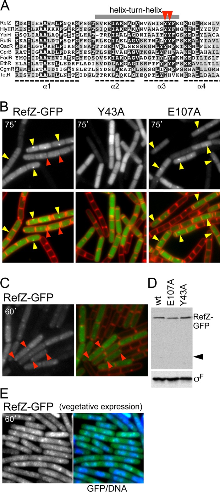
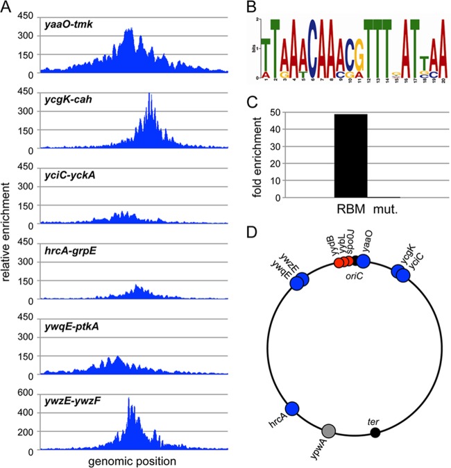
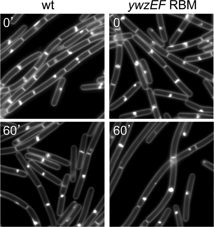
During sporulation, Bacillus subtilis redeploys the division protein FtsZ from midcell to the cell poles, ultimately generating an asymmetric septum. Here, we describe a sporulation-induced protein, RefZ, that facilitates the switch from a medial to a polar FtsZ ring placement. The artificial expression of RefZ during vegetative growth converts FtsZ rings into FtsZ spirals, arcs, and foci, leading to filamentation and lysis. Mutations in FtsZ specifically suppress RefZ-dependent division inhibition, suggesting that RefZ may target FtsZ. During sporulation, cells lacking RefZ are delayed in polar FtsZ ring formation, spending more time in the medial and transition stages of FtsZ ring assembly. A RefZ-green fluorescent protein (GFP) fusion localizes in weak polar foci at the onset of sporulation and as a brighter midcell focus at the time of polar division. RefZ has a TetR DNA binding motif, and point mutations in the putative recognition helix disrupt focus formation and abrogate cell division inhibition. Finally, chromatin immunoprecipitation assays identified sites of RefZ enrichment in the origin region and near the terminus. Collectively, these data support a model in which RefZ helps promote the switch from medial to polar division and is guided by the organization of the chromosome. Models in which RefZ acts as an activator of FtsZ ring assembly near the cell poles or as an inhibitor of the transient medial ring at midcell are discussed.


Figures

Fig 1
Fig 1
Expression of RefZ during vegetative growth blocks cell division. (A) The vegetative expression of RefZ impairs growth. Tenfold serial dilutions of early-logarithmic-phase cultures were spotted onto LB agar plates in the presence (+) and absence (−) of 1 mM IPTG. The wild type (wt) (PY79) and strains harboring the IPTG-inducible promoter Phy (BJW304) and Phy fused to refZ (BJW123) are shown. (B) Expression of RefZ inhibits cell division. Representative images of filamenting cells (BJW123) after the induction of RefZ are shown. The time (minutes) following the addition of IPTG is indicated. Membranes were visualized with FM4-64 (white in top images and red in bottom images), and DNA was visualized with DAPI (green).
Fig 2
Fig 2
RefZ targets the cell division protein FtsZ. (A) An additional copy of the ftsAZ operon suppresses the growth inhibition caused by RefZ. Tenfold serial dilutions of early-logarithmic-phase cultures were spotted onto LB agar plates in the presence (+) and absence (−) of 1 mM IPTG. The wild type (PY79) and strains harboring an additional copy of the ftsAZ operon (RL3063), Phy-refZ (BJW144), or both (BJW147) are shown. (B) Representative images of cells overexpressing RefZ in the absence (left) or presence (right) of an extra copy of the ftsAZ operon. Membranes were visualized with FM4-64. The time (minutes) following the addition of IPTG is indicated. (C) Cells harboring a point mutant of FtsZ (S85G) are resistant to RefZ-dependent cell division inhibition. Representative images show cells overexpressing RefZ that have wild FtsZ (BJW472) or the FtsZ(S85G) mutant (BJW479).
Fig 3
Fig 3
Expression of RefZ during vegetative growth disrupts FtsZ rings. (A) Localization of FtsZ-GFP before induction of refZ (BJW429), 60 min after refZ induction (BJW429), and 30 min after induction of mciZ (BJW481). (Top) FtsZ-GFP images. (Bottom) Overlay of membranes stained with FM4-64 (red) and FtsZ-GFP (green). (B) RefZ facilitates the switch of the FtsZ ring from medial to polar positions during sporulation. Histograms show the quantification of FtsZ-GFP localizations (medial, shifting, or polar) from the wild type (purple) (strain RL3056), a refZ mutant (dark blue) (strain BRB455), a spoIIE mutant (light blue) (strain BRB457), and a refZ spoIIE double mutant (green) (strain BRB459) 120 min after the induction of sporulation. Representative images of FtsZ-GFP localization are shown below the histogram. Values are averages ± standard deviations from 2 independent experiments. More than 300 cells were scored for each strain in each experiment.
Fig 4
Fig 4
RefZ localization requires an intact helix-turn-helix motif. (A) Alignment of the amino-terminal domain of RefZ with DNA binding domains from TetR superfamily protein members for which structures have been determined. The alignment was made by using ClustalW and cartooned by using BoxShade. Conserved (gray boxes) and highly conserved (black boxes) residues are indicated. The gray bar above the alignment highlights the helix-turn-helix (HTH) motif. The positions of tyrosine 43 and tyrosine 44 in the recognition helix are highlighted (red carets). Aligned proteins are HlyIIR (Bacillus cereus), FadR (B. subtilis), YbiH (Salmonella enterica serovar Typhimurium), RutR (E. coli), EthR (Mycobacterium smegmatis), QacR (Staphylococcus aureus), CprB (Streptomyces coelicolor), CgmR (Corynebacterium glutamicum), and TetR (E. coli). (B) RefZ-GFP localizes in discrete foci that require an intact HTH motif. Cells harboring refZ-gfp (strain BRB672), refZ(Y43A)-gfp (strain BRB698), or refZ(E107A)-gfp (strain BRB697) were visualized by fluorescence microscopy 75 min after the induction of sporulation. Images show RefZ-GFP (top) and an overlay (bottom) of RefZ-GFP (green) and membranes stained with TMA-DPH (red). Midcell foci (yellow carets) and more subtle foci present near the cell poles (red carets) are indicated. (C) RefZ-GFP localizes as weak polar foci at early stages of sporulation. Cells harboring refZ-gfp (strain BJW190) were visualized by fluorescence microscopy 60 min after the induction of sporulation. Images show RefZ-GFP (left) and an overlay of RefZ-GFP (green) and membranes stained with FM4-64 (red) (right). RefZ-GFP foci (red carets) present near the cell poles are indicated. (D) Immunoblot analysis shows that RefZ-GFP and point mutants remain intact during sporulation. RefZ-GFP was analyzed by using anti-GFP antibodies, and the caret identifies the predicted size of free GFP. σF was used to control for loading. RefZ-GFP forms foci that colocalize with the nucleoid when expressed during vegetative growth. (E) RefZ-GFP was induced in cells (strain BJK001) harboring Phy-refZ-gfp. Images show representative fields obtained 60 min after the induction of RefZ-GFP with 10 μM IPTG. Shown is RefZ-GFP alone and merged with DAPI-stained DNA (blue).
Fig 5
Fig 5
RefZ binds to specific sites on the chromosome. (A) RefZ binding sites identified by ChIP-seq. Plots show relative enrichment values (normalized over the input control DNA) for regions (1 to 3 kb) of interest. (B) Position-weighted matrix of the RefZ binding motif (RBM) identified in the RefZ binding regions. The height of each letter represents the relative degree of conservation of each base in all the sequences identified. (C) RefZ binds to the 20-bp RBM when moved to an ectopic location. The RBM from the ywzF locus (BRB789) and a mutated version (RBM mut.) (BRB790) were inserted at 87° (into the yhdG locus) on the B. subtilis chromosome. Binding was assessed by using ChIP-qPCR. Plots show relative enrichment values for a site adjacent to the RBM. (D) Schematic diagram showing the locations of the RefZ binding sites on the B. subtilis chromosome. Sharp peaks (blue circles) and broad peaks (red circles) are indicated. An additional RBM in the ypwA locus (gray circle) identified through bioinformatics is also shown. The replication origin (oriC) and terminus (ter) are indicated.
Fig 6
Fig 6
RefZ is a more potent division inhibitor when bound to DNA. Shown are representative images of cells expressing low levels of RefZ harboring an empty plasmid (left) (strain BJW538) or a plasmid harboring the RBM from the ywzF locus (right) (strain BJW537). Cells were grown to the mid-log phase and induced with 7.5 μM IPTG. The time (in minutes) before and after the addition of IPTG is indicated. Images show membranes stained with TMA-DPH. Before induction, strains harboring the empty plasmid and the RBM-containing plasmid had average cell lengths of 3.2 μm and 3.8 μm, respectively. Following induction, the average cell lengths were 4.0 μm and 6.0 μm, respectively.

References

    1. Adams DW, Errington J. 2009. Bacterial cell division: assembly, maintenance and disassembly of the Z ring. Nat. Rev. Microbiol. 7:642–653 - PubMed
    1. Aparicio O, Geisberg JV, Struhl K. 2004. Current protocols in molecular biology. Wiley, New York, NY
    1. Bailey TL, Elkan C. 1994. Fitting a mixture model by expectation maximization to discover motifs in biopolymers. Proc. Int. Conf. Intell. Syst. Mol. Biol. 2:28–36 - PubMed
    1. Baumeister R, Helbl V, Hillen W. 1992. Contacts between Tet repressor and tet operator revealed by new recognition specificities of single amino acid replacement mutants. J. Mol. Biol. 226:1257–1270 - PubMed
    1. Baumeister R, Muller G, Hecht B, Hillen W. 1992. Functional roles of amino acid residues involved in forming the alpha-helix-turn-alpha-helix operator DNA binding motif of Tet repressor from Tn10. Proteins 14:168–177 - PubMed

Publication types

Associated data

LinkOut - more resources