. 2012 Aug;79(8):553-63.
doi: 10.1002/mrd.22060. Epub 2012 Jul 9.

Autoregulation of Musashi1 mRNA translation during Xenopus oocyte maturation

Affiliations

Autoregulation of Musashi1 mRNA translation during Xenopus oocyte maturation

Karthik Arumugam et al. Mol Reprod Dev. 2012 Aug.

Abstract

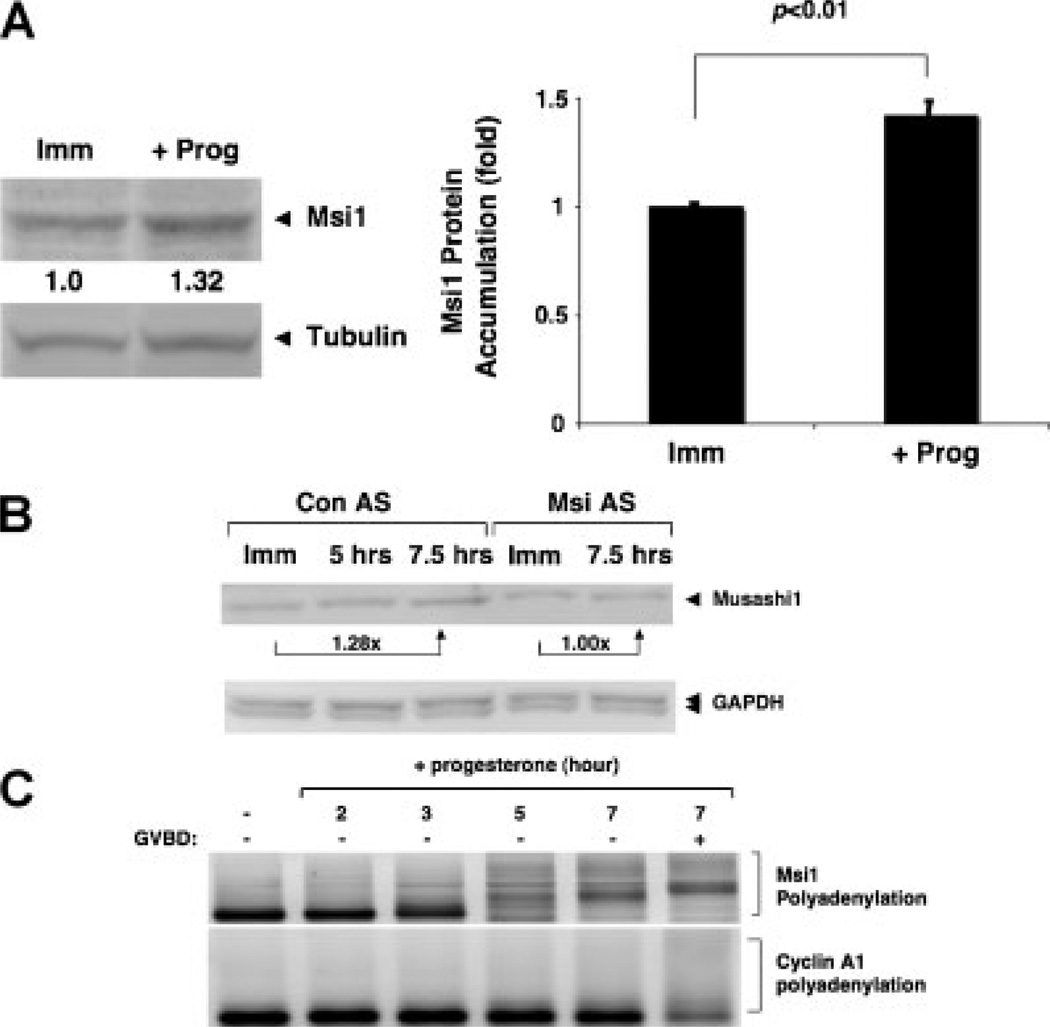
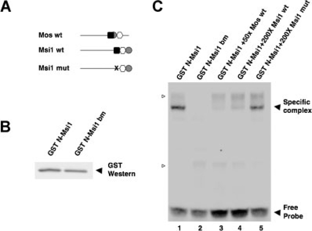
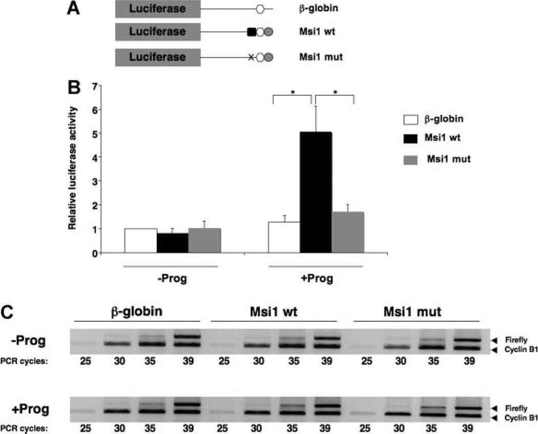
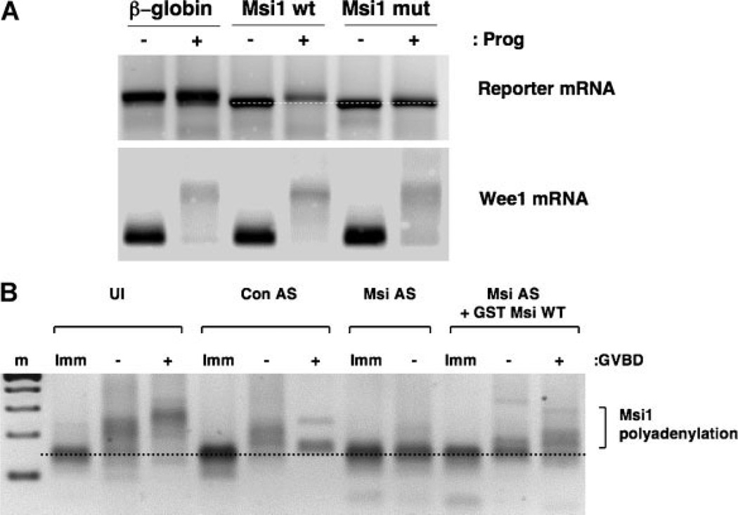
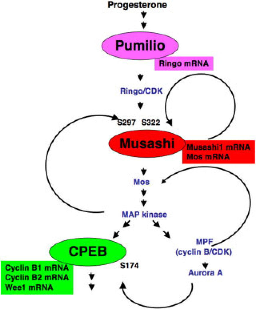
The mRNA translational control protein, Musashi, plays a critical role in cell fate determination through sequence-specific interactions with select target mRNAs. In proliferating stem cells, Musashi exerts repression of target mRNAs to promote cell cycle progression. During stem cell differentiation, Musashi target mRNAs are de-repressed and translated. Recently, we have reported an obligatory requirement for Musashi to direct translational activation of target mRNAs during Xenopus oocyte meiotic cell cycle progression. Despite the importance of Musashi in cell cycle regulation, only a few target mRNAs have been fully characterized. In this study, we report the identification and characterization of a new Musashi target mRNA in Xenopus oocytes. We demonstrate that progesterone-stimulated translational activation of the Xenopus Musashi1 mRNA is regulated through a functional Musashi binding element (MBE) in the Musashi1 mRNA 3' untranslated region (3' UTR). Mutational disruption of the MBE prevented translational activation of Musashi1 mRNA and its interaction with Musashi protein. Further, elimination of Musashi function through microinjection of inhibitory antisense oligonucleotides prevented progesterone-induced polyadenylation and translation of the endogenous Musashi1 mRNA. Thus, Xenopus Musashi proteins regulate translation of the Musashi1 mRNA during oocyte maturation. Our results indicate that the hierarchy of sequential and dependent mRNA translational control programs involved in directing progression through meiosis are reinforced by an intricate series of nested, positive feedback loops, including Musashi mRNA translational autoregulation. These autoregulatory positive feedback loops serve to amplify a weak initiating signal into a robust commitment for the oocyte to progress through the cell cycle and become competent for fertilization.


Figures

Figure 1
Figure 1
Xenopus Musashi1 mRNA is translationally activated in response to progesterone stimulation. A: Immature, stage-VI oocytes were left untreated (Imm) or stimulated with progesterone (+prog), and were analysed for endogenous Musashi1 (Msi1) protein accumulation by Western blot. When 50% of the progesterone-treated oocyte population reached GVBD (GVBD50), the oocytes were segregated and those that had not yet completed GVBD were analysed as representative of early pre-GVBD events (Charlesworth et al., 2002). Quantitations of fold-changes in Musashi1 levels (as indicated below Western blot) were normalized to tubulin from the same sample, and levels in time-matched, immature oocytes (Imm) were arbitrarily set to 1.0. The bar graph represents data from three independent experiments, with SEM indicated. Student’s t-test confirmed the significance of the differences between the sample sets (P < 0.01). B: Immature, stage-VI oocytes were injected with control antisense oligonucleotides (Con AS) or antisense oligonucleotides targeting both endogenous Musashi1 and Musashi2 mRNAs (Msi AS), and cultured overnight. Oocytes were then either left untreated (Imm) or stimulated with progesterone. In this experiment, oocytes reached GVBD50 at 7.5 hr and were segregated based on whether or not they completed GVBD. Those oocytes that had not completed GVBD were analysed, along with oocytes harvested 2.5 hr earlier. Msi AS-injected oocytes did not mature in response to progesterone, and time-matched samples were prepared at the Con AS 7.5 hr time point. Levels of endogenous Musashi1 (upper panel) and GAPDH (lower panel) protein were analysed by Western blot. Fold-change in Musashi1 protein between immature and 7.5 hr of progesterone is indicated. Similar results were seen in two additional experiments. C: Oocytes treated with or without progesterone for the indicated times were analysed for endogenous Musashi1 mRNA polyadenylation by RNA ligation-coupled PCR. In this experiment, oocytes reached GVBD50 at 7 hr and were segregated into those that had not (−) or had (+) completed GVBD. An increase in size of the PCR products is indicative of polyadenylation (Charlesworth et al., 2002; Charlesworth et al., 2004). Polyadenylation of the endogenous late class cyclin A1 mRNA in the same samples occurred after completion of GVBD, as expected.
Figure 2
Figure 2
The Musashi1 protein binds specifically to the MBE in the Xenopus Musashi1 mRNA 3′ UTR. A: Schematic representation of the Mos and Musashi1 3′ UTR constructs employed. Within the wildtype (wt) 3′ UTRs, elements are indicated by a black square (consensus Musashi binding site); white circle (consensus CPE); and grey hexagon (consensus polyadenylation hexanucleotide). The disrupted mutant MBE (AUAGU → AUccU) in the Musashi binding mutant (mut) UTR is shown as an “X”. B: The N-terminal mRNA binding domain of Musashi1 (N-Msi) or an RNA binding mutant variant (N-Msi bm) were expressed as GST fusion proteins in rabbit reticulocyte lysates for use in the EMSA reactions. A GST Western blot of the programmed lysates confirmed that each protein was expressed to comparable levels. C: RNA electrophoretic mobility shift assays using the indicated, unlabelled RNA probes to compete Mos 3′ UTR interaction with Musashi1. The Mos 3′ UTR MBE has been shown previously to be bound specifically by the N-terminal domain of Musashi1 (N-Msi) but not to an RNA-binding mutant of this protein (Charlesworth et al., 2006) as reproduced here (lanes 1 and 2, respectively). The wildtype Musashi1 3′ UTR (lane 4), like the wildtype Mos 3′ UTR (lane 3), efficiently competed with the biotinylated Mos 3′ UTR probe to prevent formation of a specific complex with N-Msi. By contrast, the MBE mutant Musashi 3′ UTR (lane 5) was not able to efficiently compete for the biotinylated Mos 3′ UTR probe binding to N-Msi. Several non-specific complexes, detected with unprogrammed reticulocyte lysate are indicated by open arrowheads. A representative experiment is shown.
Figure 3
Figure 3
The MBE is necessary for the Musashi1 3′ UTR to direct progesterone-dependent translational activation. A: Schematic representation of 3′ UTR constructs fused to a Firefly luciferase reporter mRNA. Symbols represent elements as described in the legend to Figure 2A. B: Oocytes were injected with mRNA encoding Renilla luciferase, and the indicated Musashi1 (Msi1) 3′ UTR Firefly luciferase reporter constructs and incubated for 16 hr. Subsequently, time matched immature (−Prog) and progesterone treated (+Prog) oocytes were lysed when progesterone-treated samples had reached GVBD and analysed for Renilla and Firefly luciferase activity. The plot shows an average ratio of Firefly luciferase activity derived from the Musashi1 3′ UTR reporter mRNAs relative to the co-injected Renilla luciferase mRNA, from three independent experiments. All ratios were normalized to the Firefly reporter mRNA fused to the unregulated β-globin UTR without progesterone (arbitrarily set to 1.0). Error bars indicate the SEM and differences were significant, as assessed by a Bonferroni test (*P < 0.01). C: The levels of each Firefly reporter mRNA were determined using semi-quantitative PCR. PCR-amplification of Firefly luciferase and cyclin B1 mRNA in the same samples was performed for different cycle numbers, as indicated. The PCR products were visualized after separation through a 2% agarose gel. No significant differences in stability of the different constructs were detected with or without progesterone treatment.
Figure 4
Figure 4
The MBE is necessary for progesterone-dependent polyadenylation of the Musashi1 3′ UTR. A: The indicated β-globin or Musashi1 3′ UTRs (see Fig. 3A) were fused downstream of the GST open reading frame and in vitro transcribed mRNA prepared. The GST reporter constructs were injected into immature oocytes and incubated overnight. Half of the injected oocytes were stimulated with progesterone (+) for 6 hr. Progesterone-treated oocytes were lysed at GVBD, total RNA prepared, analysed for polyadenylation of reporter mRNA constructs using RNA-ligation coupled RT-PCR, and compared to time-matched, untreated samples (−). The forward primer targeted the GST coding region to specifically amplify the reporter constructs. An increase in PCR product size above that seen in immature oocytes (dotted reference line) is indicative of polyadenylation. Polyadenylation of the endogenous, late class Wee1 mRNA(Charlesworth et al., 2000; Charlesworth et al., 2004) was used as a control. B: Immature, stage-VI oocytes were injected with control antisense oligonucleotides (Con AS) or antisense oligonucleotides targeting both endogenous Musashi1 and Musashi2 mRNAs (Msi AS), and cultured overnight. The next morning, a portion of the Msi AS-injected oocytes were re-injected with RNA encoding a GST tagged form of the wildtype Musashi1 (Msi AS + GST Msi WT). Oocytes were then either left unstimulated (Imm) or treated with progesterone. When 50% of the progesterone-treated population matured, oocytes were segregated into those that had not (−) or had (+) completed GVBD, and total RNA was isolated. Time-matched samples were also prepared from progesterone-stimulated Msi AS oocytes (no rescue), which did not mature, and from immature oocytes. Samples were analysed for endogenous Musashi1 mRNA polyadenylation by RNA ligation-coupled PCR. Uninjected (UI) control oocytes were also analysed as indicated. An increase in PCR product size above that seen in immature oocytes (dotted reference line) is indicative of polyadenylation.
Figure 5
Figure 5
Multiple feedback loops contribute to commitment and progression through meiotic cell-cycle progression. A schematic representation of the hierarchy of translational regulatory pathways and feedback loops that function sequentially to control commitment and progression through Xenopus oocyte maturation. Major translational control proteins (Pumilio, Musashi and CPEB) are represented by ovals while their specific target mRNAs are represented by rectangles. These mRNAs represent a selection of known target mRNAs, and so should not be considered an inclusive list for each factor. Key signalling components and their relative position with regard to activation timing are shown within the network. MPF activation usually coincides with oocyte GVBD. Within this network, translation of Ringo and activation of Ringo/CDK trigger phosphorylation of Musashi1 on serine-297 and -322 (S297 and S322, respectively) (Arumugam et al., 2012). Musashi then activates translation of the endogenous Musashi1 mRNA to establish a positive feedback loop (this study), as indicated. Musashi-dependent translation of the Mos mRNA leads to activation of MAP kinase signalling, which occupies a multi-nodal hub in the pathway. MAP kinase signalling can trigger phosphorylation of additional Musashi1 protein in a positive feedback loop (Arumugam et al., 2012), can phosphorylate CPEB to prime it for activation by a serine-174 (S174) kinase (Keady et al., 2007), and can phosphorylate and inhibit Myt1, leading toMPF activation (Palmer et al., 1998). MPF activation leads to activation of Aurora A (Frank-Vaillant et al., 2000; Maton et al., 2003), which can further phosphorylate CPEB S174 (Mendez et al., 2000). MPF has also been reported to phosphorylate and stabilize Mos protein (Castro et al., 2001). For the sake of clarity, two additional pathways where Ringo/CDK phosphorylates and inhibits Myt1 (Ruiz et al., 2008) and MPF phosphorylates and targets degradation of CPEB (Mendez et al., 2002) have been omitted. See text for further details.

References

    1. Arumugam K, MacNicol MC, Wang Y, Cragle CE, Tackett AJ, Hardy LL, MacNicol AM. Ringo/CDK and MAP kinase regulate the activity of the cell fate determinant Musashi to promote cell cycle re-entry in Xenopus oocytes. J Biol Chem. 2012;287:10639–10649. - PMC - PubMed
    1. Arumugam K, Wang Y, Hardy LL, MacNicol MC, MacNicol AM. Enforcing temporal control of maternal mRNA translation during oocyte cell cycle progression. EMBO J. 2010;29:387–397. - PMC - PubMed
    1. Ballantyne S, Daniel DLJ, Wickens M. A dependent pathway of cytoplasmic polyadenylation reactions linked to cell cycle control by c-mos and CDK1 activation. Mol Biol Cell. 1997;8:1633–1648. - PMC - PubMed
    1. Barnard DC, Ryan K, Manley JL, Richter JD. Symplekin and xGLD-2 are required for CPEB-mediated cytoplasmic polyadenylation. Cell. 2004;119:641–651. - PubMed
    1. Battelli C, Nikopoulos GN, Mitchell JG, Verdi JM. The RNA-binding protein Musashi-1 regulates neural development through the translational repression of p21(WAF-1) Mol Cell Neurosci. 2006;31:85–96. - PubMed

Publication types

MeSH terms

LinkOut - more resources