. 2012 Jun;24(6):2546-61.
doi: 10.1105/tpc.112.100107. Epub 2012 Jun 22.

A plasma membrane receptor kinase, GHR1, mediates abscisic acid- and hydrogen peroxide-regulated stomatal movement in Arabidopsis

Affiliations

A plasma membrane receptor kinase, GHR1, mediates abscisic acid- and hydrogen peroxide-regulated stomatal movement in Arabidopsis

Deping Hua et al. Plant Cell. 2012 Jun.

Abstract

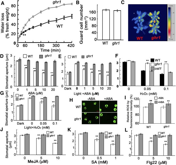
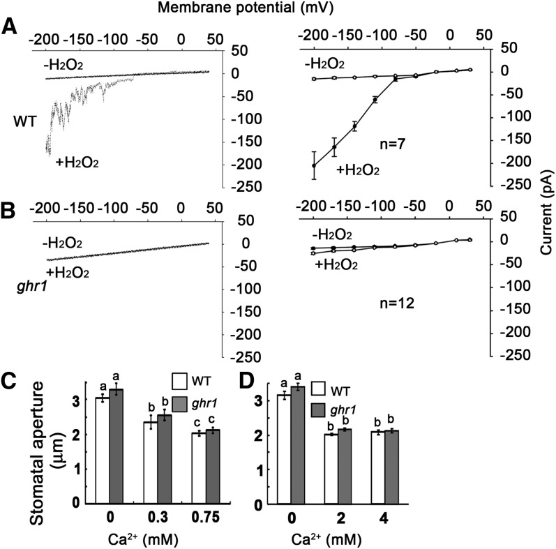
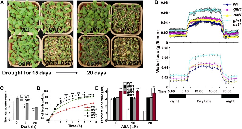
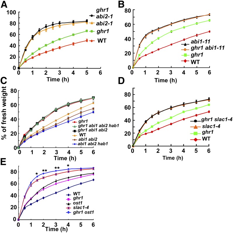
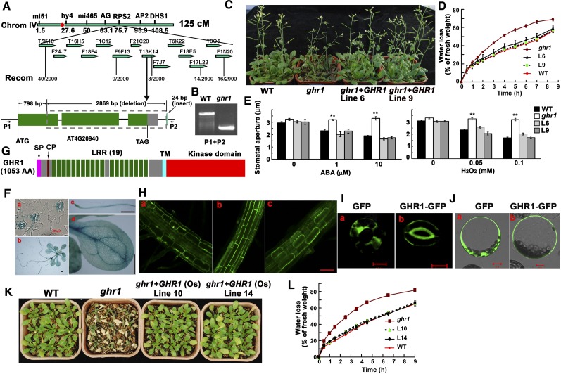
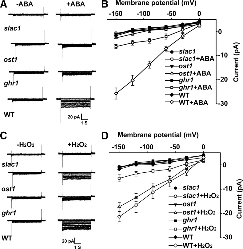
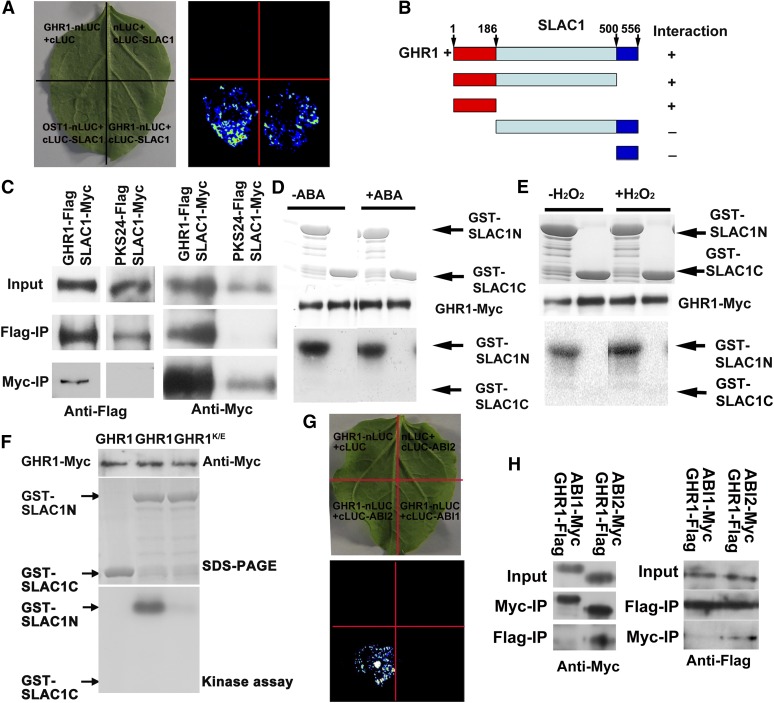
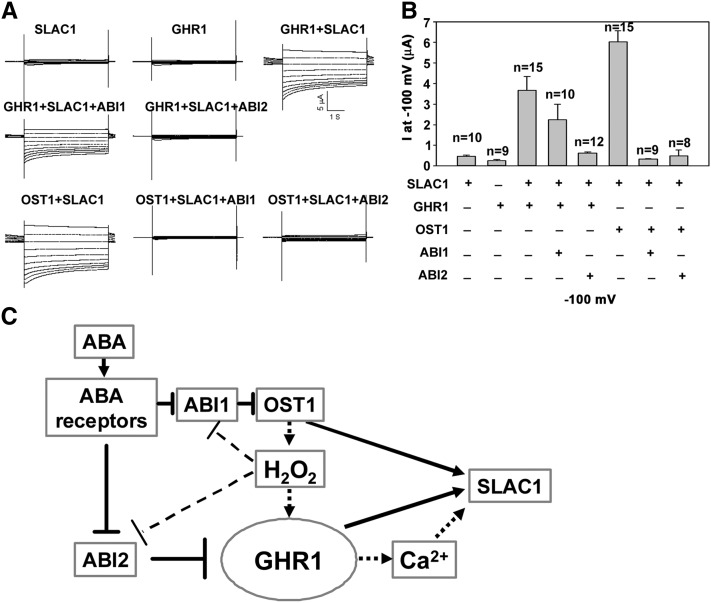
The plant hormone abscisic acid (ABA) regulates stomatal movement under drought stress, and this regulation requires hydrogen peroxide (H2O2). We isolated GUARD CELL HYDROGEN PEROXIDE-RESISTANT1 (GHR1), which encodes a receptor-like kinase localized on the plasma membrane in Arabidopsis thaliana. ghr1 mutants were defective ABA and H2O2 induction of stomatal closure. Genetic analysis indicates that GHR1 is a critical early component in ABA signaling. The ghr1 mutation impaired ABA- and H2O2-regulated activation of S-type anion currents in guard cells. Furthermore, GHR1 physically interacted with, phosphorylated, and activated the S-type anion channel SLOW ANION CHANNEL-ASSOCIATED1 when coexpressed in Xenopus laevis oocytes, and this activation was inhibited by ABA-INSENSITIVE2 (ABI2) but not ABI1. Our study identifies a critical component in ABA and H2O2 signaling that is involved in stomatal movement and resolves a long-standing mystery about the differential functions of ABI1 and ABI2 in this process.


Figures

Figure 1.
Figure 1.
The ghr1 Mutation Impairs ABA- and H2O2-Mediated Stomatal Movement. (A) Water loss of detached leaves of the wild type (WT) and the ghr1 mutant. Values are means ± se of three replicates (40 leaves from one pot per replicate) for one experiment (**P < 0.01 from the fourth time point), and three experiments were performed with similar results. (B) Number of guard cells in the leaf abaxial epidermis of the wild type (WT) and the ghr1 mutant. (C) False-color infrared image of the wild type and the ghr1 mutant. As calculated by IRWIN REPORTER version 5.31 software, the leaf temperature of ghr1 was lower than that of the wild type. (D) Stomatal closing in the wild type and the ghr1 mutant as affected by ABA. (E) Inhibition of light-induced stomatal opening by ABA in the wild type and the ghr1 mutant. (F) Stomatal closing in the wild type, ghr1, and ost1 as affected by H2O2. (G) Light-induced stomatal opening in the wild type and the ghr1 mutant as affected by H2O2. (H) Representative images of ROS production as indicated by the fluorescent dye H2DCF-DA and confocal microscopy. Epidermal peels were loaded with H2DCF-DA for 20 min before 50 µM ABA was added. After 5 min, the peels were photographed. Bar = 10 μm. (I) Quantification of relative ROS production in guard cells of the wild type and ghr1 without and with ABA treatment. ROS production in each pair of guard cells was quantified based on pixel intensity. The intensities in the wild type without ABA were taken as 100%, and the others were compared with those of the wild type. Values are means ± se from three independent experiments; n = 60 for each genotype per experiment. **P < 0.01. There was no significant difference in H2O2 production between the wild type and ghr1 with or without ABA treatment. (J) to (L) Methyl jasmonate (MeJA [J]), salicylic acid (SA [K]), and Flagellin 22 (Flg22 [L]) induced stomatal closing in the wild type and the ghr1 mutant. For the determination of stomatal apertures in (D), (F), and (J) to (L), the epidermal peels were first incubated in MES buffer (10 mM MES-KOH [pH 6.15], 10 mM KCl, and 50 μM CaCl2) under light (90 μmol/m2/s) for ∼2.5 h to fully open stomata. Different concentrations of chemicals were then added to the solution for ∼2.5 h. Values are means ± se of three replicates (30 stomata from one seedling per replicate) for one experiment, and three independent experiments were done with similar results. **P < 0.01, *P < 0.05.
Figure 2.
Figure 2.
The ghr1 Mutation Impairs the H2O2-Activated Ca2+ Channel. (A) and (B) Patch-clamp whole-cell recording of Ca2+ channel currents in guard cell protoplasts of the wild type (WT; [A], right) or ghr1 ([B], right) with or without the addition of 5 mM H2O2 in the bath solution. Current density-voltage data of the wild type ([A], left) or ghr1 ([B], left) were derived from the recordings shown to the right and are presented as means ± se (wild type, n = 7; ghr1, n = 12). Error bars are smaller than symbols when not visible. (C) and (D) Ca2+ induced similar stomatal closure in the wild type and ghr1. Epidermal peels with preopened stomata were treated with different concentrations of Ca2+ for ∼2.5 h before the apertures were measured. Values are means ± se of three replicates (30 stomata from one seedling per replicate) for one experiment, and three independent experiments were done with similar results. There was no significant difference in stomatal apertures between the wild type and ghr1 after Ca2+ treatment. Different letters (a, b, or c) indicate P < 0.01.
Figure 3.
Figure 3.
Genetic Analysis of ghr1 with ost1 Suggests That GHR1 Is in Parallel with OST1 and/or Acts Downstream of OST1. (A) Drought phenotypes of wild-type (WT), ost1, ghr1, and ghr1 ost1 seedlings in soil after water was withheld for 15 d (left) or 20 d (right). (B) Water loss of wild-type, ghr1, ost1, and ghr1 ost1 seedlings growing in soil as determined by the weighing of pots over a 24-h period during the drought treatment in (A). The values in the top panel are means of three replicates in one experiment; each replicate was represented by one pot, and each pot was weighed every 5 min. The values in the bottom panel are means of three replicate pots that were weighed each hour (by reducing the data, the bottom panel more clearly reveals the trends). At all time points, water loss was significantly greater (P < 0.01) for ghr1 ost1 than for the wild-type, ghr1, and ost1 seedlings. At all time points of night (without light), water loss was significantly greater (P < 0.05) for ghr1 than for the wild type or ost1, while water loss did not differ between the wild type and ost1. In the period simulating day (with light), water loss was significantly greater (P < 0.05) for ghr1 than for the wild type. (C) Sizes of stomatal apertures in the dark. Epidermal peels with preopened stomata were kept in the dark for different times before the apertures were measured. Values are means ± se of three replicates (30 stomata from one seedling per replicate) for one experiment (**P < 0.01, *P < 0.05). (D) Water loss of detached leaves of the wild type, ghr1, ost1, and ghr1 ost1. For the quantification of water loss from detached leaves, three independent experiments were done with similar results. Values are means ± se of three replicates (40 leaves from one pot were measured per replicate) for one experiment. Water loss was significantly greater for ghr1 ost1 than for ost1 or ghr1 from 2 to 6 h (**P < 0.01, *P < 0.05). (E) ABA-induced stomatal closure in the wild type, ghr1, ost1, and ghr1 ost1. Values are means ± se of three replicates (30 stomata from one seedling per replicate) from one experiment; three independent experiments were performed with similar results. Stomatal apertures were significantly greater (**P < 0.01) for ghr1 ost1 than for the wild type, ghr1, or ost1.
Figure 4.
Figure 4.
Genetic Analysis of ghr1 with abi1-11; abi2-1 Dominant Negative Mutants; abi1, abi2, and hab1 Null Mutants; and the slac1 Mutant. (A) Water loss of detached leaves of the wild type (WT), ghr1, abi2-1 (a dominant negative mutation; Landsberg erecta), and ghr1 abi2-1. (B) Water loss of detached leaves of the wild type, ghr1, abi1-11 (a dominant negative mutation; Col-0), and ghr1 abi1-11. (C) Water loss of detached leaves of the wild type, ghr1, abi1 abi2 double mutant, ghr1 abi1 abi2 triple mutant, abi1 abi2 hab1 triple mutant, and ghr1 abi1 abi2 hab1 quadruple mutant. abi1 (Salk_072009), abi2 (Salk_015166), and hab1 (Salk_002104) are loss-of-function mutants with a T-DNA insertion in each gene. (D) Water loss of detached leaves of the wild type, ghr1, slac1-4, and ghr1 slac1-4. (E) Water loss of detached leaves of the wild type, ghr1, ost1, slac1-4, and ghr1 ost1. For the quantification of water loss from detached leaves, three independent experiments were done with similar results. Values are means ± se of three replicates (40 leaves from one pot were measured per replicate). Water loss was not significantly different between abi1-11 ghr1 and abi1-11 (A), between abi2-1 ghr1 and abi2-1 (B), between ghr1 and ghr1 abi1 abi2 hab1 or ghr1 abi1 abi2 (C), or between slac1-4 and ghr1 slac1-4 (D). In (E), water loss was significantly greater for ghr1 ost1 than for slac1-4 from 1.5 to 4 h (*P < 0.05, **P < 0.01). Error bars are smaller than symbols when not visible.
Figure 5.
Figure 5.
GHR1 Encodes an LRR-RLK Localized on the Plasma Membrane. (A) Positional cloning of GHR1. GHR1 was mapped to chromosome 4 between BAC clones F9F13 and T13K14. The AT4G20940 gene of the ghr1 mutant lacks a 2869-bp fragment starting from 798 bp (counting from the putative ATG) and contains a 24-bp insert that is not found in the Arabidopsis genome. The red dot is the centromere of chromosome 4. (B) PCR amplification of the GHR1 genomic fragment from the wild type (WT) and ghr1 using primers P1 and P2 (shown in [A]). (C) Drought phenotypes of the wild type, ghr1, and ghr1 complemented with GHR1 genomic DNA (lines 6 and 9) grown in soil. The seedlings in this experiment were ∼6 weeks old in a growth room with 12 h of light/12 h of dark and then exposed to drought stress for ∼2 weeks before photographs were taken. (D) Water loss of detached leaves of the wild type, ghr1, and ghr1 complemented with GHR1 genomic DNA (lines 6 and 9). (E) ABA-induced (left) and H2O2-induced (right) stomatal closure in the wild type, ghr1, or ghr1 complemented with GHR1 genomic DNA line 6 (L6) and line 9 (L9). Epidermal peels with preopened stomata were treated with different concentrations of ABA or H2O2 for ∼2.5 h before apertures were measured. Values are means ± se of three replicates (30 stomata from one seedling were measured per replicate) for one experiment. After treatment with ABA or H2O2, stomatal apertures were significantly smaller for line 6, line 9, and the wild type than for ghr1 (**P < 0.01). (F) ProGHR1:GUS expression patterns in guard cells ([a]; bar = 20 μm), a whole seedling ([b]; bar = 0.5 mm), a root ([c]; bar = 0.5 mm), and a leaf ([d]; bar = 0.5 mm). (G) The protein structure of GHR1. AA, Amino acids; SP, a plasma membrane signal peptide; CP, Cys pair; TM, transmembrane domain. (H) GHR1-GFP localization in roots of transgenic plants. (a) GFP localization in the mature root zone (control). (b) GHR1-GFP localization in the mature root zone. (c) GHR1-GFP localization in plasmolyzed cells of the mature root zone (root cells treated with 0.8 M mannitol for 10 min). Bar = 60 μm. (I) GFP (a) and GHR1-GFP (b) localization in guard cells of transgenic plants. Bars = 10 μm. (J) GHR1-GFP localization in a leaf protoplast cell in a transient assay. Left, GFP; right, GHR1-GFP. Bars = 10 μm. (K) Water-loss phenotypes of the wild type, ghr1, and ghr1 transformed with the cDNA of a rice GHR1 homolog (OsGHR1; lines 10 and 14). Seedlings were grown in soil. (L) Water loss of detached leaves of the wild type, ghr1, and ghr1 transformed with OsGHR1 (lines 10 and 14). To quantify water loss from detached leaves in (D) and (L), three experiments were done with similar results. Values are means ± se of three replicates (40 leaves from one pot were measured per replicate) for one experiment. Water loss did not significantly differ between the wild type and ghr1 complemented with GHR1 or OsGHR1.
Figure 6.
Figure 6.
ghr1 Impairs ABA and H2O2 Activation of the S-Type Anion Channel. (A) Patch-clamp whole-cell recording of S-type anion channel currents in guard cell protoplasts of the wild type (WT) and slac1-4, ost1, or ghr1 mutants with or without the addition of 50 μM ABA in the bath solution. (B) Current density-voltage data derived from the recordings as shown in (A). Data are presented as means ± se (slac1, n = 5; ost1, n = 7; ghr1, n = 9; wild type, n = 6). (C) Patch-clamp whole-cell recording of S-type anion channel currents in guard cell protoplasts of the wild type, ghr1, slac1-4, and ost1 with or without the addition of 5 mM H2O2 in the bath solution. (D) Current density-voltage data derived from the recordings as shown in (C). Data are presented as means ± se (slac1-4, n = 4; ost1, n = 6; ghr1, n = 8; wild type, n = 7).
Figure 7.
Figure 7.
GHR1 Interacts with SLAC1 and ABI2. (A) Interaction of SLAC1 with GHR1 as revealed by the firefly luciferase complementation imaging assay in N. benthamiana leaves. OST1-nLUC and cLUC-SLAC1 were used as positive controls. GHR1-nLUC and cLUC as well as nLUC and cLUC-SLAC1 were used as negative controls. (B) Interaction of different parts of SLAC1 with GHR1 as indicated by the firefly luciferase complementation imaging assay in N. benthamiana leaves. (C) Coimmunoprecipitation (IP) of GHR1 with SLAC1. Arabidopsis protoplasts transiently coexpressing GHR1-Flag and SLAC1-Myc or PKS24-Flag (PKS24 is a membrane-targeted protein kinase that was used as a negative control) and SLAC1-Myc were immunoprecipitated with anti-Flag or anti-Myc antibodies, and the immunoblot was probed with anti-Myc or anti-Flag antibodies. (D) Phosphorylation analysis of GHR1. GHR1 phosphorylates the N terminus but not the C terminus of SLAC1, and GHR1 activity was not enhanced by ABA. GHR1-Myc transiently expressed in Arabidopsis protoplasts was purified and used for phosphorylation analysis. GST-SLAC1-N and GST-SLAC1-C proteins were expressed in E. coli. (E) H2O2 does not activate GHR1 activity. The assay was similar to that in (D) except that H2O2 was used in place of ABA. (F) A K798E mutation of GHR1, which changes the ATP binding domain of the GHR1 kinase motif, cannot phosphorylate SLAC1. (G) Interaction of GHR1 with ABI2 as indicated by the firefly luciferase complementation imaging assay in N. benthamiana leaves. GHR1 did not interact with ABI1 under the same conditions. GHR1-nLUC and cLUC as well nLUC and cLUC-ABI2 were used as negative controls. (H) Coimmunoprecipitation of GHR1 with ABI2 or ABI1. The experiment was similar to that in (C).
Figure 8.
Figure 8.
GHR1 Activates SLAC1 and Is Negatively Regulated by ABI2. (A) Typical whole-cell current traces recorded from oocytes injected with cRNA of SLAC1, GHR1, GHR1+SLAC1, GHR1+SLAC1+ABI1, GHR1+SLAC1+ABI2, OST1+SLAC1, OST1+SLAC+ABI1, and OST1+SLAC+ABI2. No prominent current responses of oocytes expressing SLAC1, OST1, or GHR1 alone were recorded (OST1 is not shown). (B) Average current of S-type anion channel recorded at −100 mV (μA) as shown in (A). Data are presented as means ± se (SLAC1, n = 10; GHR1, n = 9; GHR1+SLAC1, n = 15; GHR1+SLAC1+ABI1, n = 10; GHR1+SLAC1+ABI2, n = 12; OST1+SLAC1, n = 15; OST1+SLAC+ABI1, n = 9; and OST1+SLAC+ABI2, n = 8). (C) A working model for the ABA signaling pathway in stomatal movement. In the absence of ABA, GHR1 or OST1 is inhibited by ABI2 or ABI1, respectively. In the presence of ABA, the ABA receptor PYR1 binds to ABI1/2 and releases the inhibition of OST1/GHR1 by ABI1/2. In this model, SLAC1 is coordinately regulated by OST1, GHR1, and other Ca2+-dependent kinases. This model explains the genetic and biochemical data obtained previously and in this study.

References

    1. Acharya B.R., Assmann S.M. (2009). Hormone interactions in stomatal function. Plant Mol. Biol. 69: 451–462 - PubMed
    1. Allen G.J., Kuchitsu K., Chu S.P., Murata Y., Schroeder J.I. (1999). Arabidopsis abi1-1 and abi2-1 phosphatase mutations reduce abscisic acid-induced cytoplasmic calcium rises in guard cells. Plant Cell 11: 1785–1798 - PMC - PubMed
    1. Chen H., Zou Y., Shang Y., Lin H., Wang Y., Cai R., Tang X., Zhou J.M. (2008). Firefly luciferase complementation imaging assay for protein-protein interactions in plants. Plant Physiol. 146: 368–376 - PMC - PubMed
    1. Clough S.J., Bent A.F. (1998). Floral dip: a simplified method for Agrobacterium-mediated transformation of Arabidopsis thaliana. Plant J. 16: 735–743 - PubMed
    1. Delaunay A., Isnard A.D., Toledano M.B. (2000). H2O2 sensing through oxidation of the Yap1 transcription factor. EMBO J. 19: 5157–5166 - PMC - PubMed

Publication types

MeSH terms