. 2012 Jun 25;2(1):23.
doi: 10.1186/2045-3701-2-23.

PDLIM2 restricts Th1 and Th17 differentiation and prevents autoimmune disease

Affiliations

PDLIM2 restricts Th1 and Th17 differentiation and prevents autoimmune disease

Zhaoxia Qu et al. Cell Biosci. .

Abstract

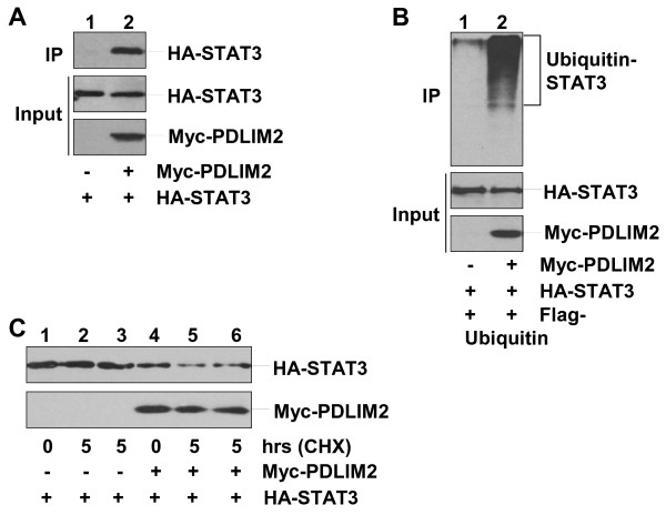
Background: PDLIM2 is essential for the termination of the inflammatory transcription factors NF-κB and STAT but is dispensable for the development of immune cells and immune tissues/organs. Currently, it remains unknown whether and how PDLIM2 is involved in physiologic and pathogenic processes.

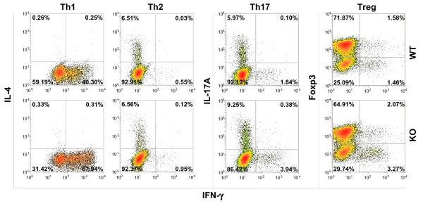
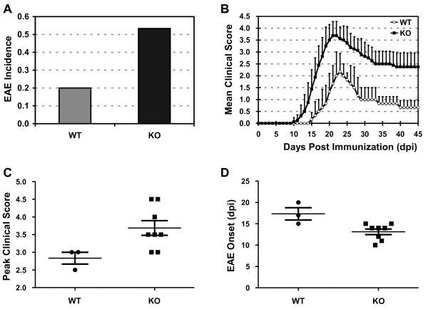
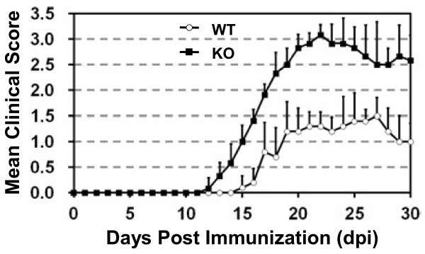
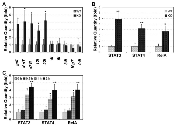
Results: Here we report that naive PDLIM2 deficient CD4+ T cells were prone to differentiate into Th1 and Th17 cells. PDLIM2 deficiency, however, had no obvious effect on lineage commitment towards Th2 or Treg cells. Notably, PDLIM2 deficient mice exhibited increased susceptibility to experimental autoimmune encephalitis (EAE), a Th1 and/or Th17 cell-mediated inflammatory disease model of multiple sclerosis (MS). Mechanistic studies further indicate that PDLIM2 was required for restricting expression of Th1 and Th17 cytokines, which was in accordance with the role of PDLIM2 in the termination of NF-κB and STAT activation.

Conclusion: These findings suggest that PDLIM2 is a key modulator of T-cell-mediated immune responses that may be targeted for the therapy of human autoimmune diseases.


Figures

Figure 1
Figure 1
Enhanced Th1 and Th17 differentiation of PDLIM2 deficient CD4+Th cells. Naive CD4+ Th cells isolated from PDLIM2+/+ (WT) or PDLIM2−/− (KO) mice were stimulated for 72 hours with anti-CD3/anti-CD28 under Th1, Th2, Th17 or Treg polarizing condition, followed by intracellular cytokine staining and flow cytometry. The data are representative of at least three independent experiments with similar results.
Figure 2
Figure 2
Increased susceptibility to EAE in PDLIM2 deficient mice.A) Incidence, B) disease progression, C) severity and D) onset of EAE in PDLIM2+/+ and PDLIM2−/− mice (n = 15). Mice were immunized with PLP180–199 peptide and monitored daily for EAE disease symptoms. The p values between the PDLIM2+/+ (WT) and PDLIM2−/− (KO) groups are at least smaller than 0.05 by two tailed t-test.
Figure 3
Figure 3
Increased severity of adoptive transfer EAE in recipients of PDLIM2 deficient CD4+T cells. CD4+ T cells were isolated from PDLIM2+/+ and PDLIM2−/− mice immunized with PLP180–199 peptide and transferred i.v. into SCID recipients (n = 20). One day after the cell transfer, recipient mice also received an injection of pertussis. Mice were then monitored for the symptoms of EAE as described in Figure .2
Figure 4
Figure 4
Enhanced nuclear expression of STAT3/4 and RelA proteins and augmented production of Th1 and Th17 cytokines in PDLIM2 deficient Teff cells. Splenic T cells from day 10 PLP180–199-immunized PDLIM2+/+ (WT) or PDLIM2−/− (KO) mice were subjected to QRT-PCR to detect the relative expression levels of the indicated cytokines genes (A) or ELISA to detect the nuclear expression levels of STAT3, STAT4 and RelA (B). The expression levels of the indicated genes and proteins were represented as fold induction relative to their WT controls. C) Naive PDLIM2−/− or PDLIM2+/+ CD4+ Th cells were stimulated for the indicated time points with anti-CD3/anti-CD28 under Th1 or Th17 polarizing condition, followed by ELISA to detect the nuclear expression levels of STAT3 (in response to Th17 stimulation), STAT4 and RelA (in response to Th1 stimulation). In A-C, n = 3, *, p < 0.03; **, p < 0.003 by two tailed t-test.
Figure 5
Figure 5
Ubiquitination and proteasomal degradation of STAT3 by PDLIM2.A) Physical interaction between PDLIM2 and STAT3. Nuclear extracts of 293 cells transfected with HA-STAT3 alone or together with Myc-PDLIM2 were subjected to immunoprecipitation (IP) using Myc antibody and immunoblotting (IB) using HA antibody. The expression levels of HA-STAT3 and Myc-PDLIM2 were examined by IB. B) Polyubiquitination of STAT3 by PDLIM2. 293 cells were transfected with HA-STAT3 plus Flag-ubiquitin in the presence or absence of Myc-PDLIM2, followed by nuclear fractionation. The nuclear extracts were subjected to IP using HA antibody and IB using Flag antibody. The expression levels of HA-STAT3 and Myc-PDLIM2 were examined by IB. C) Proteasomal degradation of STAT3 by PDLIM2. 293 cells transfected with HA-STAT3 alone or together with Myc-PDLIM2 were cycloheximide (CHX)-chased for the indicated time, followed by nuclear extractions and IB using HA or Myc antibody. In lanes 3 and 6, the cells were chased in the presence of 10 μM MG132.

Similar articles

Cited by

References

    1. O'Shea JJ, Paul WE. Mechanisms underlying lineage commitment and plasticity of helper CD4+ T cells. Science. 2010;327:1098–1102. doi: 10.1126/science.1178334. - DOI - PMC - PubMed
    1. Jäger A, Kuchroo VK. Effector and regulatory T-cell subsets in autoimmunity and tissue inflammation. Scand J Immunol. 2010;72:173–184. doi: 10.1111/j.1365-3083.2010.02432.x. - DOI - PMC - PubMed
    1. Fletcher JM, Lalor SJ, Sweeney CM, Tubridy N, Mills KH. T cells in multiple sclerosis and experimental autoimmune encephalomyelitis. Clin Exp Immunol. 2010;162:1–11. doi: 10.1111/j.1365-2249.2010.04143.x. - DOI - PMC - PubMed
    1. Toh ML, Miossec P. The role of T cells in rheumatoid arthritis: new subsets and new targets. Curr Opin Rheumatol. 2007;19:284–288. doi: 10.1097/BOR.0b013e32805e87e0. - DOI - PubMed
    1. Xiao G, Rabson A, Young W, Qing G, Qu Z. Alternative pathways of NF-κB activation: a double-edged sword in health and disease. Cytokine Growth Factor Rev. 2006;17:281–293. doi: 10.1016/j.cytogfr.2006.04.005. - DOI - PubMed