

Increased tumor homing and tissue penetration of the filamentous plant viral nanoparticle Potato virus X
- PMID: 22731633
- PMCID: PMC3482416
- DOI: 10.1021/mp300240m
Increased tumor homing and tissue penetration of the filamentous plant viral nanoparticle Potato virus X
Abstract
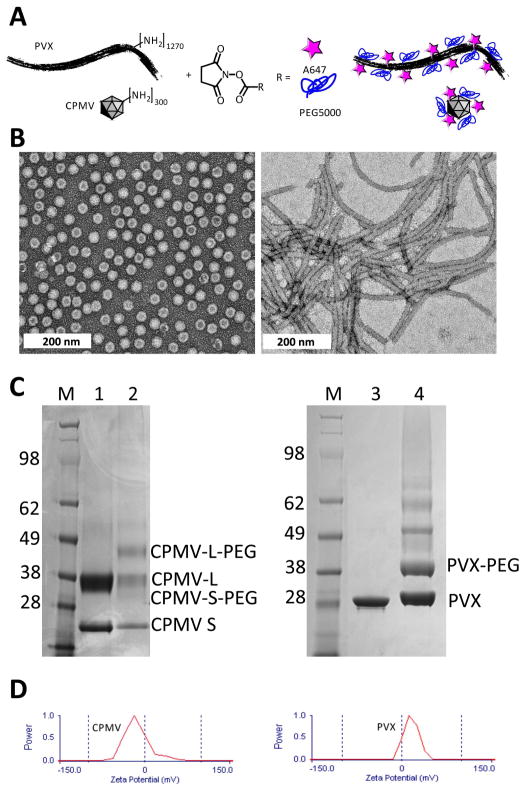
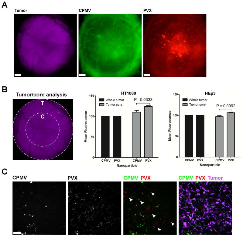
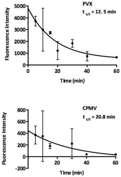
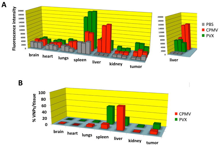
Nanomaterials with elongated architectures have been shown to possess differential tumor homing properties compared to their spherical counterparts. Here, we investigate whether this phenomenon is mirrored by plant viral nanoparticles that are filamentous (Potato virus X) or spherical (Cowpea mosaic virus). Our studies demonstrate that Potato virus X (PVX) and Cowpea mosaic virus (CPMV) show distinct biodistribution profiles and differ in their tumor homing and penetration efficiency. Analogous to what is seen with inorganic nanomaterials, PVX shows enhanced tumor homing and tissue penetration. Human tumor xenografts exhibit higher uptake of PEGylated filamentous PVX compared to CPMV, particularly in the core of the tumor. This is supported by immunohistochemical analysis of the tumor sections, which indicates greater penetration and accumulation of PVX within the tumor tissues. The enhanced tumor homing and retention properties of PVX along with its higher payload carrying capacity make it a potentially superior platform for applications in cancer drug delivery and imaging applications.
Figures

Similar articles
-
Viral nanoparticles for in vivo tumor imaging.J Vis Exp. 2012 Nov 16;(69):e4352. doi: 10.3791/4352. J Vis Exp. 2012. PMID: 23183850 Free PMC article.
-
Potato virus X, a filamentous plant viral nanoparticle for doxorubicin delivery in cancer therapy.Nanoscale. 2017 Feb 9;9(6):2348-2357. doi: 10.1039/c6nr09099k. Nanoscale. 2017. PMID: 28144662 Free PMC article.
-
Plant Virus Intratumoral Immunotherapy with CPMV and PVX Elicits Durable Antitumor Immunity in a Mouse Model of Diffuse Large B-Cell Lymphoma.Mol Pharm. 2024 Dec 2;21(12):6206-6219. doi: 10.1021/acs.molpharmaceut.4c00507. Epub 2024 Nov 11. Mol Pharm. 2024. PMID: 39526560 Free PMC article.
-
Small, Smaller, Nano: New Applications for Potato Virus X in Nanotechnology.Front Plant Sci. 2019 Feb 19;10:158. doi: 10.3389/fpls.2019.00158. eCollection 2019. Front Plant Sci. 2019. PMID: 30838013 Free PMC article. Review.
-
Application of Plant Viruses in Biotechnology, Medicine, and Human Health.Viruses. 2021 Aug 26;13(9):1697. doi: 10.3390/v13091697. Viruses. 2021. PMID: 34578279 Free PMC article. Review.
Cited by
-
Utilizing Viral Nanoparticle/Dendron Hybrid Conjugates in Photodynamic Therapy for Dual Delivery to Macrophages and Cancer Cells.Bioconjug Chem. 2016 May 18;27(5):1227-35. doi: 10.1021/acs.bioconjchem.6b00075. Epub 2016 Apr 27. Bioconjug Chem. 2016. PMID: 27077475 Free PMC article.
-
Prediction of Anti-cancer Nanotherapy Efficacy by Imaging.Nanotheranostics. 2017 Jul 6;1(3):296-312. doi: 10.7150/ntno.20564. eCollection 2017. Nanotheranostics. 2017. PMID: 29071194 Free PMC article. Review.
-
The Plant Viruses and Molecular Farming: How Beneficial They Might Be for Human and Animal Health?Int J Mol Sci. 2023 Jan 12;24(2):1533. doi: 10.3390/ijms24021533. Int J Mol Sci. 2023. PMID: 36675043 Free PMC article. Review.
-
Injectable Hydrogel Containing Cowpea Mosaic Virus Nanoparticles Prevents Colon Cancer Growth.ACS Biomater Sci Eng. 2022 Jun 13;8(6):2518-2525. doi: 10.1021/acsbiomaterials.2c00284. Epub 2022 May 6. ACS Biomater Sci Eng. 2022. PMID: 35522951 Free PMC article.
-
Tobacco mosaic virus delivery of mitoxantrone for cancer therapy.Nanoscale. 2018 Aug 30;10(34):16307-16313. doi: 10.1039/c8nr04142c. Nanoscale. 2018. PMID: 30129956 Free PMC article.
References
-
- Cai S, Vijayan K, Cheng D, Lima EM, Discher DE. Micelles of Different Morphologies—Advantages of Worm-like Filomicelles of PEO-PCL in Paclitaxel Delivery. Pharm Res. 2007;24:2099–2109. - PubMed
-
- Decuzzi P, et al. Size and shape effects in the biodistribution of intravascularly injected particles. Journal of Controlled Release. 2010;141:320–327. - PubMed
Publication types
MeSH terms
Substances
Grants and funding
LinkOut - more resources
Full Text Sources
Other Literature Sources
