

Pulse Q-band EPR and ENDOR spectroscopies of the photochemically generated monoprotonated benzosemiquinone radical in frozen alcoholic solution
- PMID: 22731760
- PMCID: PMC3432317
- DOI: 10.1021/jp304555u
Pulse Q-band EPR and ENDOR spectroscopies of the photochemically generated monoprotonated benzosemiquinone radical in frozen alcoholic solution
Abstract
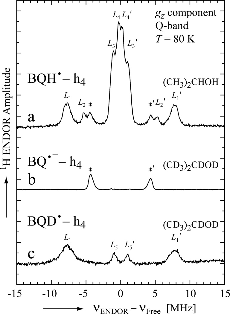
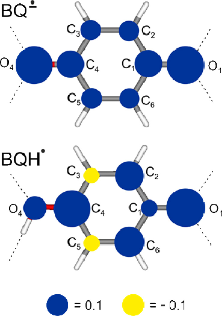
Quinones are essential cofactors in many physiological processes, among them proton-coupled electron transfer (PCET) in photosynthesis and respiration. A key intermediate in PCET is the monoprotonated semiquinone radical. In this work we produced the monoprotonated benzosemiquinone (BQH(•)) by UV illumination of BQ dissolved in 2-propanol at cryogenic temperatures and investigated the electronic and geometric structures of BQH(•) in the solid state (80 K) using EPR and ENDOR techniques at 34 GHz. The g-tensor of BQH(•) was found to be similar to that of the anionic semiquinone species (BQ(•-)) in frozen solution. The peaks present in the ENDOR spectrum of BQH(•) were identified and assigned by (1)H/(2)H substitutions. The experiments reconfirmed that the hydroxyl proton (O-H) on BQH(•), which is abstracted from a solvent molecule, mainly originates from the central CH group of 2-propanol. They also showed that the protonation has a strong impact on the electron spin distribution over the quinone. This is reflected in the hyperfine couplings (hfc's) of the ring protons, which dramatically changed with respect to those typically observed for BQ(•-). The hfc tensor of the O-H proton was determined by a detailed orientation-selection ENDOR study and found to be rhombic, resembling those of protons covalently bound to carbon atoms in a π-system (i.e., α-protons). It was found that the O-H bond lies in the quinone plane and is oriented along the direction of the quinone oxygen lone pair orbital. DFT calculations were performed on different structures of BQH(•) coordinated by four, three, or zero 2-propanol molecules. The O-H bond length was found to be around 1.0 Å, typical for a single covalent O-H bond. Good agreement between experimental and DFT results were found. This study provides a detailed picture of the electronic and geometric structures of BQH(•) and should be applicable to other naturally occurring quinones.
Figures


Similar articles
-
EPR and ENDOR investigation of the primary electron acceptor radical anion QA.- in iron-depleted photosystem II membrane fragments.Biochemistry. 1995 Jun 27;34(25):8144-56. doi: 10.1021/bi00025a021. Biochemistry. 1995. PMID: 7794928
-
Hydrogen bond geometries from electron paramagnetic resonance and electron-nuclear double resonance parameters: density functional study of quinone radical anion-solvent interactions.J Am Chem Soc. 2004 Mar 17;126(10):3280-90. doi: 10.1021/ja0392014. J Am Chem Soc. 2004. PMID: 15012159
-
Hydrogen bonding and spin density distribution in the Qb semiquinone of bacterial reaction centers and comparison with the Qa site.J Am Chem Soc. 2011 Apr 13;133(14):5525-37. doi: 10.1021/ja2001538. Epub 2011 Mar 18. J Am Chem Soc. 2011. PMID: 21417328 Free PMC article.
-
Spin distribution and the location of protons in paramagnetic proteins.Annu Rev Biophys Biomol Struct. 2004;33:441-68. doi: 10.1146/annurev.biophys.33.110502.140344. Annu Rev Biophys Biomol Struct. 2004. PMID: 15139821 Review.
-
Coupling of electron transfer to proton uptake at the Q(B) site of the bacterial reaction center: a perspective from FTIR difference spectroscopy.Biochim Biophys Acta. 2008 Oct;1777(10):1229-48. doi: 10.1016/j.bbabio.2008.06.012. Epub 2008 Jul 11. Biochim Biophys Acta. 2008. PMID: 18671937 Review.
Cited by
-
Q-Band Electron-Nuclear Double Resonance Reveals Out-of-Plane Hydrogen Bonds Stabilize an Anionic Ubisemiquinone in Cytochrome bo3 from Escherichia coli.Biochemistry. 2016 Oct 11;55(40):5714-5725. doi: 10.1021/acs.biochem.6b00669. Epub 2016 Sep 28. Biochemistry. 2016. PMID: 27622672 Free PMC article.
-
The photoreactive free radical in eumelanin.Sci Adv. 2018 Mar 28;4(3):eaaq1293. doi: 10.1126/sciadv.aaq1293. eCollection 2018 Mar. Sci Adv. 2018. PMID: 29600273 Free PMC article.
-
Cationic penetrating antioxidants switch off Mn cluster of photosystem II in situ.Photosynth Res. 2019 Nov;142(2):229-240. doi: 10.1007/s11120-019-00657-2. Epub 2019 Jul 13. Photosynth Res. 2019. PMID: 31302832
References
-
- Trumpower BL. Function of Quinones in Energy Conserving Systems. New York: Academic Press; 1982.
-
- Lenaz G. Coenzyme Q: Biochemistry, Bioenergetics and Clinical Applications of Ubiquinone. Suffolk: John Wiley & Sons; 1985.
-
- Blankenship RE. Molecular Mechanisms of Photosynthesis. Oxford: Blackwell Science Ltd; 2002.
-
- Cramer WA, Knaff DB. In: Energy transduction in biological membranes: a textbook of bioenergetics. Cantor CR, editor. New York: Springer-Verlag; 1990. p. 193.
-
- Nohl H, Kozlov AV, Staniek K, Gille L. Bioinorg. Chem. 2001;29:1–13. - PubMed
Publication types
MeSH terms
Substances
Grants and funding
LinkOut - more resources
Full Text Sources
Research Materials
Miscellaneous
