. 2012 Jun 25:10:59.
doi: 10.1186/1741-7007-10-59.

Population dynamics and habitat sharing of natural populations of Caenorhabditis elegans and C. briggsae

Affiliations

Population dynamics and habitat sharing of natural populations of Caenorhabditis elegans and C. briggsae

Marie-Anne Félix et al. BMC Biol. .

Abstract

Background: The nematode Caenorhabditis elegans is a major model organism in laboratory biology. Very little is known, however, about its ecology, including where it proliferates. In the past, C. elegans was mainly isolated from human-made compost heaps, where it was overwhelmingly found in the non-feeding dauer diapause stage.

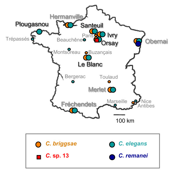
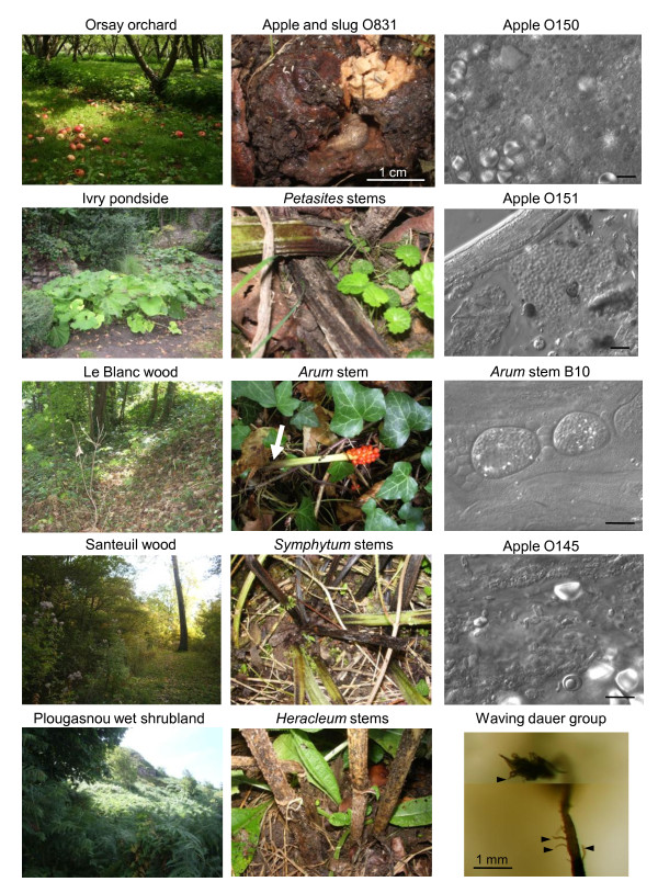
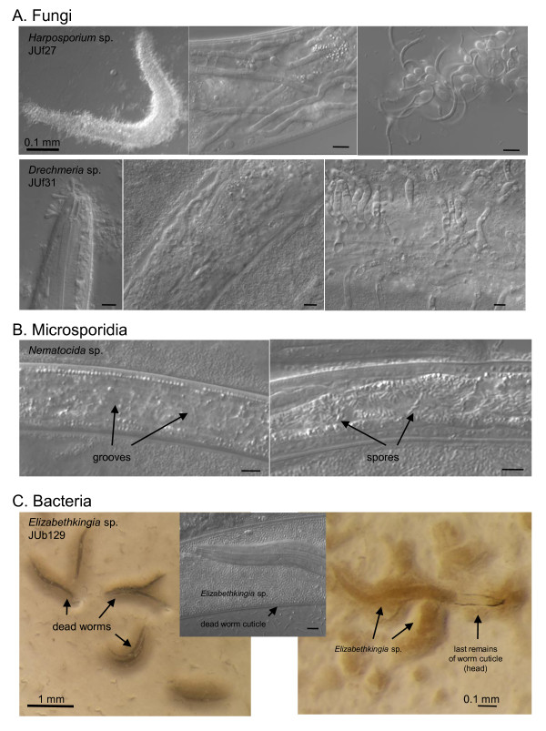
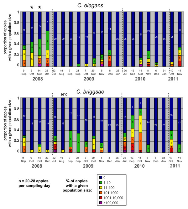
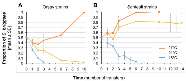
Results: C. elegans and C. briggsae were found in large, proliferating populations in rotting plant material (fruits and stems) in several locations in mainland France. Both species were found to co-occur in samples isolated from a given plant species. Population counts spanned a range from one to more than 10,000 Caenorhabditis individuals on a single fruit or stem. Some populations with an intermediate census size (10 to 1,000) contained no dauer larvae at all, whereas larger populations always included some larvae in the pre-dauer or dauer stages. We report on associated micro-organisms, including pathogens. We systematically sampled a spatio-temporally structured set of rotting apples in an apple orchard in Orsay over four years. C. elegans and C. briggsae were abundantly found every year, but their temporal distributions did not coincide. C. briggsae was found alone in summer, whereas both species co-occurred in early fall and C. elegans was found alone in late fall. Competition experiments in the laboratory at different temperatures show that C. briggsae out-competes C. elegans at high temperatures, whereas C. elegans out-competes C. briggsae at lower temperatures.

Conclusions: C. elegans and C. briggsae proliferate in the same rotting vegetal substrates. In contrast to previous surveys of populations in compost heaps, we found fully proliferating populations with no dauer larvae. The temporal sharing of the habitat by the two species coincides with their temperature preference in the laboratory, with C. briggsae populations growing faster than C. elegans at higher temperatures, and vice at lower temperatures.


Figures

Figure 1
Figure 1
Sampling locations in France. Both C. elegans and C. briggsae were found in the locations where extensive sampling was performed (large disks), except in the Western France (Plougasnou) location. Locations with names in black are those in Table 1. Others are from [9,52] or new locations that have been only occasionally and sparsely sampled (small symbols and fonts) (Credits: Bergerac: Victor Nigon [53]; Buzançais: Jean-Baptiste Pénigault; Marseille, Nice, Antibes: Christian Braendle).
Figure 2
Figure 2
Landscapes and substrates with proliferating Caenorhabditis populations. The left panels depict landscapes in mainland France locations, as referenced in Table 1 and shown on the map on Figure 1. The central panels show examples of the corresponding sampled substrates that yielded Caenorhabditis. The right panels, except the bottom-most one, are Nomarski pictures of samples, showing a very heterogeneous composition, with bacteria, fungi and unidentified structures. A young Caenorhabditis juvenile is visible in the second panel from the top, labeled "Apple O151". The bottom panel is a juxtaposition of two focal planes of a large waving group of thousands of dauers, standing on rotten apple O673 (at the bottom of the picture). A few individual dauer larvae are indicated by arrowheads. The corresponding movie is available on request. Bars: 10 μm, unless otherwise indicated. On the Arum panel, the white arrow designates the rotting part of the stem. On the upperleft of the apple O831 panel, the small white animals are springtails.
Figure 3
Figure 3
Alimentary tracts of freshly sampled animals. (A) C. elegans late L3 larva, freshly isolated from a rotting apple (#7) in Santeuil, with bacteria and yeast in the intestinal lumen. (B) C. elegans adult in the same sample, with empty fungal cell walls in the intestinal lumen. (C). C. elegans adult freshly isolated from a rotting apple (O145) from Orsay. (D) C. briggsae adult in a rotting peach from Le Blanc. (E, F) C. briggsae adults in a rotting apple (O635) from Orsay, showing bacterial proliferation in the intestinal lumen, with total obstruction in (F). (G) C. elegans adult in a rotting Arum stem from Plougasnou, with defects in feeding, accumulating bacteria in the pharyngeal lumen. The posterior pharyngeal bulb is visible in (E-G). The ventral side of the animals is down in all pictures.
Figure 4
Figure 4
Pathogens in natural C. elegans and C. briggsae populations. (A) Fungal pathogens. Top three panels: The nematophagous fungus Harposporium sp. JUf27 was isolated from this dead C. elegans individual O143.12 (29 October 2008). JUf27 can infect the intestine of C. elegans N2 and produce hyphae that invade the whole body, resulting in death within six to eight days (middle panel). New spores are then produced at the surface of the dead nematode (right panel). The host response that it provokes has been characterized transcriptionally [23]. Bottom three panels: Fungal pathogen JUf31 in Orsay apple O641 on C. briggsae, Drechmeria coniosporia. Another D. coniosporia strain JUf28 was isolated from apple O567 on a Pristionchus sp. and deposited as CBS129433 at the CBS Fungal Biodiversity Centre, Utrecht, The Netherlands by Nathalie Pujol. These spores also develop into hyphae that invade the nematode body (middle panel) and produce a new generation of spores at the surface of the dead nematode (right panel). (B) Microsporidial infection in C. elegans in Orsay apple O695. Two infected dauer juveniles, in the microsporidial groove stage (left) and in the spore stage (right) (see [21] for microsporidial stages). Both groove and spore stages were also seen in C. elegans L2 larvae in apple O575. (C) Bacterial pathogen Elizabethkingia sp. (strain JUb129), found on C. elegans in Orsay apple O675, here shown on C. elegans N2. The bacteria induce worm bagging and then dissolve their cuticle. On the right, a larva is seen within her mother's corpse. Bars: 10 μm, except otherwise indicated.
Figure 5
Figure 5
Temporal distribution of C. elegans (top) and C. briggsae (bottom) abundance in Orsay apples along four consecutive years. The graphs show the proportion of apples with a given census size (expressed on a log scale) of C. elegans or C. briggsae populations. The number of apples with a given census size is indicated in each color-coded bar. The two time points labeled by stars (6 and 14 October 2008) were sampled under a single tree (see Additional File 4) and were not used with the others in the statistical analyses. 19 August 2008 was particularly hot (36°C maximum temperature, labeled above the graph). See statistical analyses in the Results.
Figure 6
Figure 6
Experimental evolution of the proportion of C. briggsae individuals in competition with C. elegans at different growth temperatures. Two wild strains of C. elegans and C. briggsae from Orsay (A) or Santeuil (B) were competed against each other in the laboratory for several generations at three different temperatures (15°C, 21°C and 27°C). Starting from a frequency of 50% (ten C. elegans L4 larvae and ten C. briggsae L4 larvae), the proportion of C. briggsae individuals was quantified at different time points with five replicate populations per treatment. The mean proportion across replicates is indictaed as a thick line, and error bars indicate standard errors over replicates. Replicates are indicated as thin dotted lines. Time is represented as the number of transfers of each population to a fresh culture plate from the beginning of the experiment. For each treatment, the experiment was continued until all replicates reached fixation of either C. briggsae or C. elegans. With the Santeuil strains at 21°C, C. briggsae outcompeted C. elegans in four out of five replicates.

Comment in

Similar articles

Cited by

References

    1. Félix M-A, Braendle C. The natural history of Caenorhabditis elegans. Curr Biol. 2010;20:R965–R969. doi: 10.1016/j.cub.2010.09.050. - DOI - PubMed
    1. Kammenga JE, Phillips PC, de Bono M, Doroszuk A. Beyond induced mutants: using worms to study natural variation in genetic pathways. Trends Genet. 2008;24:178–185. doi: 10.1016/j.tig.2008.01.001. - DOI - PubMed
    1. Cutter AD, Dey A, Murray RL. Evolution of the Caenorhabditis elegans genome. Mol Biol Evol. 2009;26:1199–1234. doi: 10.1093/molbev/msp048. - DOI - PubMed
    1. Gaertner BE, Phillips PC. Caenorhabditis elegans as a platform for molecular quantitative genetics and the systems biology of natural variation. Genet Res. 2010;92:331–348. doi: 10.1017/S0016672310000601. - DOI - PubMed
    1. Riddle DL. In: The Nematode Caenorhabditis elegans. Wood WB, editor. Cold Spring Harbor: Cold Spring Harbor Press; 1988. The dauer larva; pp. 393–412.

Publication types

MeSH terms