. 2012 Sep;73(9):871-8.
doi: 10.1016/j.humimm.2012.06.008. Epub 2012 Jun 22.

Central and overlapping role of Cathepsin B and inflammasome adaptor ASC in antigen presenting function of human dendritic cells

Affiliations

Central and overlapping role of Cathepsin B and inflammasome adaptor ASC in antigen presenting function of human dendritic cells

Xiuyang Guo et al. Hum Immunol. 2012 Sep.

Abstract

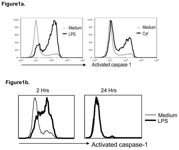
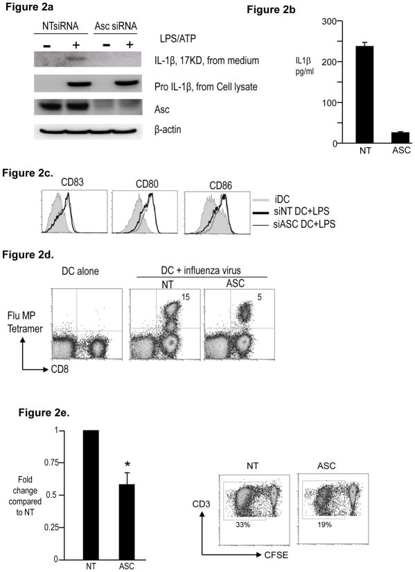
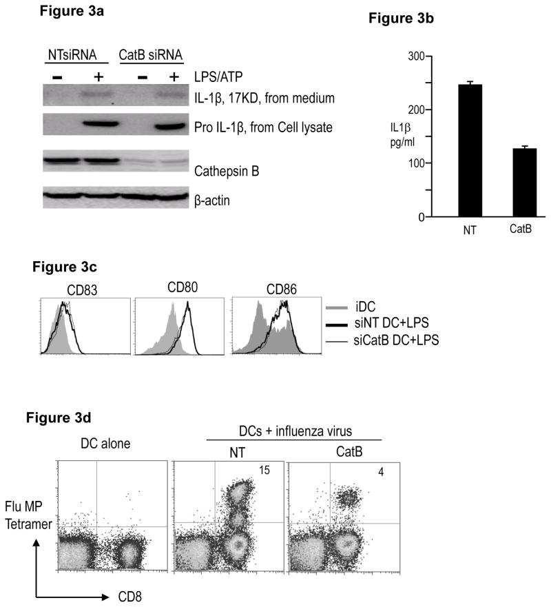
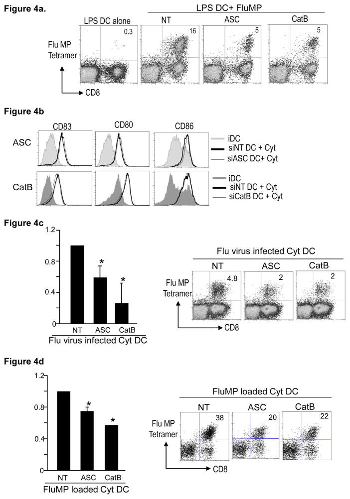
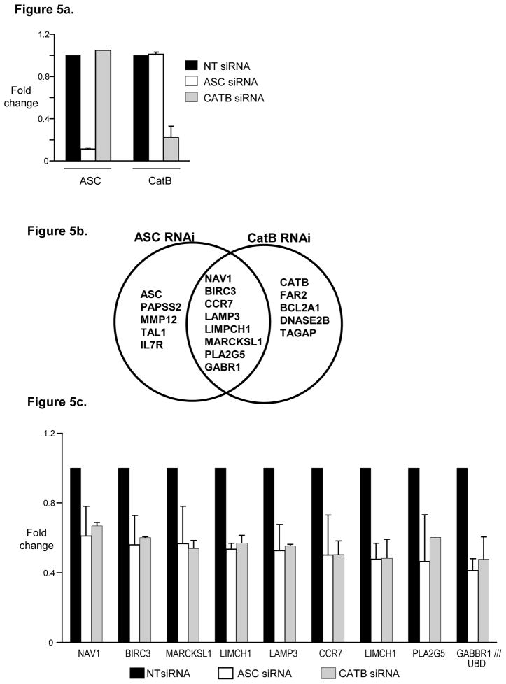
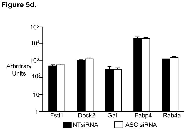
Inflammasomes are increasingly implicated in regulating immunity, but how their activation relates to function of human dendritic cells (DCs) is unknown. Here we show that DC maturation stimuli lead to rapid activation of caspase-1 in human monocyte-derived DCs. RNAi mediated inhibition of the inflammasome component ASC leads to marked inhibition of the capacity of lipopolysachharide (LPS)-matured DCs to stimulate antigen-specific T cells. RNAi mediated inhibition of Cathepsin B (CatB) also similarly inhibits the capacity of human DCs to stimulate immunity. The defective ability of ASC or CatB deficient DCs to stimulate T cells is independent of inflammasome-mediated processing of inflammatory cytokines and also includes DCs loaded with pre-processed peptide. Gene expression profiles of ASC or CatB deficient human DCs show marked overlap with downregulation of genes implicated in DC function. These data demonstrate an important role for ASC and CatB in regulating function of human DCs with overlapping effects on gene expression.


Figures

Figure 1
Figure 1. Activation of caspase 1 with maturation of human Mo-DCs
Monocyte derived immature DCs were cultured alone (Medium) or in the presence of lypopolysaccharide (LPS) or inflammatory cytokines (Cyt). Induction of activated caspase-1 was detected using flow cytometry. a. Figure shows induction of activated caspase-1 in DCs at 2 hrs after treatment with both LPS as well as inflammatory cytokines. b. Figure shows induction of activated caspase-1 in DCs at 2 and 24 hrs after treatment with LPS.
Figure 2
Figure 2. Effect of RNAi mediated inhibition of ASC on maturation and function of human Mo-DCs
HLA A2.1+ CD14+ monocytes obtained from peripheral blood were electroporated using ASC siRNA or a control non-targeting (NT) siRNA. The cells were cultured in 1% plasma supplemented with IL4 and GMCSF. On day 5, DCs were matured as indicated. a. NT or ASC siRNA treated DCs were cultured alone or in the presence of LPS and ATP. The cells were examined for the presence of ASC and pro- IL1β protein.β-actin was used as loading control. The cell supernatant was examined for the presence of 17KD active form of IL-1β b. Some of the supernatant as in Figure 2a was also examined for the presence of IL1β by luminex. c. Day 5 immature NT or ASC siRNA treated DCs were infected with influenza virus for one hour and matured overnight using LPS. Cell culture supernatants were harvested at 24 hrs and assayed for the presence of IL1β using the luminex assay. d. NT or ASC siRNA treated DCs were cultured in medium alone (iDC) or in the presence of LPS (LPS). After 24 hours, the expression of CD80, CD83, CD86 and HLADR was monitored using flow cytometry. Figure is representative of 5 similar experiments. e. NT or ASC siRNA treated DCs were infected with influenza virus, matured with LPS and then used to stimulate autologous T cells. Some T cells were cultured with uninfected autologus DCs (DC alone). The presence of Flu MP specific CD8+ T cells was examined using the HLA A2.1 specific Flu MP tetramer by flow cytometry. The numbers in the figure represent tetramer positive CD3+ T cells. The figure is representative of 3 different donors. f. NT or ASC siRNA treated day 5 iDCs were co-cultured with CFSE labeled allogenic T cells at a ratio of 1:30. 5 days later proliferating CD3+ T cells were enumerated using flow cytometry analysis of the CFSE diluting CD3+ T cells. Figure shows the ability of ASC siRNA treated DCs to stimulate Allo-MLR as compared to those treated with control NT siRNA. Figure represents 4 different experiments. * p<0.05
Figure 3
Figure 3. Effect of RNAi mediated inhibition of Cathepsin B on maturation and function of human Mo-DCs
HLA A2.1+ CD14+ monocytes were electroporated with either Cathepsin B (CatB) or control NT siRNA. The cells were cultured in 1% plasma supplemented with IL4 and GMCSF. On day 5, DCs were matured as indicated. a. NT or Cathepsin B (CatB) siRNA treated DCs were cultured alone or in the presence of LPS and ATP. The cells were examined for the presence of cathepsin B and pro- IL1β protein. β-actin was used as loading control. The cell supernatant was examined for the presence of 17KD active form of IL-1β b. Some of the supernatant as in Figure 3a was also examined for the presence of IL1β by luminex. c. Day 5 immature NT or CatB siRNA treated DCs were infected with influenza virus for one hour and matured overnight using LPS. Cell culture supernatants were harvested at 24 hrs and assayed for the presence of IL1β using the luminex assay. d. NT or CatB siRNA treated DCs were cultured in medium alone (iDC) or in the presence of LPS (LPS). After 24 hours, the expression of CD80, CD83, CD86 and HLADR was monitored using flow cytometry. Figure is representative of 4 similar experiments. e. NT or CatB siRNA treated DCs were infected with influenza virus, matured with LPS and then used to stimulate autologous T cells. The presence of Flu MP specific CD8+ T cells was examined using the HLA A2.1 specific Flu MP tetramer using flow cytometry. The numbers in the figure represent tetramer positive CD3+ T cells. The figure is representative of 3 different donors.
Figure 4
Figure 4. Effect of exogenous IL1B on the maturation and function of ASC and CatB RNAi treated DCs
HLA A2.1 positive CD14+ monocytes were electroporated with either ASC, CathepsinB (CatB) or non targeting control (NT) siRNA. The cells were cultured in 1% plasma supplemented with IL4 and GMCSF and used for the following experiments on day 5 of culture. a. NT, ASC or CatB electroporated day 5 immature DCs were matured using LPS. After 24 hrs in culture, the DCs were loaded with the HLA A2.1 specific influenza matrix peptide and used to stimulate autologous T cells at a 1:30 ratio. After 10 days in culture in the presence of IL2, the expansion of the Flu MP specific T cells was examined using the Flu MP tetramer using flowcytometry. The numbers in the figure represent tetramer positive CD3+ T cells. Figure represents 3 different donors *p<0.05 compared to NT treated DCs. b. NT, ASC or CatB siRNA treated immature DCs were cultured alone or in the presence of inflammatory cytokine cocktail (IL1B, TNFα, PGE2 and IL6). After 24 hours the DCs were examined for their expression of CD83, CD80, CD86 and HLADR using flowcytometry. The figure is representative of 4 similar experiments. c. NT, ASC or CatB siRNA treated DCs were infected with influenza virus, matured with inflammatory cytokines and then used to stimulate autologous T cells. The presence of Flu MP specific CD8+ T cells was examined using a HLA A2.1 Flu MP specific tetramer by flow cytometry. The figure is representative of 3 different donors. *p<0.05 compared to NT treated DCs. d. NT, ASC or CatB siRNA treated Day5 immature DCs were matured using inflammatory cytokines. After overnight culture, the DCs were loaded with the A2.1 fluMP peptide and then cocultured with autologus T cells. The presence of Flu specific T cells was examined using the FluMP tetramer using flowcytometry. The figure is representative of 3 different donors.*p<0.05 compared to NT treated DCs.
Figure 5
Figure 5. SiRNA inhibition of ASC as well as Cathepsin B lead to changes in the gene expression of the treated DCs
CD14+ monocytes were treated with either ASC, Cathepsin B or non targeting control (NT) siRNA. The CD14+ cells were cultured in 1% plasma supplemented with IL4 and GMCSF. On day 5 the DCs were harvested, RNA extracted and analyzed using the affymetrix U133 plus 2 chips. The experiments shown in figure 5 were done using two different donors. a. Expression of ASC and CatB in DCs treated with NT, ASC or CatB siRNA. b. Figure shows overlap between genes affected by treatment with ASC siRNA and those affected by treatment with CatB siRNA. c. Expression of genes that are affected by both ASC as well as CatB siRNA treatment. d. Expression in our data set of genes previously shown to be affected in ASC knockout mice in a study by Ippagunta et al[21].

Similar articles

Cited by

References

    1. Steinman RM, Banchereau J. Taking dendritic cells into medicine. Nature. 2007;449(7161):419. - PubMed
    1. Schroder K, Tschopp J. The inflammasomes. Cell. 2010;140(6):821. - PubMed
    1. Trombetta ES, Ebersold M, Garrett W, Pypaert M, Mellman I. Activation of lysosomal function during dendritic cell maturation. Science. 2003;299(5611):1400. - PubMed
    1. Shen L, Sigal LJ, Boes M, Rock KL. Important role of cathepsin S in generating peptides for TAP-independent MHC class I crosspresentation in vivo. Immunity. 2004;21(2):155. - PubMed
    1. Rock KL, Farfan-Arribas DJ, Shen L. Proteases in MHC class I presentation and cross-presentation. J Immunol. 2010;184(1):9. - PMC - PubMed

Publication types

MeSH terms