. 2012 Sep 15;369(2):177-90.
doi: 10.1016/j.ydbio.2012.06.012. Epub 2012 Jun 23.

Xenopus laevis zygote arrest 2 (zar2) encodes a zinc finger RNA-binding protein that binds to the translational control sequence in the maternal Wee1 mRNA and regulates translation

Affiliations

Xenopus laevis zygote arrest 2 (zar2) encodes a zinc finger RNA-binding protein that binds to the translational control sequence in the maternal Wee1 mRNA and regulates translation

Amanda Charlesworth et al. Dev Biol. .

Abstract

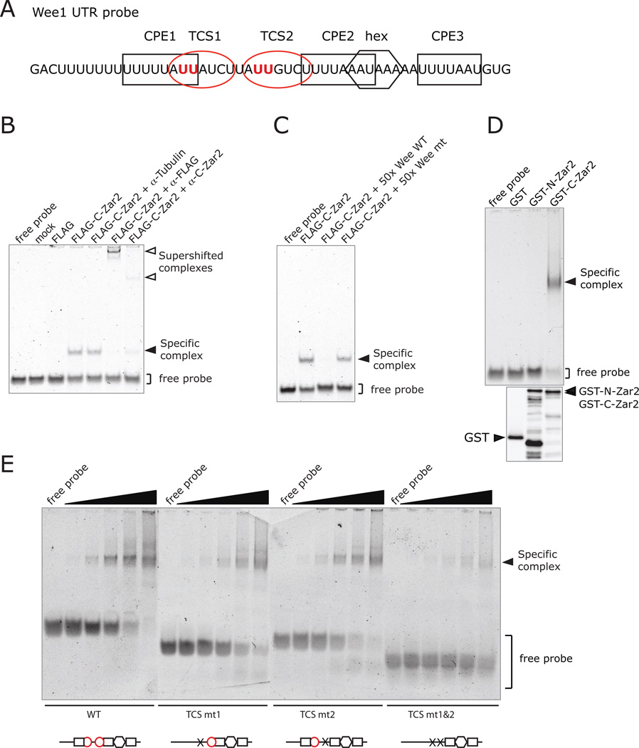
Zygote arrest (Zar) proteins are crucial for early embryonic development, but their molecular mechanism of action is unknown. The Translational Control Sequence (TCS) in the 3' untranslated region (UTR) of the maternal mRNA, Wee1, mediates translational repression in immature Xenopus oocytes and translational activation in mature oocytes, but the protein that binds to the TCS and mediates translational control is not known. Here we show that Xenopus laevis Zar2 (encoded by zar2) binds to the TCS in maternal Wee1 mRNA and represses translation in immature oocytes. Using yeast 3 hybrid assays and electrophoretic mobility shift assays, Zar2 was shown to bind specifically to the TCS in the Wee1 3'UTR. RNA binding required the presence of Zn(2+) and conserved cysteines in the C-terminal domain, suggesting that Zar2 contains a zinc finger. Consistent with regulating maternal mRNAs, Zar2 was present throughout oogenesis, and endogenous Zar2 co-immunoprecipitated endogenous Wee1 mRNA from immature oocytes, demonstrating the physiological significance of the protein-RNA interaction. Interestingly, Zar2 levels decreased during oocyte maturation. Dual luciferase reporter tethered assays showed that Zar2 repressed translation in immature oocytes. Translational repression was relieved during oocyte maturation and this coincided with degradation of Zar2 during maturation. This is the first report of a molecular function of zygote arrest proteins. These data show that Zar2 contains a zinc finger and is a trans-acting factor for the TCS in maternal mRNAs in immature Xenopus oocytes.


Figures

Figure 1
Figure 1
Characterization of a TCS binding protein by yeast three hybrid analysis. A. Cartoon of the RNA hybrids used: white oval, MBE; square, CPE; hexagon, polyadenylation hexanucleotide; red oval, TCS; X, disrupted CPE; dotted line, deleted sequences; bold line, Iron Response Element (IRE). B. Yeast three hybrid colony lift assay for lacZ expression. Blue color indicates RNA-protein interaction. Yeast were transformed with pACT2-A8 and pIIIA plasmids containing the indicated RNA hybrids and three independent colonies streaked for each analysis. A8 interacts with sequences 3’ of the polyadenylation hexanucleotide in the Mos 3’ UTR, the TCS region in the Wee1 3’ UTR, and a TCS inserted into the β-globin 3’ UTR. C. Yeast three hybrid liquid culture assay for lacZ expression. Yeast were transformed with the indicated RNA and protein hybrids. Chart shows β-galactosidase activity. The IRE/Iron Response Protein (IRP) interaction serves as a positive control (SenGupta et al., 1996). More β-galactosidase activity was observed when the TCS was present in the β-globin 3’ UTR.
Figure 2
Figure 2
A8 is the C-terminal of Zar2. A. Alignment of Xenopus laevis Zar1 and Zar2 amino acid sequences was performed using MacVector 11.1.2. A blue box and arrow below the sequences designates the start of A8. Shaded areas indicate identical amino acid residues. Residues targeted by antibodies are highlighted in yellow and labeled below the sequence. Cysteine pairs conserved in both Zar1 and Zar2 proteins are highlighted in red. Cysteines targeted for mutation are indicated below the residue of interest in bold text. Genbank accession numbers are: Zar1, AAP37038; Zar2a, BAH36746; and Xzar2b (Zar2b), JQ776638. For purposes of discussion, the putative loop 1 is between C215 and C242, and the putative loop 2 is between C259 and C287. B. Proposed schematic of Zar protein regions and functions. Shaded areas (in grey) represent regions of high sequence homology between Zar1 and Zar2 while red lines indicate conserved cysteine pairs of interest. The box and arrow designate the boundary of the A8, depicted as a blue line.
Figure 3
Figure 3
Zar2 protein expression during oogenesis and oocyte maturation. A–D. Characterization by western blot of the antibodies used in this study. A and B Lysates from uninjected (−) or GST-Zar2-expressing (+) immature oocytes were analyzed by the purified C-terminal Zar2 antibody (A), or N-terminal antibody (B), or GST antibody as indicated. The immunizing peptide 29–44 (A), or 267–286 (B), was used to block specific antibody sites. Both antibodies recognize GST-Zar2. C. Lysates from uninjected immature (I) or progesterone-stimulated mature (P) oocytes were analyzed with both Zar2 antibodies. A common band at 39 kDa is seen that is endogenous Zar2. D. Lysates from immature oocytes expressing GST-C-Zar2 or GST-C-Zar1 were analyzed by anti C-Zar2 or GST antibodies. The C-Zar2 antibody does not recognize GST-C-Zar1. E–G. Endogenous Zar2 expression. Western blots using Zar2 C-terminal antibody (solid arrowhead) and β-Tubulin (open arrowhead) as a loading control. Representative blots are shown. Similar results were obtained in oocytes from 4–5 different frogs. E. Pools of oocytes were lysed and protein equivalent to a single oocyte from stage I to VI or a single in vitro matured oocyte (egg) (E) was loaded. F. 3.75 µg of total protein was loaded. G. Pools of oocytes were lysed at different times after progesterone stimulation and protein equivalent to 0.5 oocyte was loaded.
Figure 4
Figure 4
Zar2 binds to TCSs in Wee1 3’UTR in vitro. A. Diagram showing the Wee1 UTR probe used in these studies: box, CPE; red oval, TCS; hexagon, polyadenylation hexanucleotide. The red “U” s were mutated to “g” in the TCS mutants. B. EMSA showing that Sf9-expressed FLAG-C-Zar2 forms specific complexes with Wee1 3’ UTR that can be supershifted with specific antibodies to FLAG or C-Zar2. The RNA probe (the last 50 nt of the Wee1 3’ UTR) was incubated with mock purified, FLAG peptide, or FLAG-C-Zar2 proteins. Antibodies against β̃Tubulin, FLAG or C-terminal Zar2 were added where indicated. C. EMSA showing that specific complex formation can be competed with unlabelled RNA containing TCSs. The Wee1 RNA probe was incubated FLAG-C-Zar2 protein, and the unlabeled RNAs were used to compete for labeled probe binding. A 50-fold molar excess of wild type (WT) or TCS-disrupted (mt) Wee1 3’ UTR was used. D. Upper panel, EMSA showing bacterially expressed GST-C-Zar2, but not GST-N-Zar2, binds to Wee1 3’UTR. The Wee1 probe was incubated with GST alone, GST-N-Zar2 or GST-C-Zar2 proteins. Lower panel, western blot with GST antibodies showing that equivalent amounts of proteins were expressed. E. EMSA showing that Zar2 directly binds to the TCSs in the Wee1 UTR. Wee1 probes with mutations in TCS 1 (TCS mt1), TCS 2 (TCS mt2) or both TCSs (TCS mt1&2) were used. These mutant probes were incubated with decreasing amounts of GST-C-Zar2 from 15 µg to 24 ng in a 5-fold dilution series. Below each gel is a diagram of the probe that was used showing which TCSs or CPEs were present. Mutating either TCS alone did not affect binding, but mutating both TCSs markedly reduced binding of GST-C-Zar2.
Figure 5
Figure 5
Zar2 binds RNA via a zinc finger. A. EMSA showing reduced specific complex formation in reduced Zn2+ conditions. The Wee1 probe was incubated with GST-C-Zar2 protein purified from bacteria in the presence of EDTA. Binding reactions were performed in buffer with (+ Zn2+) or without (− Zn2+) zinc chloride. B. Upper panel, EMSA showing no specific complex formation with mutant C-Zar2 proteins that contained cysteine to alanine mutations within the predicted zinc finger domain. A series of cysteine to alanine mutations were made in the C-terminal domain of Zar2 (as shown in Fig. 2). Mutant proteins were bacterially expressed, purified and mixed with the Wee1 RNA probe. Lower panel, western blot showing equivalent amounts of mutant proteins were used in the assay.
Figure 6
Figure 6
Endogenous Zar2 interacts with endogenous mRNAs in immature oocytes. A. Western blot showing immunoprecipitation of Zar2. Immature oocytes were lysed and Zar2 immunoprecipitated with the Zar2 N-terminal antibody (α-Zar2) (see Fig. 2, aa 29–44 and Fig. 3) and protein A/G-agarose beads. As a specificity control, an anti-GST antibody (α-GST) was used to mock immunoprecipitate. As a positive control, total cell lysate was run on the gel. Only samples that contained lysate, beads and N-Zar2 antibodies immunoprecipitated Zar2. B. Semi-quantitative PCR showing selective co-precipitation of maternal mRNAs. PCR conditions were adjusted for each mRNA so that a 1/100 and 1/1000 oocyte equivalent of total RNA showed a 10-fold difference. Wee1 and Mos mRNAs were present in Zar2 immunoprecipitates and were not detected in the mock immunoprecipitation pellets.
Figure 7
Figure 7
The N-terminal of Zar2 represses translation in immature oocytes. A. Cartoon representing the tethered assay constructs used in this experiment. Right panel shows the MS2 fusion proteins. Left panel shows the luciferase reporter mRNAs, the luciferase coding region is fused to the β-globin UTR. The firefly stem-loop reporter contained two MS2 stem-loops (fluc-2x-SL) or no stem-loops (fluc). Renilla luciferase (rluc) had no stem-loops. B. Bar chart showing N-Zar2-MS2 represses translation when tethered to the reporter mRNA. Oocytes were injected with RNA encoding MS2 fusion proteins or not injected (−), and incubated overnight. N-Xzar2-MS2 RNA was injected at 5 ng, 20 ng and 50 ng. Oocytes were then injected with a mixture of firefly with stem-loops (fluc-2x-SL) and Renilla (rluc) reporter constructs. Half the oocytes were stimulated with progesterone (P) and all oocytes were harvested when the progesterone-stimulated oocytes had reached meiosis II. Relative luciferase activity was calculated as described in materials and methods. Bars show mean relative luciferase activity normalized to MS2 alone for immature (black) and progesterone-treated (white) oocytes. Error bars represent S.D. and differences in mean were considered significant with p<0.01 (**) as analyzed by one way ANOVA. MS2-Xp54 shows repression of translation in both immature and mature oocytes. N-Zar2-MS2 represses translation in immature oocytes and this repression is relieved upon maturation (n=5). D. Bar chart showing N-Zar2-MS2 does not repress translation when not tethered. Oocytes were injected with RNA encoding MS2 fusion proteins and incubated overnight. N-Xzar2-MS2 RNA was injected at 50 ng. Oocytes were then injected with a mixture of firefly without stem-loops (fluc) and Renilla (rluc) reporter constructs. Oocytes were harvested when control oocytes had reached meiosis II (n=4). C, E. Western blots showing expression of MS2 fusion proteins at the end of the experiments. MS2, MS2-Xp54 and N-Zar2-MS2 (50 ng) were expressed at equivalent levels. N-Zar2 levels were markedly lower in progesterone treated samples. F. Left panel, semi-quantitative PCR to show that the firefly reporter (fluc-2x-SL) is stable in immature oocytes. The detection limits of the PCR are demonstrated by the lower product formed when ½ or ¼ of the cDNA was added to the PCR reaction. Reporter RNA levels were not lower when tethered to N-Zar2-MS2 than when tethered to MS2 alone. Right panel, ethidium bromide stained gel of total RNA after extraction from oocytes showing equal recovery of rRNA.

References

    1. Arumugam K, Wang Y, Hardy LL, MacNicol MC, MacNicol AM. Enforcing temporal control of maternal mRNA translation during oocyte cell-cycle progression. Embo J. 2010;29:387–397. - PMC - PubMed
    1. Balzer E, Moss EG. Localization of the developmental timing regulator Lin28 to mRNP complexes, P-bodies and stress granules. RNA Biol. 2007;4:16–25. - PubMed
    1. Bardwell VJ, Wickens M. Purification of RNA and RNA-protein complexes by an R17 coat protein affinity method. Nucleic Acids Res. 1990;18:6587–6594. - PMC - PubMed
    1. Bernstein DS, Buter N, Stumpf C, Wickens M. Analyzing mRNA-protein complexes using a yeast three-hybrid system. Methods. 2002;26:123–141. - PubMed
    1. Bienz M. The PHD finger, a nuclear protein-interaction domain. Trends Biochem Sci. 2006;31:35–40. - PubMed

Publication types

MeSH terms

LinkOut - more resources