. 2012 Aug 1;189(3):1459-66.
doi: 10.4049/jimmunol.1200484. Epub 2012 Jun 25.

Helminth infection impairs autophagy-mediated killing of bacterial enteropathogens by macrophages

Affiliations

Helminth infection impairs autophagy-mediated killing of bacterial enteropathogens by macrophages

Chien-wen Su et al. J Immunol. .

Abstract

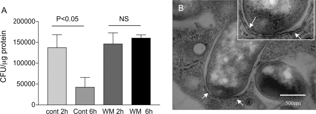
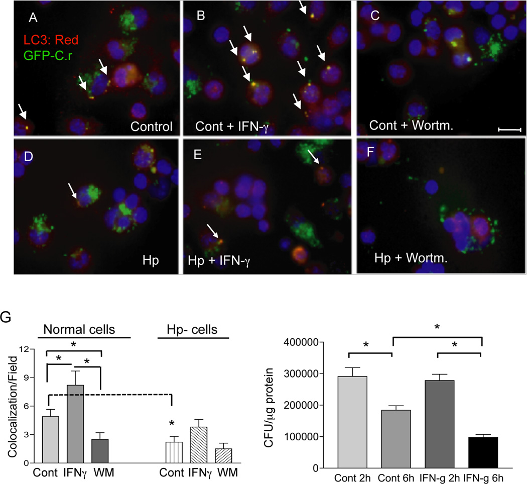
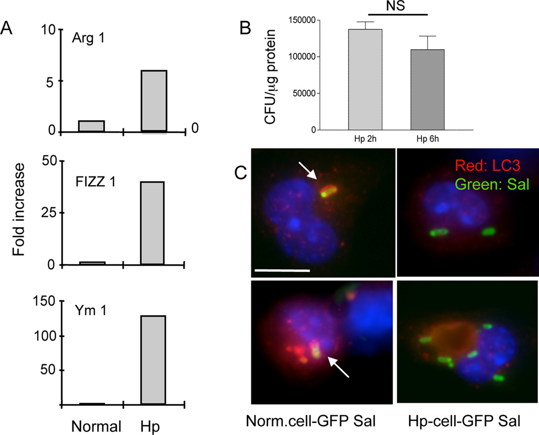
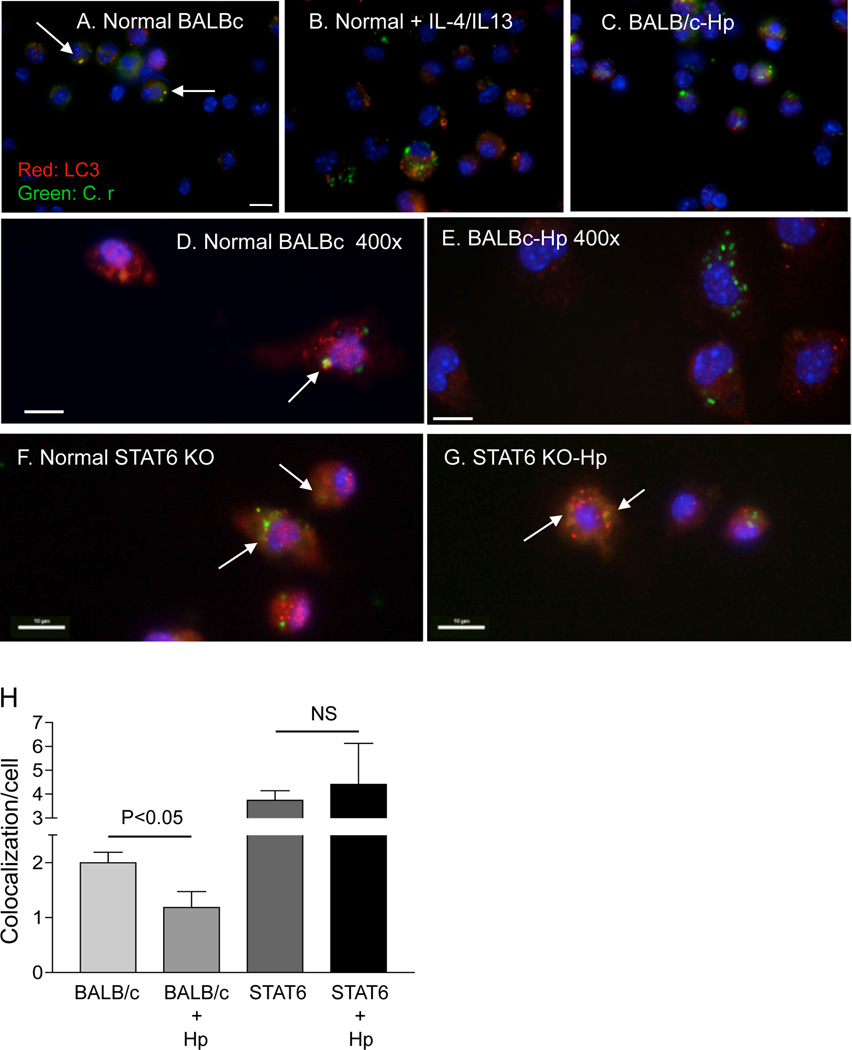
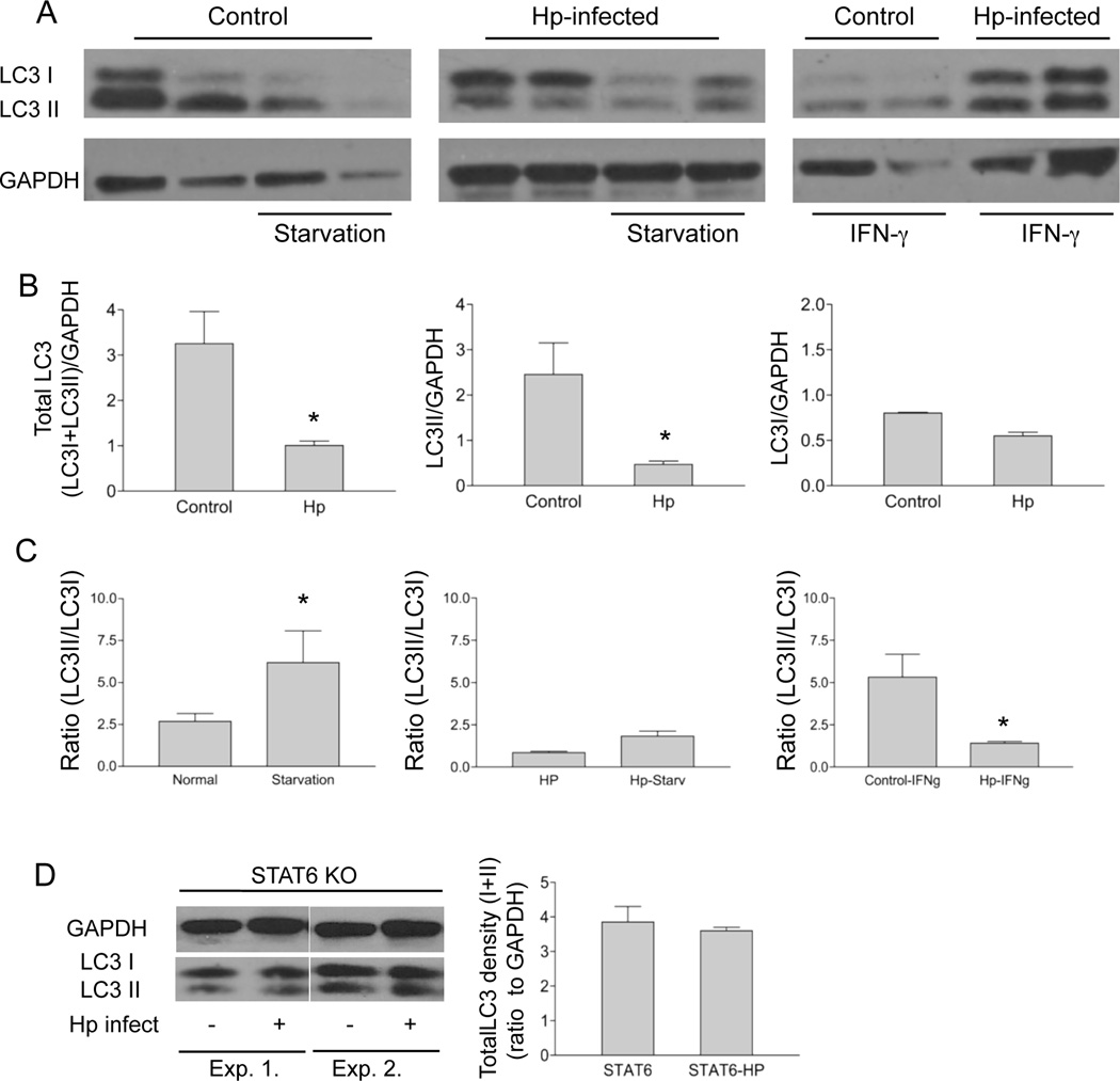
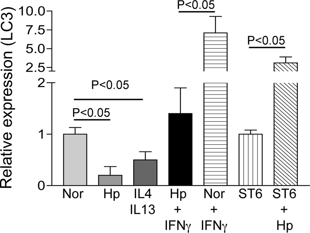
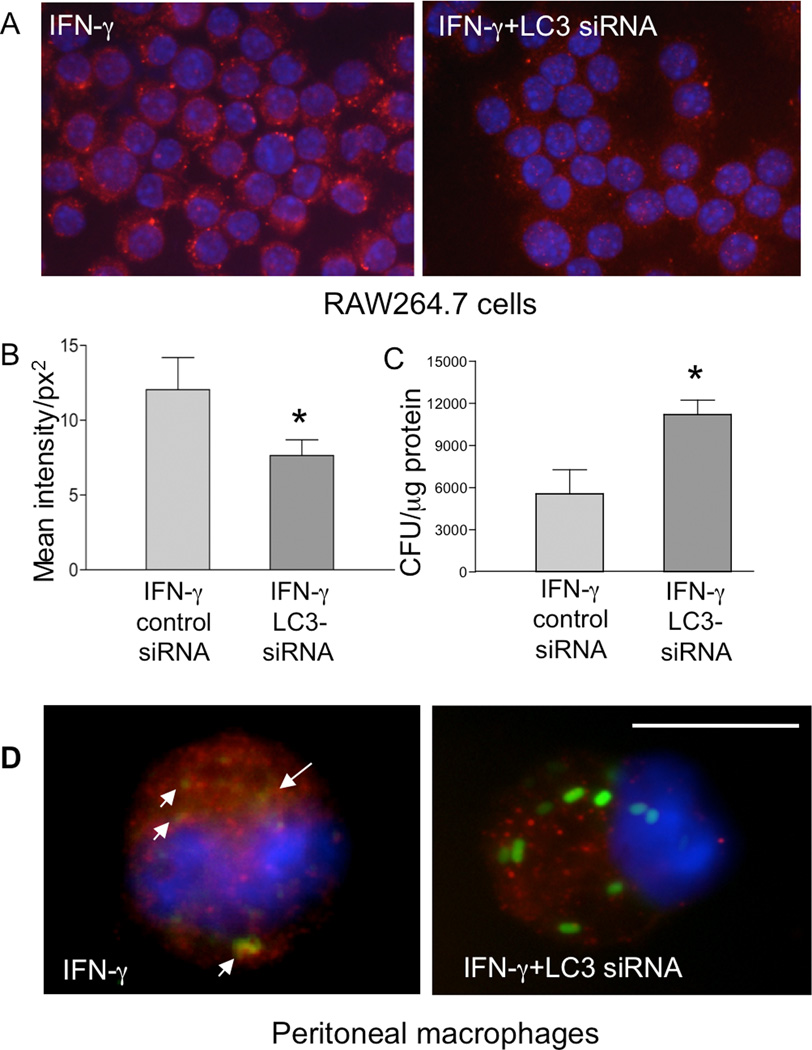
Autophagy is an important mechanism used by macrophages to kill intracellular pathogens. The results reported in this study demonstrate that autophagy is also involved in the macrophage killing of the extracellular enteropathogen Citrobacter rodentium after phagocytosis. The process was significantly impaired in macrophages isolated from mice chronically infected with the helminth parasite Heligmosomoides polygyrus. The H. polygyrus-mediated inhibition of autophagy was Th2 dependent because it was not observed in macrophages isolated from helminth-infected STAT6-deficient mice. Moreover, autophagy of Citrobacter was inhibited by treating macrophages with IL-4 and IL-13. The effect of H. polygyrus on autophagy was associated with decreased expression and processing of L chain protein 3 (LC3), a key component of the autophagic machinery. The helminth-induced inhibition of LC3 expression and processing was STAT6 dependent and could be recapitulated by treatment of macrophages with IL-4 and IL-13. Knockdown of LC3 significantly inhibited autophagic killing of Citrobacter, attesting to the functional importance of the H. polygyrus-mediated downregulation of this process. These observations reveal a new aspect of the immunosuppressive effects of helminth infection and provide mechanistic insights into our earlier finding that H. polygyrus significantly worsens the in vivo course of Citrobacter infection.


Figures

Figure 1
Figure 1
Autophagy-mediated bacterial killing is impaired in macrophages from helminth-infected mice. A: Treatment of macrophages with wortmannin, an autophagy inhibitor, resulted in impaired bacterial killing. Peritoneal macrophages were isolated from normal BALB/c mice and exposed to C. rodentium (10 bacteria/cell) for 1 h. At different time points (2 and 6 h) after infection, the number of viable intracellular bacteria was determined by plating the cell lysates onto LB plates. The data shown represent the mean ± SD of triplicate cultures, and are from one of three experiments showing similar results. Additional cultures of macrophages without infection by C. rodentium showed no detection of viable bacteria (data not show). B: Autophagic double-membrane structures are present in macrophages with phagocytosed C. rodentium. Peritoneal macrophages were isolated from normal BALB/c mice, pre-treated with IFNγ (100 U/ml) overnight and exposed to C. rodentium for 1 h. The ultra-structure of the cells was examined by transmission electron microscopy (images shown are 60,000×). Arrows (and the inset) indicate the presence of double-membrane structures surrounding the internalized bacteria. Scale bar represents 500 nm.
Figure 2
Figure 2
Helminth-infection resulted in reduced co-localization of LC3 and C. rodentium in macrophages. A–F: Immunofluorescence microscopy data show the detection of LC3, an autophagy marker, internalized GFP-expressing C. rodentium and colocalization of LC3 and GFP-C. rodentium in peritoneal macrophages (stained with anti-LC3 and Cy3: red) from Normal (A, B and C) and H. polygyrus-infected (D, E and F) mice at 6 h after C. rodentium-GFP infection. Wortmannin-treated cells displayed reduced LC3-GFP bacterial colocalization (C and F). IFNγ treatment resulted in an increase in LC3 expression and colocalization of LC3 with GFP-C. rodentium (B and E). Scale bar represents 10 µm. G: Frequency of LC3-GFP-C. rodentium colocalization within macrophages isolated from normal and helminth-infected BALB/c mice with and without wortmannin and IFNγ. The data shown are from one of three experiments showing similar results. Arrows indicate the presence of co-localization of LC3 and GFP-expressing C. rodentium. *p < 0.05 for a comparison of two groups. H: IFNγ treatment of the cells results in enhanced bacterial killing. Peritoneal macrophages were isolated from normal BALB/c mice, and then exposed to C. rodentium for 1 h with or without overnight IFNγ pretreatment. The number of viable intracellular bacteria recovered from the macrophages was determined by plating the cell lysates onto LB plates 6 h after gentamicin treatment. The data represent the mean ± SD of triplicate cultures. *p < 0.05 for a comparison of two groups.
Figure 3
Figure 3
A. H. polygyrus infection up-regulates Ym1, Arg1 and Fizz1 expression in peritoneal macrophages and impairs bacterial killing by macrophages. Peritoneal macrophages were isolated from normal and helminth-infected BALB/c mice at 7 days post infection. Total RNA was isolated from the cells. Ym1, Arg1 and Fizz1 expression was determined using real-time RT-PCR. Values are the fold increase compared with baseline obtained from uninfected control mice. B. H. polygyrus infection impairs bacterial killing by macrophages. Peritoneal macrophages were isolated from H. polygyrus infected mice and used for gentamicin protection assay. The results show similar numbers of viable internalized bacteria recovered in macrophages isolated from heminth-infected mice at 2 and 6 h post bacterial infection in vitro. P > 0.05. C. Immunofluorescence microscopy shows LC3-Samonella co-localization in macrophages from control and helminth-infected mice. Scale bar indicates 10 µm. The data shown are from one of three experiments with similar results.
Figure 4
Figure 4
The effect of H. polygyrus on autophagy of C. rodentium is mediated by a Th2 dependent mechanism. A–G: Immunofluorescence microscopy data show the detection of LC3, an autophagy marker, internalized GFP-expressing C. rodentium and colocalization of LC3 and GFP-C. rodentium in peritoneal macrophages isolated from normal BALB/c (A & D), H. polygyrus-infected BALB/c (C & E), normal STAT6 KO (F) or H. polygyrus-infected STAT6 KO mice (G). B: Peritoneal macrophages were isolated from normal BALB/c mice, pre-treated with IL-4/IL-13 (3 ng/ml) overnight and then exposed to GFP-C. rodentium for 1 h. Cells were collected 2 h after infection, fixed and stained for LC3. Scale bar represents 10 µm. For a detailed description, see Figure 1 C–H. H. Frequency of LC3-GFP-C. rodentium colocalization within macrophages isolated from normal and helminth-infected BALB/c and STAT6 KO mice. The data represent the mean (co-localization of LC3 and GFP-C. rodentium/cell) ± SD obtained from 30–50 cells per slide (1 slide from each of the two experiments was examined).
Figure 5
Figure 5
H. polygyrus infection reduces LC3 expression and LC3 II conversion in macrophages via a STAT6 dependent mechanism. A. Peritoneal macrophages were isolated from normal and H. polygyrus infected BALB/c mice (2 weeks after infection). Autophagy was induced in macrophages by incubating the cells overnight in the presence and absence of recombinant IFNγ (100u/ml) or by amino acid and serum starvation at 37°C for 3 h. Cell lysates were collected for detection of LC3 (LC3I and LC3II) by western-blotting. GAPDH was used as the loading control. Each lane represents the sample from an individual mouse. B and C. The bar graphs show the total LC3 density (LC3I+LC3II), LC3I, LC3II density and the LC3II/LC3I ratio (mean density + SD from 3 experiments) based on the western blotting analysis of LC3 expression (in A). D. Western blotting analysis of LC3 expression in peritoneal macrophages isolated from normal and H. polygyrus infected STAT 6 KO mice.
Figure 6
Figure 6
H. polygyrus infection down-regulates LC3 gene expression in wild-type mice but not in STAT6 KO mice. Peritoneal macrophages were isolated from normal or H. polygyrus-infected wild-type and STAT6 KO BALB/c mice. After overnight incubation in vitro in the presence or absence of Th1 or Th2 cytokines, total RNA was isolated. LC3 expression was determined using real-time RT-PCR. Values are the fold decrease or increase compared with baseline obtained from uninfected control mice. The data shown are from one of two experiments showing similar results.
Figure 7
Figure 7
siRNA-mediated knock-down of LC3 leads to impaired bacterial killing in RAW264.7 cells and peritoneal macrophages in vitro. A. RAW264.7 cells, with or without overnight IFNγ pretreatment, were transfected with or without an siRNA specific for LC3. The cells were then stained with anti-LC3 antibody as described in Figure 1. Results show that IFNγ-induced LC3 expression was effectively decreased by the siRNA (400 ×). B. Quantification of LC3 fluorescence intensity in RAW264.7 cells transfected with either a control or LC3-specific siRNA. The LC3 fluorescence intensity per cell was measured digitally using the Graph digitizing software (Nikon Imaging Software Elements). The data shown represent the mean values of fluorescence intensity obtained from 50–60 cells per slide (1 slide from each of the two experiments was examined). *p < 0.05 for a comparison of two groups. C. After knocking down LC3 expression with the siRNA, the RAW264.7 cells were used for gentamicin protection assay. The data shown represent the mean ± SD of triplicate cultures. *p < 0.05 for a comparison of two groups. D: Peritoneal macrophages were isolated from normal BALB/c mice and treated with IFNγ overnight in vitro. The cells were transfected with siRNA specific for LC3 and infected 48 h later with C. rodentium for 1 h. LC3, GFP-Citrobacter and colocalization of LC3 and bacteria were examined (1000 ×). Scale bar represents 10 µm.

Similar articles

Cited by

References

    1. Nokes C, Cooper E, Robinson B, Bundy D. Geohelminth infection and academic assessment in Jamaican children. Trans R Soc Trop Med Hyg. 1991;2:272–273. - PubMed
    1. Crompton D, Nesheim M. Nutritional impact of intestinal helminthiasis during the human life cycle. Annual Review of Nutrition. 2002;22:35–59. - PubMed
    1. Babu S, Bhat SQ, Kumar NP, Anuradha R, Kumaran P, Gopi PG, Kolappan C, Kumaraswami V, TB N. Attenuation of toll-like receptor expression and function in latent tuberculosis by coexistent filarial infection with restoration following antifilarial chemotherapy. PLoS One. 2009;3:e489. - PMC - PubMed
    1. Helmby H. Gastrointestinal nematode infection exacerbates malaria-induced liver pathology. J Immunol. 2009;182:5663–5671. - PMC - PubMed
    1. Chen C-C, Louie Steve, McCormick Beth A., W WA, Shi HN. Concurrent infection of an intestinal helminth parasite impairs host resistance to enteric Citrobacter rodentium and enhances Citrobacter-induced colitis in mice. Infection and Immunity. 2005;73:5468–5481. - PMC - PubMed

Publication types

MeSH terms

Substances