Review
. 2012 Aug;8(8):423-34.
doi: 10.1038/nrneurol.2012.117. Epub 2012 Jun 26.

Advances in understanding the molecular basis of frontotemporal dementia

Affiliations
Review

Advances in understanding the molecular basis of frontotemporal dementia

Rosa Rademakers et al. Nat Rev Neurol. 2012 Aug.

Erratum in

  • Nat Rev Neurol. 2013 May;9(5):240

Abstract

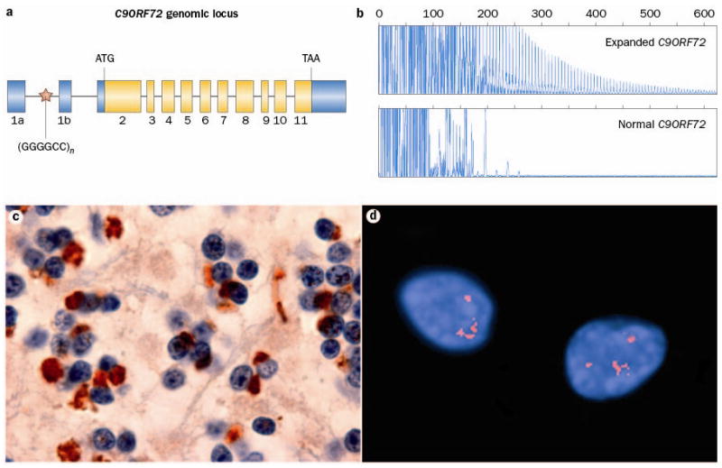
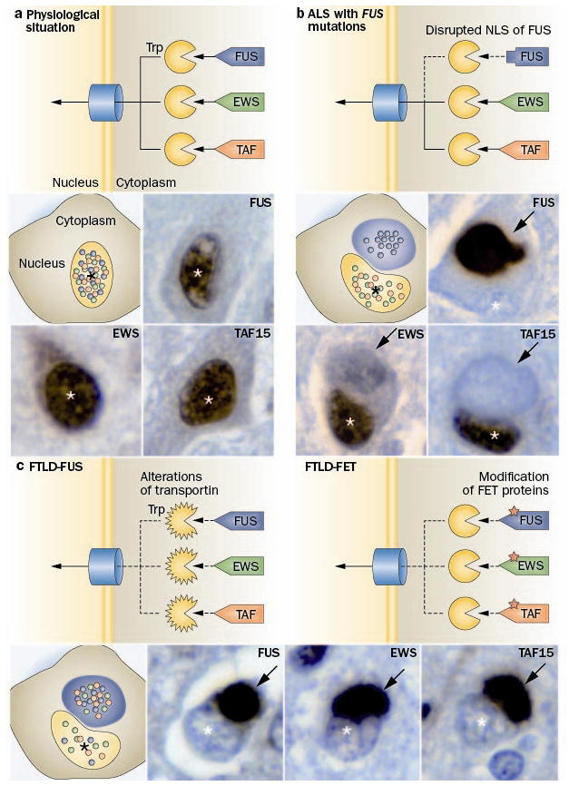
Frontotemporal dementia (FTD) is a clinical syndrome with a heterogeneous molecular basis. Until recently, the underlying cause was known in only a minority of cases that were associated with abnormalities of the tau protein or gene. In 2006, however, mutations in the progranulin gene were discovered as another important cause of familial FTD. That same year, TAR DNA-binding protein 43 (TDP-43) was identified as the pathological protein in the most common subtypes of FTD and amyotrophic lateral sclerosis (ALS). Since then, substantial efforts have been made to understand the functions and regulation of progranulin and TDP-43, as well as their roles in neurodegeneration. More recently, other DNA/RNA binding proteins (FET family proteins) have been identified as the pathological proteins in most of the remaining cases of FTD. In 2011, abnormal expansion of a hexanucleotide repeat in the gene C9orf72 was found to be the most common genetic cause of both FTD and ALS. All common FTD-causing genes have seemingly now been discovered and the main pathological proteins identified. In this Review, we highlight recent advances in understanding the molecular aspects of FTD, which will provide the basis for improved patient care through the development of more-targeted diagnostic tests and therapies.


Figures

Figure 1
Figure 1
Expanded GGGGCC hexanucleotide repeat in non-coding region of C9ORF72 causes FTD and ALS linked to chromosome 9p. a Overview of the genomic structure of C9ORF72. Numbered boxes represent coding (white) and non-coding (gray) exons and the position of the start codon (ATG) and stop codon (TAA) are indicated. The position of the (GGGGCC)n repeat in the intronic region between exons 1a and 1b is indicated with a red star. b PCR products of repeat-primed PCR reactions separated on an ABI3730 DNA Analyzer and visualized by GENEMAPPER software. Electropherograms are zoomed to 2,000 relative fluorescence units to show stutter amplification. One FTD patient with a pathogenic expanded C9ORF72 repeat (top) and one FTD patient with a C9ORF72 normal repeat length (bottom) are shown. c In addition to FTLD-TDP and ALS pathology, all patients with the C9ORF72 mutation show a unique pattern of ubiquitin-positive (brown), TDP-43-negative neuronal inclusions in the cerebellar granule layer and other specific neuroanatomical regions. This disease-specific finding implies the mis-metabolism and accumulation of some yet unidentified protein(s). d RNA foci, visualized using a Cy3-labeled (GGCCCC)4 oligonucleotide probe (red), in the nuclei of two lower motor neurons from an FTD-ALS patient carrying the expanded GGGGCC repeat in C9ORF72.
Figure 2
Figure 2
Distinct pathomechanisms of ALS-FUS and FTLD-FUS. a The FET protein members, FUS, EWS and TAF15 contain a proline-tyrosine nuclear localization signal (PY-NLS, triangle) which is bound by the receptor protein Transportin (Trp). This mediates the transport of these proteins across the nuclear pore complex into the nucleus, resulting in a predominant nuclear localization of all three proteins under physiological conditions. b In ALS with FUS mutations, the PY-NLS of FUS is disrupted due to mutations (rectangle) leading to an impaired interaction with Trp and nuclear import of FUS, while TAF15 and EWS are normally transported to the nucleus. This results in a selective accumulation of FUS into cytoplasmic inclusion in ALS-FUS patients, with retained nuclear localization of TAF15 and EWS. c In contrast, FTLD-FUS patients show co-accumulation of all FET proteins into cytoplasmic inclusions accompanied with their nuclear depletion. This complex dysregulation of all FET proteins in FTLD-FUS might be explained by either of two broad scenarios: c-1 alterations of Trp itself, e. g. by genetic variations, reduced expression levels or posttranslational modifications; or c-2 posttranslational modifications of FET proteins, which interfere with proper Trp binding. Each photomicrograph shows a single neuron with a cytoplasmic inclusion (arrow) and the nucleus indicated by the asterisk (*), immunostained for the FET protein indicated (brown stain).

References

    1. Bird T, et al. Epidemiology and genetics of frontotemporal dementia/Pick’s disease. Ann Neurol. 2003;54 (Suppl 5):S29–31. - PubMed
    1. Feldman H, et al. A Canadian cohort study of cognitive impairment and related dementias (ACCORD): study methods and baseline results. Neuroepidemiology. 2003;22:265–274. - PubMed
    1. The Lund and Manchester Groups. Clinical and neuropathological criteria for frontotemporal dementia. J Neurol Neurosurg Psychiatr. 1994;57:416–418. - PMC - PubMed
    1. McKhann GM, et al. Clinical and pathological diagnosis of frontotemporal dementia: report of the Work Group on Frontotemporal Dementia and Pick’s Disease. Arch Neurol. 2001;58:1803–1809. - PubMed
    1. Neary D, et al. Frontotemporal lobar degeneration: a consensus on clinical diagnostic criteria. Neurology. 1998;51:1546–1554. - PubMed

Publication types

MeSH terms