. 2012 Aug;13(8):741-9.
doi: 10.1038/embor.2012.89. Epub 2012 Jun 26.

Nuclear movement during myotube formation is microtubule and dynein dependent and is regulated by Cdc42, Par6 and Par3

Affiliations

Nuclear movement during myotube formation is microtubule and dynein dependent and is regulated by Cdc42, Par6 and Par3

Bruno Cadot et al. EMBO Rep. 2012 Aug.

Abstract

Cells actively position their nucleus within the cytoplasm. One striking example is observed during skeletal myogenesis. Differentiated myoblasts fuse to form a multinucleated myotube with nuclei positioned in the centre of the syncytium by an unknown mechanism. Here, we describe that the nucleus of a myoblast moves rapidly after fusion towards the central myotube nuclei. This movement is driven by microtubules and dynein/dynactin complex, and requires Cdc42, Par6 and Par3. We found that Par6β and dynactin accumulate at the nuclear envelope of differentiated myoblasts and myotubes, and this accumulation is dependent on Par6 and Par3 proteins but not on microtubules. These results suggest a mechanism where nuclear movement after fusion is driven by microtubules that emanate from one nucleus that are pulled by dynein/dynactin complex anchored to the nuclear envelope of another nucleus.


Conflict of interest statement

The authors declare that they have no conflict of interest.

Figures
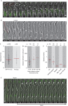
Figure 1
Figure 1
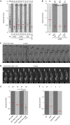
Nuclear movement after fusion requires MTs. (A) Frames from a time-lapse two-channel movie (phase contrast and fluorescence) of differentiated GFP-H1-C2 myoblasts during fusion (supplementary Movie S2 online). A myoblast (red outline) fused with a myotube (green outline) to form a new myotube (white outline; time in h:min). Note that after fusion (0:40), myoblast nucleus (arrowhead) moves towards the myotube nuclei (arrow). (B) Frames from a time-lapse movie of differentiated primary myoblasts during fusion (supplementary Movie S3 online). A myoblast (red outline) fused with a myotube (green outline) to form a new myotubes (white outline). Note that after fusion (00:40) myoblast nucleus (arrowhead) moves towards the myotube nuclei (arrow). (C) Speed of the nuclei after fusion of myoblasts into myotubes on differentiated GFP-H1-C2 and primary myoblasts. (D) Plot representing the distance between the myoblast nucleus and the nuclei in the centre of the myotube (distance between nuclei, y axis) and the time delay between fusion and initiation of myoblast nuclear movement (delay before nuclear movement, x axis). (E) Frames from a time-lapse two-channel movie of differentiated GFP-H1-C2 myoblasts during fusion of a myoblast (red outline) with a myotube (green outline) after addition of 100 nM taxol (supplementary Movie S4 online). Fusion occurred between 00:15 and 00:30, and taxol was added at 00:45. Note that myoblast nucleus (arrowhead) did not move towards the myotube nuclei (arrow). (F) Speed of the nuclei after fusion of differentiated GFP-H1-C2 cells in nontreated, non-transfected myotubes (Ctr), myotubes expressing the indicated spastin constructs as in supplementary Fig S1a online, or myotubes treated with 75 nM nocodazole (Ndz) or 100 nM taxol as in (E). Scale bar in (A,B,E), 20 μm. P-value in (C,D,F); **P<0.01, ***P<0.005. Red line indicates the median. MTs, microtubules.
Figure 2
Figure 2
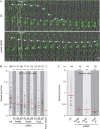
Cdc42 regulates nuclear movement after fusion. (A) Speed of the nuclei after fusion of differentiated GFP-H1-C2 cells in nontreated, treated with three different siRNA against DHC, IC2, p150 or myotubes transfected with GFP-p50. (B) Speed of the nuclei after fusion of differentiated primary myoblasts in untreated or after transfection with the indicated siRNAs. (C,D) Frames from a time-lapse movie of differentiated primary myoblasts isolated from Cdc42 flox mice (supplementary Movies S5 and S6 online). A myoblast (arrowhead) fused with a myotube (arrow) to form a new myotubes (black outline) in the absence (C) or presence (D) of a Cre-encoding adenovirus. The myoblast nucleus and the myotube nuclei are highlighted by a black circle in (C). Note that in Cre-treated cultures, nuclear movement after fusion was reduced. Scale Bar, 20 μm. (E) Speed of the nuclei after fusion in WT primary cells or primary Cdc42 flox myoblasts in the absence or presence of Cre-encoding adenovirus. (F) Speed of the nuclei after fusion of differentiated GFP-H1-C2 cells in myotubes nonmicroinjected or microinjected with constructs encoding GFP, Cdc42N17 or Cdc42V12. P-value in (A), (B), (E) and (F); *P<0.05, **P<0.01, ***P<0.005. Red line indicates the median. DHC, dynein heavy chain; GFP, green fluorescent protein; siRNA, short interfering RNA; WT, wild-type.
Figure 3
Figure 3
Par proteins and dynein/dynactin complex are involved in nuclear movement after fusion. (A) Frames from a time-lapse two-channel movie (phase contrast and fluorescence) of differentiated GFP-H1-C2 cells untreated or Par6β siRNA treated annotated as in Fig 1A (supplementary Movie S7 online). Scale Bar, 20 μm. (B) Speed of the nuclei after fusion of differentiated GFP-H1-C2 cells nontreated, treated with three different siRNA against Par6β, Par6γ, Par3 or myotubes microinjected with YFP-Par3-PDZ1. (C) Speed of the nuclei after fusion of differentiated primary myoblasts in untreated or after transfection with the indicated siRNAs. P-value in (B,C); **P<0.01, ***P<0.005. Red line indicates the median. GFP, green fluorescent protein; siRNA, short interfering RNA.
Figure 4
Figure 4
Par6 and dynactin accumulate at the NE of differentiated myoblasts and myotubes. (A) Representative epi-fluorescence images of differentiated C2C12 myoblasts immunostained for Par6β, PC and DNA (DAPI). (B) Representative epi-fluorescence images of differentiated C2C12 myoblasts immunostained for p150, PC and DNA (DAPI). (CE) Representative confocal images of differentiated C2C12 myotubes immunostained for Par6β (C), p50 (D) or p150 (E), pericentrin and DAPI. White square in left panel delineates the region showed in the right panel. Nondifferentiated myoblasts (#) and myotubes (*) nuclei are indicated based on pericentrin localization. Scale bars, 10 μm. NE, nuclear envelope; PC, pericentrin.
Figure 5
Figure 5
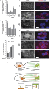
Par proteins regulate Par6 and dynactin localization at the NE of differentiated myoblasts and myotubes. (A) Quantification of nuclei with Par6β at the NE in differentiated myoblasts and myotubes transfected with the indicated siRNAs, relative to non-siRNA control. At least 1,700 nuclei were counted corresponding to three independent experiments. (B) Representative epi-fluorescence images showing Par6β, pericentrin and DNA (DAPI) staining in control, Par6γ and DHC siRNA. (C) Quantification of nuclei with p150 at the NE in differentiated myoblasts and myotubes transfected with the indicated siRNAs, relative to non-siRNA control. At least 1,600 nuclei were counted corresponding to three independent experiments. (D) Representative epi-fluorescence images showing p150, pericentrin and DNA (DAPI) staining in control, Par6β and DHC siRNA. (E) Quantification of nuclei with Par6β and p150 at the NE of differentiated myoblasts and myotubes in cells untreated (Ctr) or treated with 5 μm nocodazole during 2 h before fixation (Ndz). At least 800 nuclei were counted corresponding to three independent experiments. (F) Proposed model for nuclear movement after fusion. Before fusion, both MTs from differentiated myoblast (orange) and myotube (green) nuclei are anchored by their minus ends to the nuclei with Par6β and dynein/dynactin at the NE (brown). After fusion, MTs emanating from the myoblast nucleus contact the nuclei of the myotube (green; left inset) and can be pulled by dynein/dynactin resulting in the movement of the myoblast nucleus towards the centre of the myotube (orange arrow). In addition, or in alternative, MTs emanating from the myotube nuclei might also contact the myoblast nuclei (orange; right inset) via dynein/dynactin resulting in the movement of the myoblast nucleus on the MTs. P-value in (A), (C) and (E); *P<0.05, **P<0.01. Errors bars are s.e.m. Scale bars, 10 μm. DHC, dynein heavy chain; MTs, microtubules; NE, nuclear envelope; siRNA, short interfering RNA.

References

    1. Dauer WT, Worman HJ (2009) The Nuclear envelope as a signaling node in development and disease. Dev Cell 17: 626–638 - PubMed
    1. Starr DA, Fridolfsson HN (2010) Interactions between nuclei and the cytoskeleton are mediated by SUN-KASH nuclear-envelope bridges. Annu Rev Cell Dev Biol 26: 421–444 - PMC - PubMed
    1. Reinsch S, Gönczy P (1998) Mechanisms of nuclear positioning. J Cell Sci 111: 2283–2295 - PubMed
    1. Gomes ER, Jani S, Gundersen GG (2005) Nuclear movement regulated by Cdc42, MRCK, myosin, and actin flow establishes MTOC polarization in migrating cells. Cell 121: 451–463 - PubMed
    1. Cappello S et al. (2006) The Rho-GTPase cdc42 regulates neural progenitor fate at the apical surface. Nat Neurosci 9: 1099–1107 - PubMed

Publication types

MeSH terms