. 2012 Aug;9(4):046005.
doi: 10.1088/1741-2560/9/4/046005. Epub 2012 Jun 25.

Improved spatial targeting with directionally segmented deep brain stimulation leads for treating essential tremor

Affiliations

Improved spatial targeting with directionally segmented deep brain stimulation leads for treating essential tremor

Maureen Keane et al. J Neural Eng. 2012 Aug.

Abstract

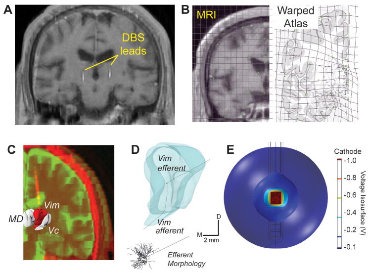
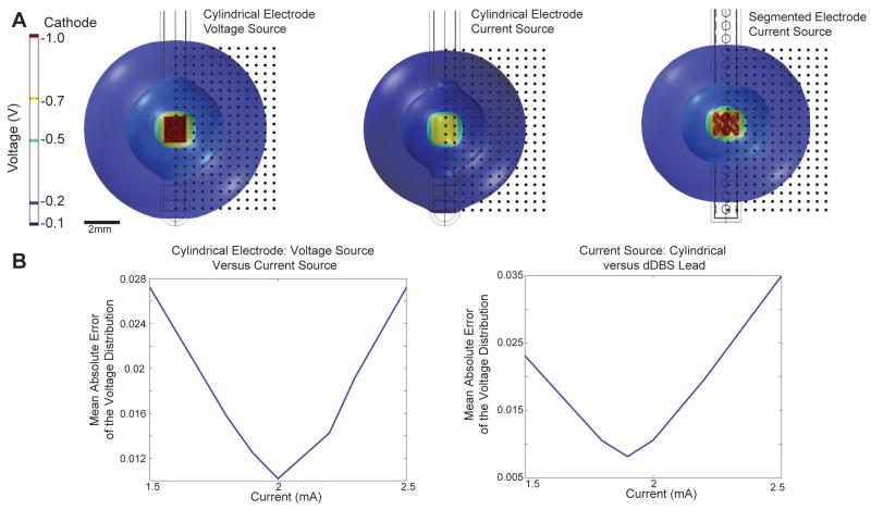
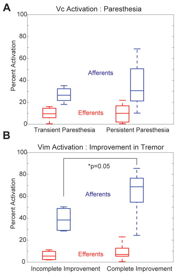
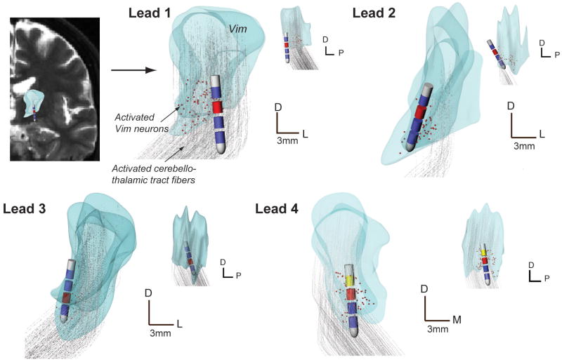
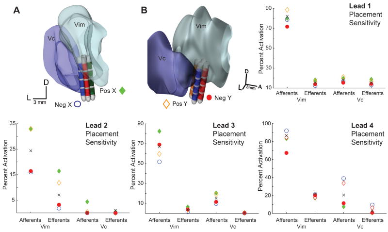
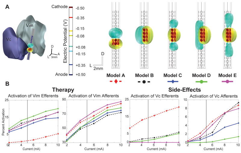
Deep brain stimulation (DBS) in the ventral intermediate nucleus of thalamus (Vim) is known to exert a therapeutic effect on postural and kinetic tremor in patients with essential tremor (ET). For DBS leads implanted near the caudal border of Vim, however, there is an increased likelihood that one will also induce paresthesia side-effects by stimulating neurons within the sensory pathway of the ventral caudal (Vc) nucleus of thalamus. The aim of this computational study was to (1) investigate the neuronal pathways modulated by therapeutic, sub-therapeutic and paresthesia-inducing DBS settings in three patients with ET and (2) determine how much better an outcome could have been achieved had these patients been implanted with a DBS lead containing directionally segmented electrodes (dDBS). Multi-compartment neuron models of the thalamocortical, cerebellothalamic and medial lemniscal pathways were first simulated in the context of patient-specific anatomies, lead placements and programming parameters from three ET patients who had been implanted with Medtronic 3389 DBS leads. The models showed that in these patients, complete suppression of tremor was associated most closely with activating an average of 62% of the cerebellothalamic afferent input into Vim (n = 10), while persistent paresthesias were associated with activating 35% of the medial lemniscal tract input into Vc thalamus (n = 12). The dDBS lead design demonstrated superior targeting of the cerebello-thalamo-cortical pathway, especially in cases of misaligned DBS leads. Given the close proximity of Vim to Vc thalamus, the models suggest that dDBS will enable clinicians to more effectively sculpt current through and around thalamus in order to achieve a more consistent therapeutic effect without inducing side-effects.


Conflict of interest statement

Conflict of Interest: None

Figures

Figure 1
Figure 1
Patient-specific DBS models for treating essential tremor. A) MR data for each patient was matched to a corresponding brain atlas plate based on anatomical landmarks. B) A human brain atlas was digitally warped to MR images from each subject. C) MRI and CT data were segmented and co-registered to determine lead location. D) Each thalamic nucleus was populated with multi-compartment thalamocortical neuron and thalamic afferent axons models. E) Finite element analysis involved simulating the voltage distribution in the brain during DBS, as shown for these voltage isosurfaces around a stimulated electrode.
Figure 2
Figure 2
Finite element model calibration with voltage isosurfaces. A) Calibration between voltage- and current-controlled DBS and between stimulation through cylindrical and directionally-segmented electrodes. B) Plots of mean error in the spatial voltage profile surrounding each electrode contact during monopolar DBS.
Figure 3
Figure 3
Grouped analysis of the neuronal pathways activated during DBS programming across the three essential tremor patients. A) Percent activation of Vc efferent and afferent projections for outcomes containing transient and persistent paresthesias (n=12 settings). B) Percent activation of Vim efferent and afferent projections for outcomes containing incomplete and complete resolution of tremor (n=10 settings).
Figure 4
Figure 4
Patient-specific modeling results for each lead implant. Vim volumes are shown in coronal and sagittal planes and include the co-registered DBS lead positions and active contacts (cathode in red, anode in yellow, IPG was the anode where not shown). Activated Vim neurons (indicated in red) and activated Vim afferents (pathways in gray) are also shown for the clinician-identified best therapy setting. All scale bars: 3mm.
Figure 5
Figure 5
Sensitivity analysis of lead placement was performed by shifting the DBS lead 1 mm in each direction of the stereotactic microdrive plane. An example of this manipulation is illustrated in A) and B) for Subject 3 (Lead 4). Plots show model predictions for percent activation of the afferent and efferent pathways after shifting the DBS lead in each direction (blue-negative x, green-positive x, orange-positive y, red-negative y). The black x marker indicates the original model predictions for the clinician-optimized bipolar DBS setting.
Figure 6
Figure 6
Five dDBS stimulation configurations were evaluated in place of the cylindrical lead for Subject 3 (Lead 4), who never achieved complete suppression of tremor. A) dDBS lead configurations with isosurfaces illustrating the electric potential surrounding the lead. Cathodic contacts are indicated in red and anodic contacts in blue. Model A represents an estimated match between the dDBS lead and the bipolar setting finalized in the clinic. Models B-E maintained eight cathodic contacts oriented towards the Vim while the anodic contacts were shifted around the lead for assessment of optimized activation. The Vim is located anterior to the lead, and the Vc is located posterior to the lead. B) Activation plots of Vim and Vc efferents and afferents versus total current injection. The vertical bar represents the estimated equivalent voltage source stimulated in the final clinical setting.

Similar articles

Cited by

References

    1. Abosch A, Yacoub E, Ugurbil K, Harel N. An assessment of current brain targets for deep brain stimulation surgery with susceptibility-weighted imaging at 7 tesla. Neurosurgery. 2010;67:1745–1756. discussion 1756. - PMC - PubMed
    1. Barbe MT, Liebhart L, Runge M, Deyng J, Florin E, Wojtecki L, Schnitzler A, Allert N, Sturm V, Fink GR, Maarouf M, Timmermann L. Deep brain stimulation of the ventral intermediate nucleus in patients with essential tremor: stimulation below intercommissural line is more efficient but equally effective as stimulation above. Experimental neurology. 2011a;230:131–137. - PubMed
    1. Barbe MT, Liebhart L, Runge M, Pauls KA, Wojtecki L, Schnitzler A, Allert N, Fink GR, Sturm V, Maarouf M, Timmermann L. Deep brain stimulation in the nucleus ventralis intermedius in patients with essential tremor: habituation of tremor suppression. Journal of neurology. 2011b;258:434–439. - PubMed
    1. Baron MS, Sidibe M, DeLong MR, Smith Y. Course of motor and associative pallidothalamic projections in monkeys. The Journal of comparative neurology. 2001;429:490–501. - PubMed
    1. Benabid AL, Lebas JF, Grand S, Benazzouz A, Pollak P, Krack P, Koudsie A, Chabardes S, Fraix V, Limousin P, Pinto S, Hoffmann D, Ardouin C, Funkiewiez A. Deep brain stimulation for movement disorders. In: Winn HR, editor. Youmans Neurological Surgery. Philadelphia: Saunders; 2004.

Publication types