

Standard and novel imaging methods for multiple myeloma: correlates with prognostic laboratory variables including gene expression profiling data
- PMID: 22733020
- PMCID: PMC3533662
- DOI: 10.3324/haematol.2012.066555
Standard and novel imaging methods for multiple myeloma: correlates with prognostic laboratory variables including gene expression profiling data
Abstract
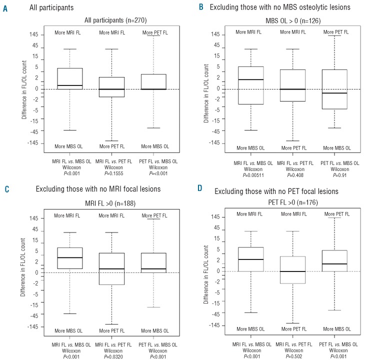
Multiple myeloma causes major morbidity resulting from osteolytic lesions that can be detected by metastatic bone surveys. Magnetic resonance imaging and positron emission tomography can detect bone marrow focal lesions long before development of osteolytic lesions. Using data from patients enrolled in Total Therapy 3 for newly diagnosed myeloma (n=303), we analyzed associations of these imaging techniques with baseline standard laboratory variables assessed before initiating treatment. Of 270 patients with complete imaging data, 245 also had gene expression profiling data. Osteolytic lesions detected on metastatic bone surveys correlated with focal lesions detected by magnetic resonance imaging and positron emission tomography, although, in two-way comparisons, focal lesion counts based on both magnetic resonance imaging and positron emission tomography tended to be greater than those based on metastatic bone survey. Higher numbers of focal lesions detected by magnetic resonance imaging and positron emission tomography were positively linked to high serum concentrations of C-reactive protein, gene-expression-profiling-defined high risk, and the proliferation molecular subgroup. Positron emission tomography focal lesion maximum standardized unit values were significantly correlated with gene-expression-profiling-defined high risk and higher numbers of focal lesions detected by positron emission tomography. Interestingly, four genes associated with high-risk disease (related to cell cycle and metabolism) were linked to counts of focal lesions detected by magnetic resonance imaging and positron emission tomography. Collectively, our results demonstrate significant associations of all three imaging techniques with tumor burden and, especially, disease aggressiveness captured by gene-expression-profiling-risk designation. (Clinicaltrials.gov identifier: NCT00081939).
Figures
References
-
- Roodman GD. Mechanisms of bone metastasis. N Engl J Med. 2004;350(16):1655-64 - PubMed
-
- Durie BG, Salmon SE. A clinical staging system for multiple myeloma. Correlation of measured myeloma cell mass with presenting clinical features, response to treatment, and survival. Cancer. 1975;36(3):842-54 - PubMed
-
- Edelstyn GA, Gillespie PJ, Grebbell FS. The radiological demonstration of osseous metastases. Experimental observations. Clin Radiol. 1967;18(2):158-62 - PubMed
-
- Wahner HW, Kyle RA, Beabout JW. Scintigraphic evaluation of the skeleton in multiple myeloma. Mayo Clin Proc. 1980;55(12):739-46 - PubMed
-
- Mundy GR, Raisz LG, Cooper RA, Schechter GP, Salmon SE. Evidence for the secretion of an osteoclast stimulating factor in myeloma. N Engl J Med. 1974;291(20):1041-6 - PubMed
Publication types
MeSH terms
Associated data
Grants and funding
LinkOut - more resources
Full Text Sources
Medical
Research Materials
