. 2012 Sep;56(9):4718-28.
doi: 10.1128/AAC.00957-12. Epub 2012 Jun 25.

Severe acute respiratory syndrome coronavirus replication inhibitor that interferes with the nucleic acid unwinding of the viral helicase

Affiliations

Severe acute respiratory syndrome coronavirus replication inhibitor that interferes with the nucleic acid unwinding of the viral helicase

Adeyemi O Adedeji et al. Antimicrob Agents Chemother. 2012 Sep.

Abstract

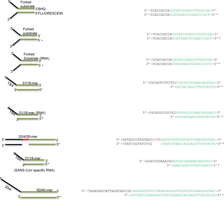
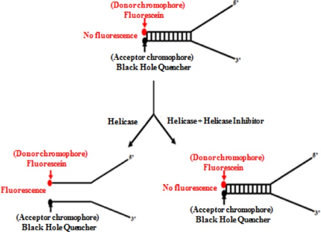
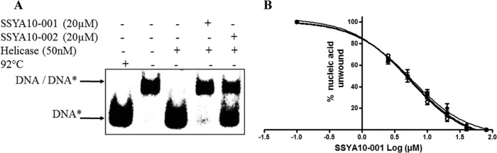
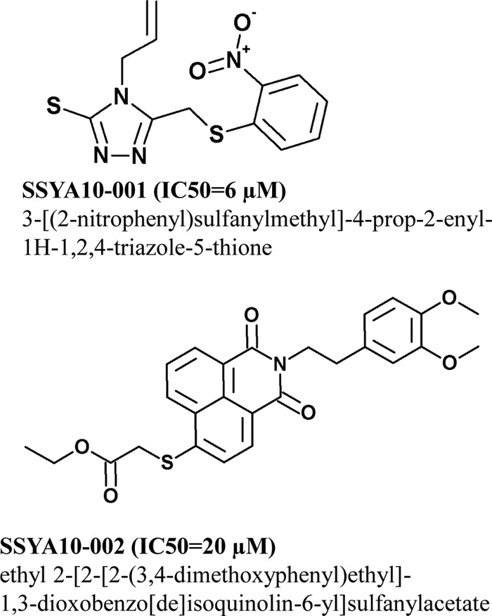
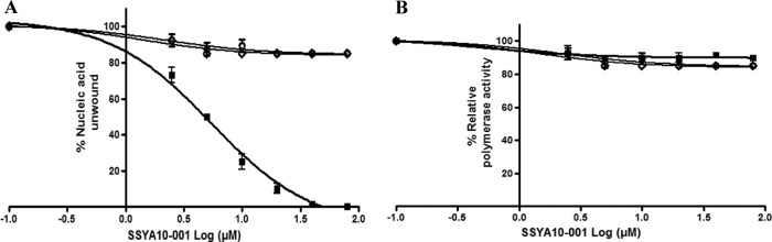
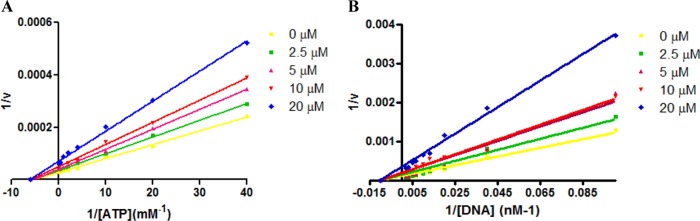
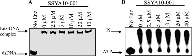
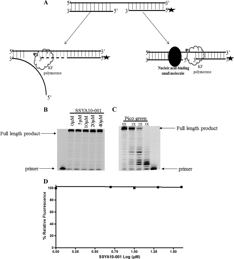
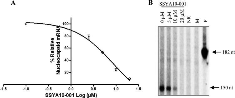
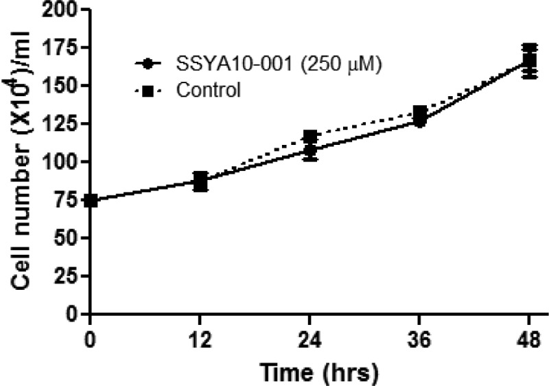
Severe acute respiratory syndrome (SARS) is a highly contagious disease, caused by SARS coronavirus (SARS-CoV), for which there are no approved treatments. We report the discovery of a potent inhibitor of SARS-CoV that blocks replication by inhibiting the unwinding activity of the SARS-CoV helicase (nsp13). We used a Förster resonance energy transfer (FRET)-based helicase assay to screen the Maybridge Hitfinder chemical library. We identified and validated a compound (SSYA10-001) that specifically blocks the double-stranded RNA (dsRNA) and dsDNA unwinding activities of nsp13, with 50% inhibitory concentrations (IC(50)s) of 5.70 and 5.30 μM, respectively. This compound also has inhibitory activity (50% effective concentration [EC(50)] = 8.95 μM) in a SARS-CoV replicon assay, with low cytotoxicity (50% cytotoxic concentration [CC(50)] = >250 μM), suggesting that the helicase plays a still unidentified critical role in the SARS-CoV life cycle. Enzyme kinetic studies on the mechanism of nsp13 inhibition revealed that SSYA10-001 acts as a noncompetitive inhibitor of nsp13 with respect to nucleic acid and ATP substrates. Moreover, SSYA10-001 does not affect ATP hydrolysis or nsp13 binding to the nucleic acid substrate. SSYA10-001 did not inhibit hepatitis C virus (HCV) helicase, other bacterial and viral RNA-dependent RNA polymerases, or reverse transcriptase. These results suggest that SSYA10-001 specifically blocks nsp13 through a novel mechanism and is less likely to interfere with the functions of cellular enzymes that process nucleic acids or ATP. Hence, it is possible that SSYA10-001 inhibits unwinding by nsp13 by affecting conformational changes during the course of the reaction or translocation on the nucleic acid. SSYA10-001 will be a valuable tool for studying the specific role of nsp13 in the SARS-CoV life cycle, which could be a model for other nidoviruses and also a candidate for further development as a SARS antiviral target.


Figures

Fig 1
Fig 1
Oligonucleotides and substrates used in this study. The Cy3-labeled strands are marked by asterisks. The sequences in green denote complementary sequences, while the sequences in black denote noncomplementary sequences.
Fig 2
Fig 2
Schematic representation of the fluorescence helicase assay. A dsDNA substrate (forked substrate) was designed with 2 chromophores at the ends of the complementary strands (i.e., fluorescein and black hole quencher). In the presence of an active helicase, the two complementary strands are separated, allowing fluorescence detection of the fluorescein chromophore, whose excitation occurs at 495 nm and emission occurs at 520 nm. In the presence of a potential helicase inhibitor, the two complementary strands will not be separated; therefore, fluorescence will not be detected.
Fig 3
Fig 3
Validation of nsp13 inhibition by use of a gel-based helicase assay. (A) nsp13 (50 nM) was incubated in the presence of 20 μM inhibitor compound, 20 mM HEPES, 20 mM NaCl, 0.01% BSA, 2 mM DTT, 5% glycerol, and 5 mM MgCl2. The helicase reaction was initiated by the addition of 100 nM 31/18-mer (13ss:18ds) as the substrate (Cy3 labeled) at 30°C, along with 0.5 mM ATP and a 2 μM concentration of unlabeled ssDNA with a sequence complementary to that of the unlabeled DNA strand. The reactions were allowed to proceed for 10 min at 30°C, and the reaction was quenched with 100 mM EDTA, 0.2% SDS, and 20% glycerol. The products were separated and analyzed by 6% nondenaturing PAGE. (B) The IC50 of SSYA10-001 was determined using the gel-based assay described for panel A with various concentrations of the compound (0, 2.5, 10, 20, 40, and 80 μM). Reaction products were resolved and quantitated using ImageQuant software (Pharmacia). The fractions of unwound DNA (○), RNA (■), and SARS-CoV-specific RNA (◆) were plotted against the concentrations of inhibitor, and the data were fit to dose-response curves by use of GraphPad Prism 5.0 (GraphPad Inc.) to determine the IC50 (mean ± standard deviation [SD]): 5.7 ± 2.3 μM with 31/18-mer RNA (13ss:18ds), 5.6 ± 0.5 μM with SARS-CoV-specific 31/18-mer RNA (13ss:18ds), and 5.3 ± 2.0 μM with 31/18-mer DNA (13ss:18ds). Experiments were performed three times, and error bars represent standard deviations for the three independent experiments.
Fig 4
Fig 4
Chemical structures, names, and IC50s of the identified nsp13 inhibitors.
Fig 5
Fig 5
Effect of SSYA10-001 on activity of other helicases and polymerases. (A) Plot of the fractions of unwound RNA for the HCV NS3h helicase (♢), dengue virus NS3 helicase (○), and SARS-CoV nsp13 helicase (■). Experiments were performed three times, and error bars represent standard deviations for three independent experiments. (B) Plot of the fractions of polymerization product for KF (♢), MoMLV RT (○), and FMDV 3D pol (■). Experiments were performed three times, and error bars represent standard deviations for three independent experiments.
Fig 6
Fig 6
Noncompetitive inhibition of nsp13 by SSYA10-001 under steady-state conditions. Kinetic experiments with nsp13 were conducted in 96-well plates by using a fluorescence-based assay (see Materials and Methods) in the presence of increasing concentrations of SSYA10-001 (0 to 20 μM), varying either the ATP substrate (0.0125 mM to 7.5 mM) (A) or the forked substrate (0.01 μM to 1 μM) (B). In both cases, the x axis intercept (−1/Km for the ATP or forked substrate) was not affected by the inhibitor concentration, and this is a feature of noncompetitive inhibition.
Fig 7
Fig 7
Investigation of the effect of SSYA10-001 on nucleic acid binding and ATP hydrolysis of nsp13. (A) nsp13 (25 nM) was preincubated with various concentrations of SSYA10-001 (0, 2.5, 10, 20, and 40 μM) followed by incubation with a 5 nM concentration of a Cy3-labeled version of the forked substrate in 20 mM HEPES (pH 7.5), 20 mM NaCl, 5 mM MgCl2, 1 mM DTT, 0.1 mg/ml BSA, and 5% glycerol at 30°C for 20 min. The reaction samples were then analyzed in a nondenaturing 6% polyacrylamide gel. (B) Preincubated nsp13 (50 nM) and various concentrations of SSYA10-001 (0, 2.5, 10, 20, and 40 μM) were mixed with 5 μM [γ-32P]ATP for 10 s. The reaction products were separated by thin-layer chromatography and visualized by autoradiography.
Fig 8
Fig 8
Determination of nucleic acid binding of SSYA10-001. (A) Klenow fragment strand displacement and polymerization assay design. A DNA substrate with a 3′-end primer is annealed with a complementary strand at the 5′ end. Primer extension can take place only in the presence of a polymerase with strand displacement activity, such as KF. The presence of a dsDNA chelator such as Pico green will prevent strand displacement and therefore prevent primer extension. (B and C) Various concentrations of SSYA10-001 (0, 5, 10, and 20 μM) (B) and Pico green (1×, 2×, and 3×) (C) were preincubated with 20 nM 13ds:4ss:18ds DNA substrate in a mixture including 50 mM Tris-HCl, pH 7.5, 50 mM NaCl, 5 mM MgCl2, 1 mM DTT, 5% glycerol, and a 50 μM concentration of each dNTP. The reaction was initiated by the addition of 20 nM KF at 37°C for 10 min, and the reactions were resolved and analyzed in a 15% polyacrylamide-7 M urea gel. (D) Various concentrations of SSYA10-001 (0, 2.5, 5, 10, 20, and 40 μM) were preincubated with dsDNA (31/18-mer) for 10 min, followed by addition of a 1× final concentration of Pico green reagent and immediate fluorescence measurement. The percent relative fluorescence was plotted against the concentration of the inhibitor, and the data were fit to dose-response curves by use of GraphPad Prism 5.0 (GraphPad Inc.). Experiments were performed in triplicate in two independent experiments.
Fig 9
Fig 9
Measurement of SARS-CoV replication in the presence of SSYA10-001. (A) HEK 293T cells were mock transfected or transfected with the SARS-CoV replicon or a nonreplicative construct by use of Lipofectamine 2000 (Invitrogen) in the presence of various concentrations of SSYA10-001 (0, 2.5, 5, 10, and 20 μM). Total RNA was isolated at 48 h posttransfection and analyzed by RT-qPCR with specific oligonucleotides to detect N gene mRNA. Triplicate RT-qPCR products were amplified in parallel. The average N gene mRNA quantity for each concentration was normalized internally by using the CT of the housekeeping gene U6. Samples were additionally normalized to the NRC controls, and the RQ value for each sample was obtained using the relative quantity method (2−ΔΔCT). The RQ value for each sample was then normalized to the RQ value of the NRC (which is 1), and the data were graphed as percent relative replicon activities against the log inhibitor concentrations (in μM) by use of a dose-response curve in GraphPad Prism 5.0 (GraphPad Inc.). Experiments were repeated three times, and error bars represent standard deviations for triplicate samples. (B) HEK 293T cells were mock transfected or transfected with the SARS-CoV replicon or a nonreplicative construct by use of Lipofectamine 2000 (Invitrogen) in the presence of various concentrations of SSYA10-001 (0, 5, 10, and 20 μM). Total RNA was isolated at 48 h posttransfection. An RNase protection assay was performed by overnight annealing of the newly generated probes with the total intracellular RNA, followed by RNase A and T1 treatment of the annealed reaction mixture. The reaction samples were analyzed in a 6% urea-polyacrylamide gel and visualized using a phosphorimager (FLA 5000; FujiFilm). Lanes: P, the probe by itself; MT, mock transfection; NR, sample from the nonreplicative construct. The probe was 182 nt long because of the extra 32 nt between the Sp6 transcription start site and the XbaI site of the pGEM-3Z vector.
Fig 10
Fig 10
Cytostatic effect of SSYA10-001. HEK 293T cells (7.5 × 10⁵) were treated with 250 μM SSYA10-001. The medium was changed and fresh compound added daily. Viability of the cells was assessed every 12 h through 48 h by trypan blue exclusion. The number of viable cells counted was plotted against time, using Graphpad Prism 5.0 (GraphPad Inc.). Experiments were performed in triplicate, and error bars represent standard deviations for triplicate samples. ●, 250 μM SSYA10-001; ■, 0.2% DMSO control.

Similar articles

Cited by

References

    1. Abdel-Megeed AM, Abdel-Rahman HM, Alkaramany GE, El-Gendy MA. 2009. Design, synthesis and molecular modeling study of acylated 1,2,4-triazole-3-acetates with potential anti-inflammatory activity. Eur. J. Med. Chem. 44:117–123 - PubMed
    1. Adedeji AO, et al. 2012. Mechanism of nucleic acid unwinding by SARS-CoV helicase. PLoS One 7:e36521 doi:10.1371/journal.pone.0036521 - DOI - PMC - PubMed
    1. Ahnert P, Patel SS. 1997. Asymmetric interactions of hexameric bacteriophage T7 DNA helicase with the 5′- and 3′-tails of the forked DNA substrate. J. Biol. Chem. 272:32267–32273 - PubMed
    1. Ali JA, Lohman TM. 1997. Kinetic measurement of the step size of DNA unwinding by Escherichia coli UvrD helicase. Science 275:377–380 - PubMed
    1. Almazan F, et al. 2006. Construction of a severe acute respiratory syndrome coronavirus infectious cDNA clone and a replicon to study coronavirus RNA synthesis. J. Virol. 80:10900–10906 - PMC - PubMed

Publication types

MeSH terms

LinkOut - more resources