

Insights into cell membrane microdomain organization from live cell single particle tracking of the IgE high affinity receptor FcϵRI of mast cells
- PMID: 22733211
- PMCID: PMC3677563
- DOI: 10.1007/s11538-012-9738-9
Insights into cell membrane microdomain organization from live cell single particle tracking of the IgE high affinity receptor FcϵRI of mast cells
Abstract
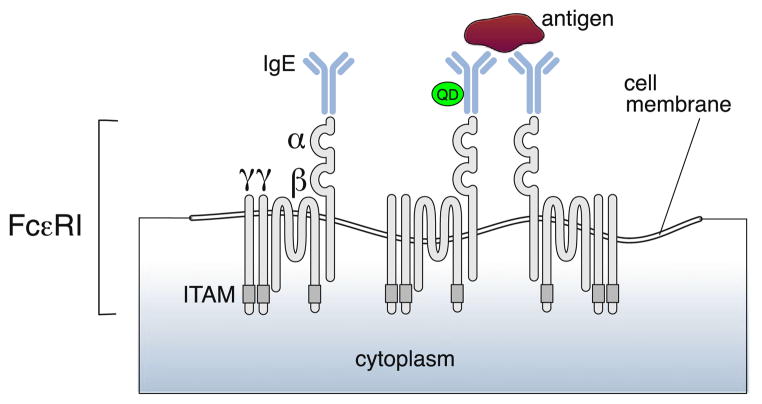
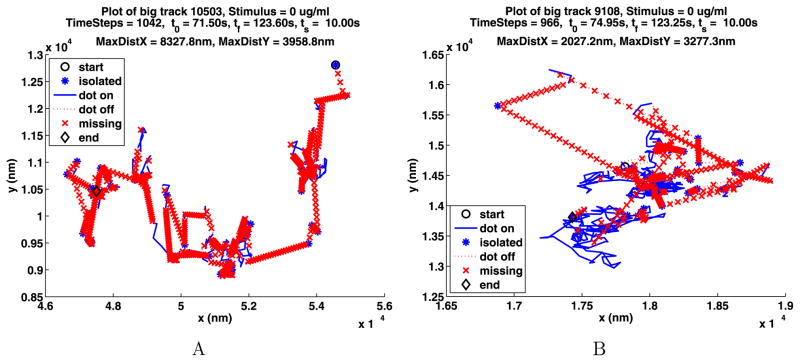
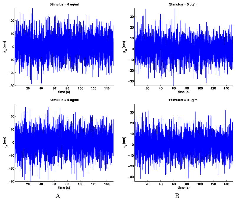
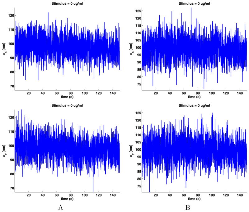
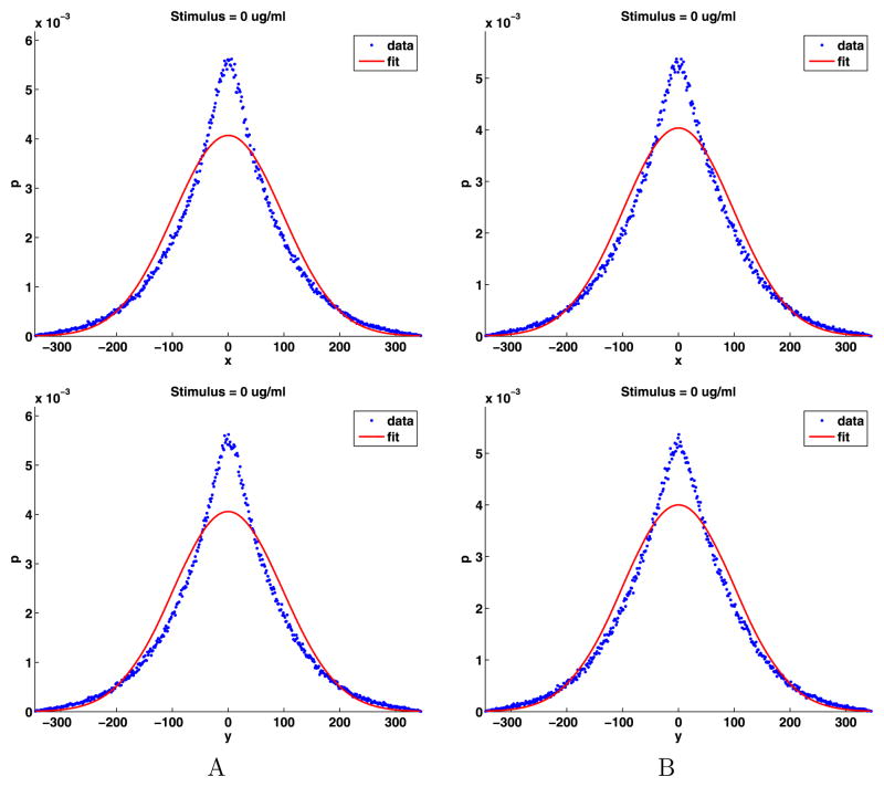
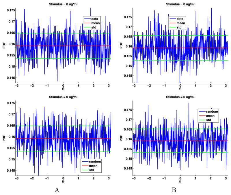
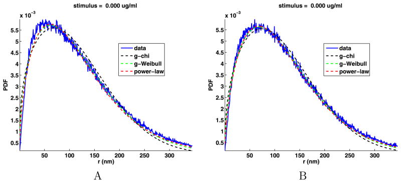
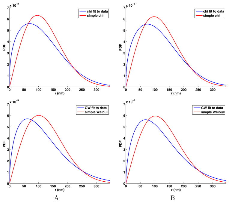
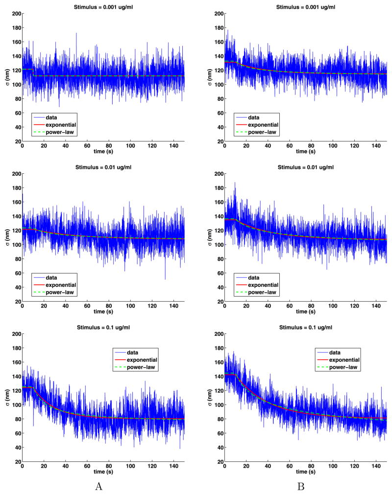
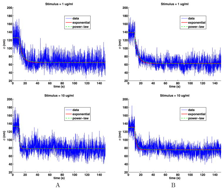
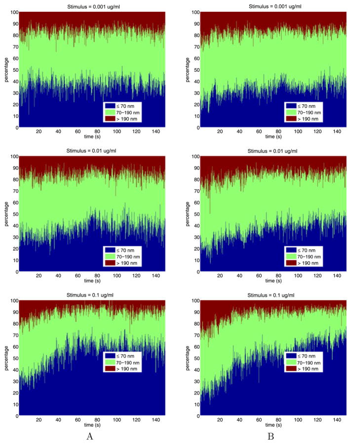
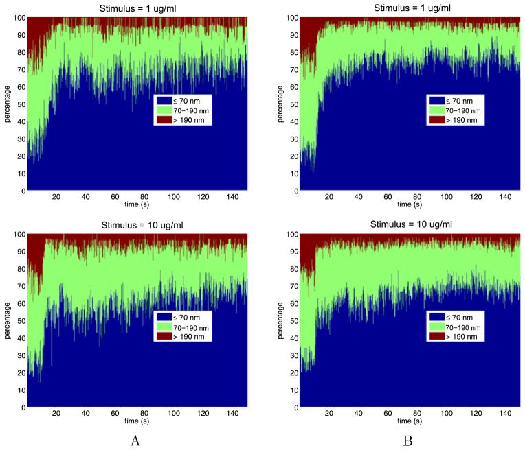
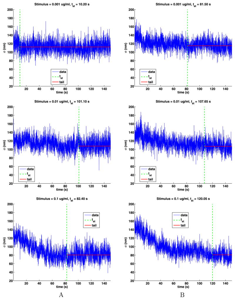
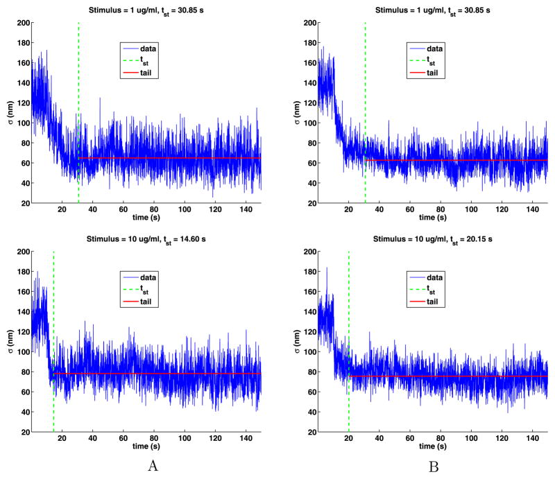
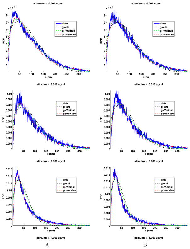
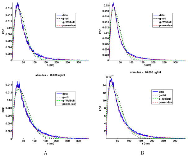
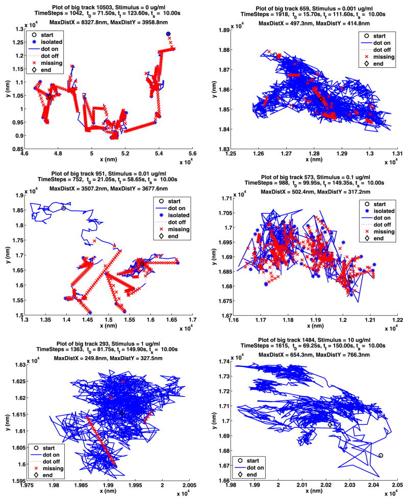
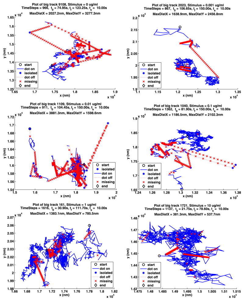
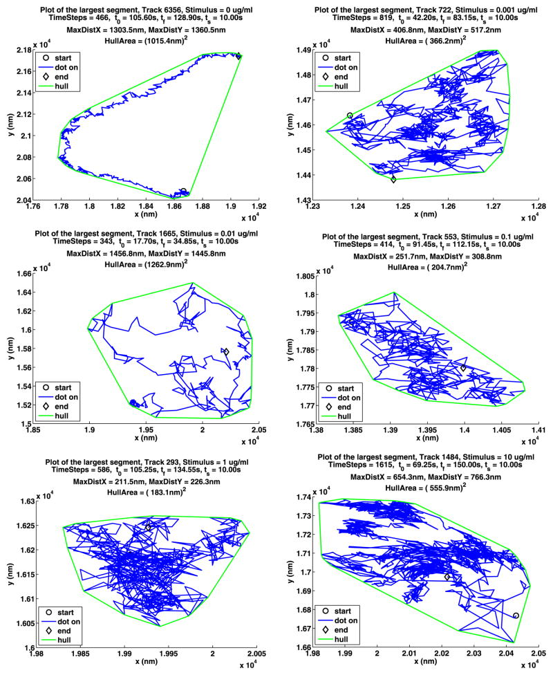
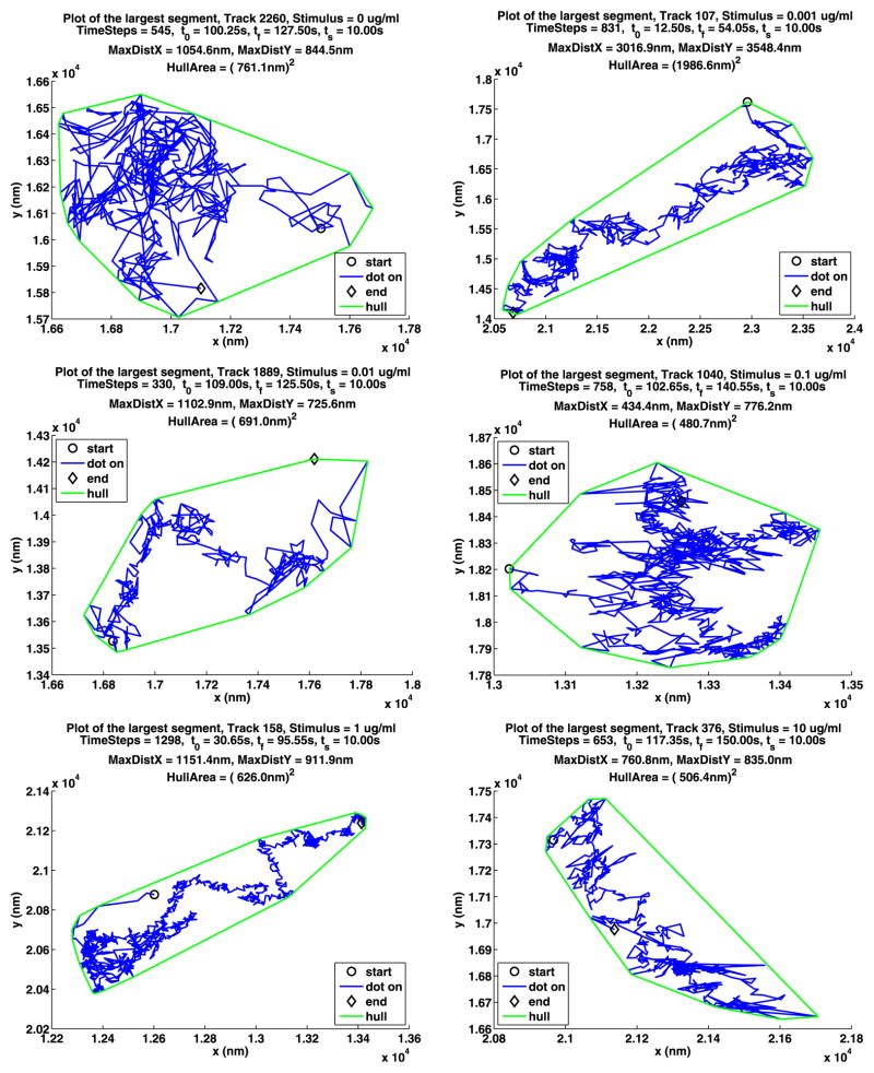
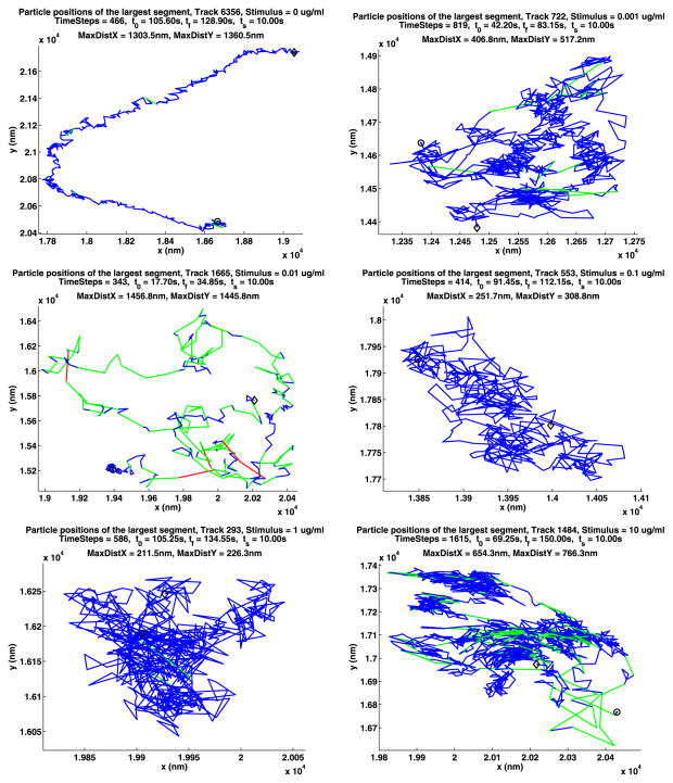
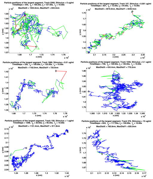
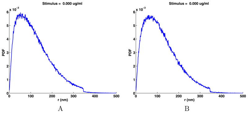
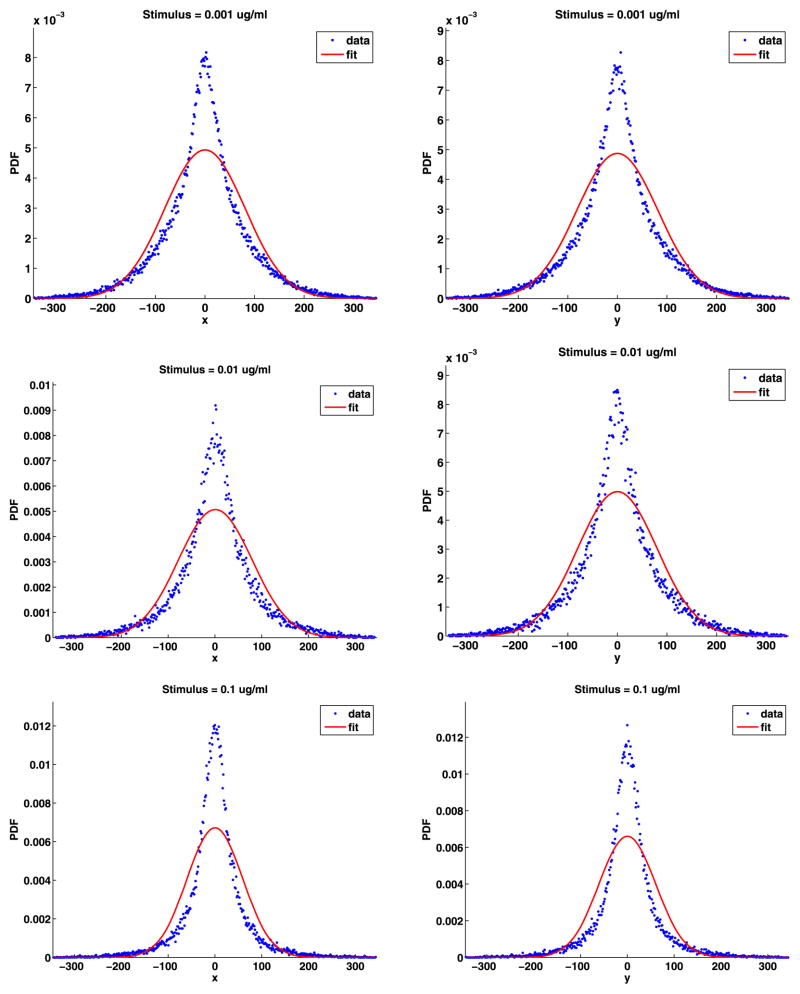
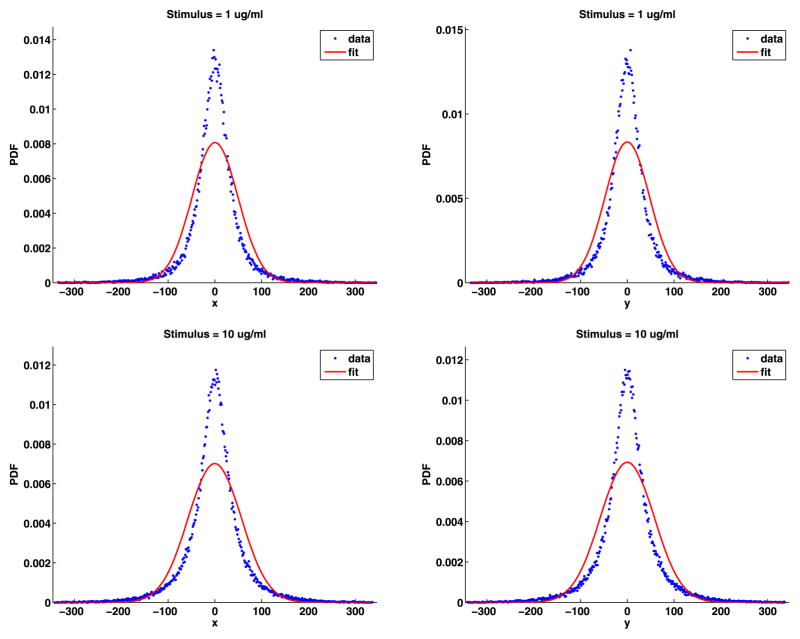
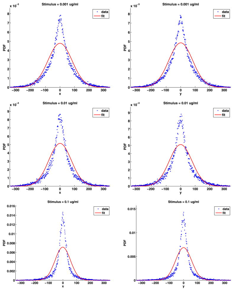
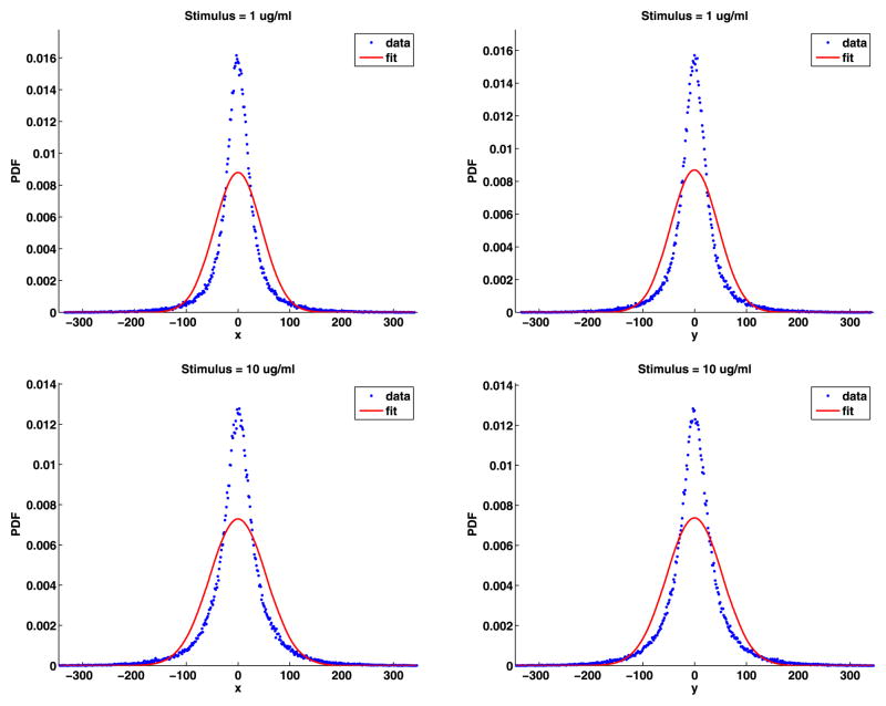
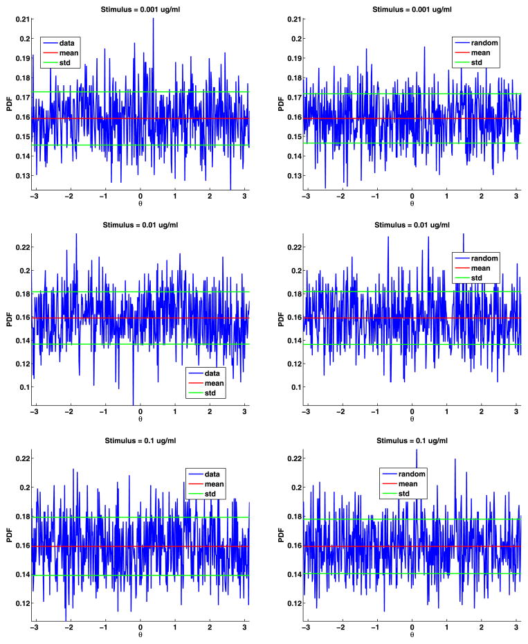
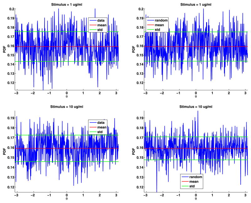
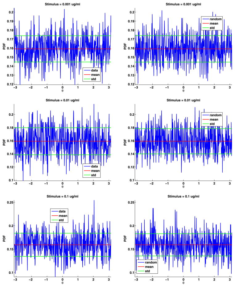
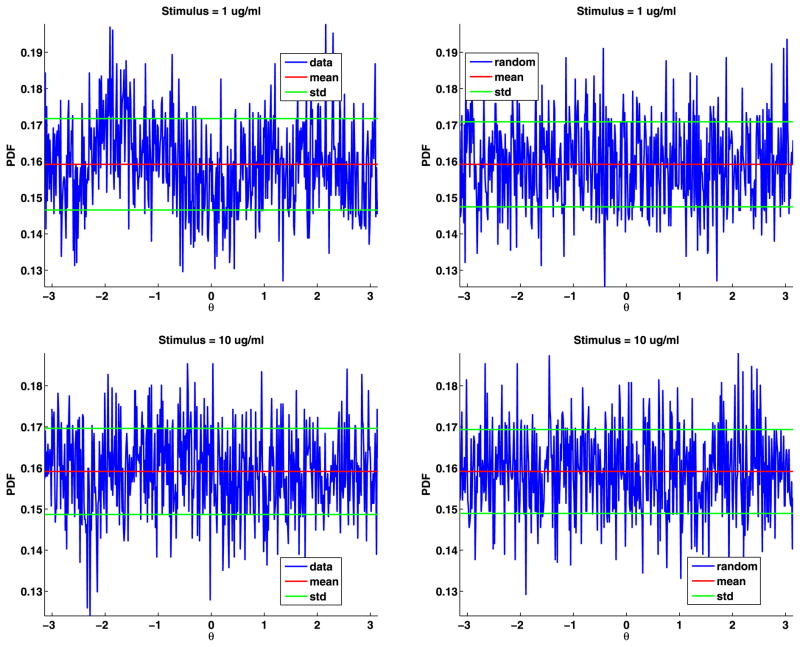
Current models propose that the plasma membrane of animal cells is composed of heterogeneous and dynamic microdomains known variously as cytoskeletal corrals, lipid rafts and protein islands. Much of the experimental evidence for these membrane compartments is indirect. Recently, live cell single particle tracking studies using quantum dot-labeled IgE bound to its high affinity receptor FcϵRI, provided direct evidence for the confinement of receptors within micrometer-scale cytoskeletal corrals. In this study, we show that an innovative time-series analysis of single particle tracking data for the high affinity IgE receptor, FcϵRI, on mast cells provides substantial quantitative information about the submicrometer organization of the membrane. The analysis focuses on the probability distribution function of the lengths of the jumps in the positions of the quantum dots labeling individual IgE FcϵRI complexes between frames in movies of their motion. Our results demonstrate the presence, within the micrometer-scale cytoskeletal corrals, of smaller subdomains that provide an additional level of receptor confinement. There is no characteristic size for these subdomains; their size varies smoothly from a few tens of nanometers to a over a hundred nanometers. In QD-IGE labeled unstimulated cells, jumps of less than 70 nm predominate over longer jumps. Addition of multivalent antigen to crosslink the QD-IgE-FcϵRI complexes causes a rapid slowing of receptor motion followed by a long tail of mostly jumps less than 70 nm. The reduced receptor mobility likely reflects both the membrane heterogeneity revealed by the confined motion of the monomeric receptor complexes and the antigen-induced cross linking of these complexes into dimers and higher oligomers. In both cases, the probability distribution of the jump lengths is well fit, from 10 nm to over 100 nm, by a novel power law. The fit for short jumps suggests that the motion of the quantum dots can be modeled as diffusion in a fractal space of dimension less than two.
Figures
Similar articles
-
Time series analysis of particle tracking data for molecular motion on the cell membrane.Bull Math Biol. 2009 Nov;71(8):1967-2024. doi: 10.1007/s11538-009-9434-6. Epub 2009 Aug 6. Bull Math Biol. 2009. PMID: 19657701 Free PMC article.
-
Molecular perspective of antigen-mediated mast cell signaling.J Biol Chem. 2008 Mar 14;283(11):7117-27. doi: 10.1074/jbc.M708879200. Epub 2007 Dec 19. J Biol Chem. 2008. PMID: 18093971
-
Determining FcεRI diffusional dynamics via single quantum dot tracking.Methods Mol Biol. 2011;748:121-32. doi: 10.1007/978-1-61779-139-0_8. Methods Mol Biol. 2011. PMID: 21701970
-
Role of immunoglobulin E and mast cells in murine models of asthma.Braz J Med Biol Res. 2003 Jul;36(7):821-7. doi: 10.1590/s0100-879x2003000700001. Epub 2003 Jun 26. Braz J Med Biol Res. 2003. PMID: 12845367 Review.
-
Interaction of IgE with its high-affinity receptor. Structural basis and requirements for effective cross-linking.Int Arch Allergy Appl Immunol. 1989;88(1-2):23-8. doi: 10.1159/000234742. Int Arch Allergy Appl Immunol. 1989. PMID: 2523358 Review.
Cited by
-
Membrane Diffusion Occurs by Continuous-Time Random Walk Sustained by Vesicular Trafficking.Biophys J. 2018 Jun 19;114(12):2887-2899. doi: 10.1016/j.bpj.2018.04.024. Biophys J. 2018. PMID: 29925025 Free PMC article.
-
Enabling surface dependent diffusion in spatial simulations using Smoldyn.BMC Res Notes. 2015 Dec 8;8:752. doi: 10.1186/s13104-015-1723-6. BMC Res Notes. 2015. PMID: 26647064 Free PMC article.
-
Orchestration of ErbB3 signaling through heterointeractions and homointeractions.Mol Biol Cell. 2015 Nov 5;26(22):4109-23. doi: 10.1091/mbc.E14-06-1114. Epub 2015 Sep 16. Mol Biol Cell. 2015. PMID: 26378253 Free PMC article.
References
-
- Adamic L. Complex systems: unzipping Zipf’s law. Nature. 2011;474(7350):164–165. - PubMed
-
- Andrews NL. PhD thesis. University of New Mexico; Albuquerque, New Mexico, USA: 2011. The role of diffusion and membrane topography in the initiation of high affinity IgE receptor signaling.
-
- Bachir AL. Characterization of blinking dynamics in quantum dots ensembles using image correlation spectroscopy. J Appl Phys. 2006;99(6) doi: 10.1063/1.2175470. - DOI
Publication types
MeSH terms
Substances
Grants and funding
LinkOut - more resources
Full Text Sources
