. 2012 Aug;4(8):914-24.
doi: 10.1039/c2ib20055d. Epub 2012 Jun 25.

Differential effects of a soluble or immobilized VEGFR-binding peptide

Affiliations

Differential effects of a soluble or immobilized VEGFR-binding peptide

Justin T Koepsel et al. Integr Biol (Camb). 2012 Aug.

Abstract

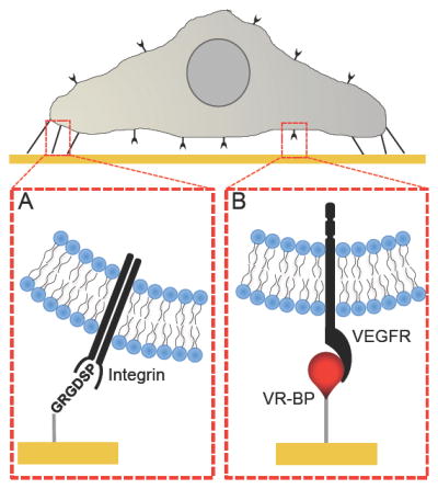
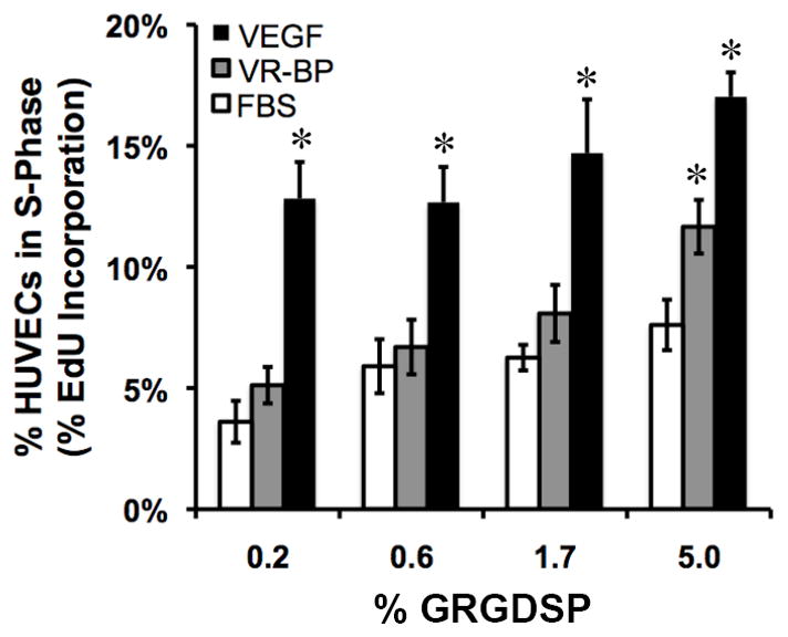
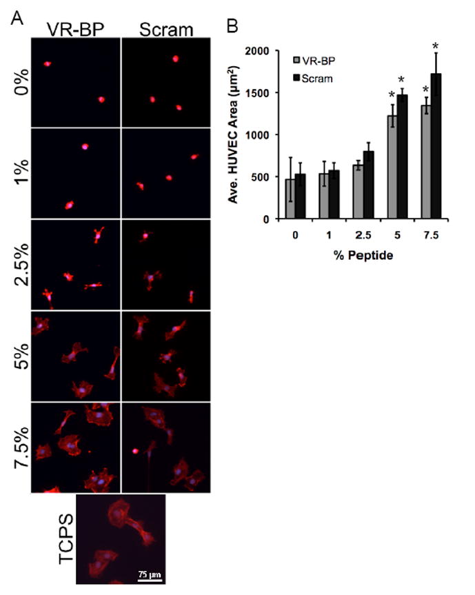
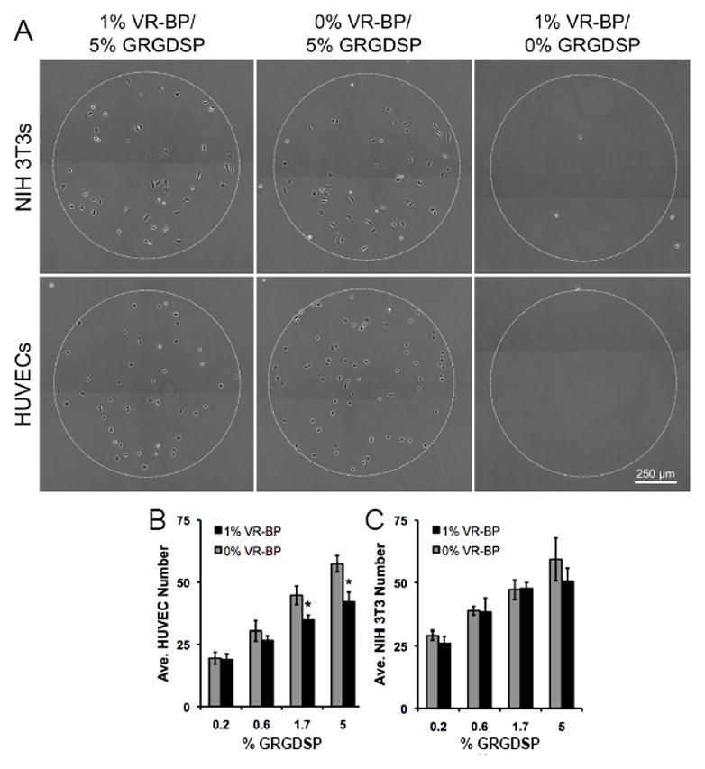
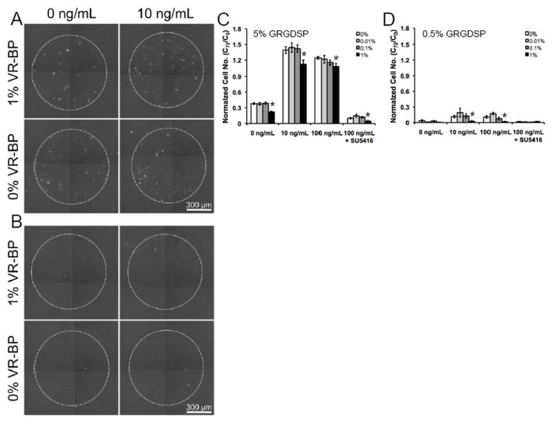
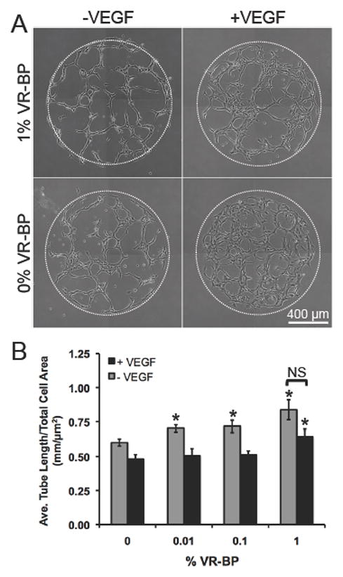
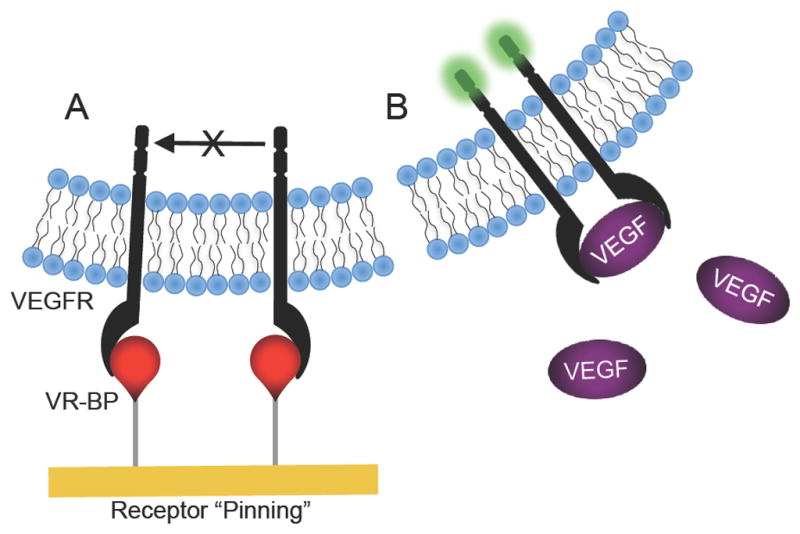
Regulating endothelial cell behavior is a key step in understanding and controlling neovascularization for both pro-angiogenic and anti-angiogenic therapeutic strategies. Here, we characterized the effects of a covalently immobilized peptide mimic of vascular endothelial growth factor, herein referred to as VEGF receptor-binding peptide (VR-BP), on human umbilical vein endothelial cell (HUVEC) behavior. Self-assembled monolayer arrays presenting varied densities of covalently immobilized VR-BP and varied densities of the fibronectin-derived cell adhesion peptide Gly-Arg-Gly-Asp-Ser-Pro (GRGDSP) were used to probe for changes in HUVEC attachment, proliferation and tubulogenesis. In a soluble form, VR-BP exhibited pro-angiogenic effects in agreement with previous studies, indicated by increases in HUVEC proliferation. However, when presented to cells in an insoluble context, covalently immobilized VR-BP inhibited several pro-angiogenic HUVEC behaviors, including attachment and proliferation, and also inhibited HUVEC response to soluble recombinant VEGF protein. Furthermore, substrates with covalently immobilized VR-BP also modulated HUVEC tubulogenesis when a matrigel overlay assay was used to provide cells with a pseudo-three dimensional environment. Taken together, these results demonstrate that the context in which ligands are presented to cell surface receptors strongly influences their effects, and that the same ligand can be an agonist or an antagonist depending on the manner of presentation to the cell.


Figures

Figure 1
Figure 1
Screening HUVEC microenvironments using alkanethiolate self-assembled monolayer arrays presenting the (A) cell adhesion ligand Gly-Arg-Gly-Asp-Ser-Pro (GRGDSP) and (B) Covalently immobilized VEGF receptor-binding peptide (VR-BP).
Figure 2
Figure 2
Characterization and immobilization of the vascular endothelial growth factor-receptor binding peptide (VR-BP) on self assembled monolayer substrates. Secondary structure of VR-BP (GGGKLTWQELYQLKYKGI) and the scrambled sequence (GGGKTKQQKEIYLLWYLG): (A) Circular dichroism (CD) spectra of 75 μM peptide in phosphate buffered saline at pH 7.4 at RT and (B) Polarization modulation-infrared reflection-absorption spectroscopy (PM-IRRAS) spectra of peptide conjugated to 2.5% HS-C11-EG6-COOH SAMs.
Figure 3
Figure 3
HUVEC response to soluble morphogens. HUVECs starved overnight in 2% FBS were seeded on arrays presenting varied densities of GRGDSP in the presence of 2% FBS alone or with the addition of 10 ng/mL VR-BP (4.8 nM) or 10 ng/mL VEGF (0.26 nM). (Error bars indicate standard error of the mean and asterisk indicate significance compared to FBS alone, p < 0.05)
Figure 4
Figure 4
Fluorescent detection of peptide incorporation. (A) VR-BP or scrambled peptide was mixed with GRGDSP at varying percentages and coupled to SAM arrays spots with a fixed density of carboxylate. After coupling to the surface, lysine side chains were labeled using Alexa Fluor 488 sulfodichlorophenol ester and imaged using a fluorescent scanner. (B) Normalized fluorescent intensity (Error bars indicate standard deviation and % peptide refers the the percent of either VR-BP or scrambled peptide present during covalent coupling).
Figure 5
Figure 5
Assaying for non-specific protein adsorption and surface fouling. HUVECs were seeded on SAMs presenting varied densities of VR-BP, scrambled peptide, or tissue culture polystyrene and (A) stained for their f-actin cytoskeleton (f-actin:red, nuclei:blue). (B) HUVEC projected cell area was used as a measurement of surface fouling. (Error bars represent standard error of the mean, asterisk indicates significant increase in projected cell area compared to 0% peptide, p < 0.05) % GRGDSP % GRGDSP
Figure 6
Figure 6
Cell attachment to surfaces presenting covalently immobilized VR-BP and varied densities of GRGDSP. (A) HUVECs and NIH 3T3 fibroblasts were seeded on SAM arrays presenting either 1% VR-BP or 0% VR-BP and varied densities of GRGDSP and allowed to attach for ~1 hr. Quantification of (B) HUVEC and (C) 3T3 fibroblast attachment. (Note: 0% VR-BP corresponds to 1% scrambled peptide and 0% GRGDSP corresponds to 5% of the mutant peptide GRGESP. Error bars represent standard error of the mean and asterisk indicates significant decrease in cell number compared to 0% VR-BP at a specific GRGDSP density, p < 0.05)
Figure 7
Figure 7
HUVEC proliferation and survival on surfaces presenting covalently immobilized VR-BP. HUVECs were seeded onto SAM arrays presenting (A) 5% or (B) 0.5% GRGDSP with varied densities of VR-BP including 1%, 0.1%, 0.01% and 0% and allowed to attach for 3 hours. After attachment, HUVECs were stimulated with VEGF concentrations of 0 ng/mL, 10ng/mL, 100 ng/mL. As indicated, 10 μM SU5416 was included during cell seeding and washed out before stimulation with VEGF. Normalized cell number after 72 hours of culture in (C) 5% or (D) 0.5% GRGDSP. (Error bars indicate standard error of the mean. Asterisk indicate significance decrease compared to 0% VR-BP at p < 0.05)
Figure 8
Figure 8
HUVEC tubulogenesis on surfaces presenting 5% GRGDSP and covalently immobilized VR-BP. (A) HUVECs on SAM arrays were placed on a thin layer of matrigel with and without 100 ng/mL VEGF and imaged for over 24 hrs (See Supplemental Movies 1A–D). (B) Tubulogenesis was quantified by averaging the length of all objects in each array spot and normalizing by total area occupied by cells. (Error bars represent standard error of the mean. Asterisk indicates significant increase compared to 0% VR-BP and “NS” indicates no significance between soluble VEGF conditions, p < 0.05)
Figure 9
Figure 9
Schematic of proposed VEGFR “pinning” mechanism. We hypothesize that (A) covalently immobilized VR-BP may bind VEGFRs and prevent them from dimerizing or interacting with (B) soluble VEGF and activating intracellular signaling cascades.

References

    1. Cross MJ, et al. VEGF-receptor signal transduction. Trends Biochem Sci. 2003;28(9):488–94. - PubMed
    1. Ferrara N, Gerber HP, LeCouter J. The biology of VEGF and its receptors. Nat Med. 2003;9(6):669–76. - PubMed
    1. Folkman J. Angiogenesis: an organizing principle for drug discovery? Nat Rev Drug Discov. 2007;6(4):273–86. - PubMed
    1. D’Andrea LD, et al. Peptide-based molecules in angiogenesis. Chem Biol Drug Des. 2006;67(2):115–26. - PubMed
    1. Soker S, et al. Inhibition of vascular endothelial growth factor (VEGF)-induced endothelial cell proliferation by a peptide corresponding to the exon 7-encoded domain of VEGF165. J Biol Chem. 1997;272(50):31582–8. - PubMed

Publication types