. 2012 Dec;228(4):554-64.
doi: 10.1002/path.4068. Epub 2012 Sep 28.

Endoplasmic reticulum stress disrupts placental morphogenesis: implications for human intrauterine growth restriction

Affiliations
Free PMC article

Endoplasmic reticulum stress disrupts placental morphogenesis: implications for human intrauterine growth restriction

Hong Wa Yung et al. J Pathol. 2012 Dec.
Free PMC article

Abstract

We recently reported the first evidence of placental endoplasmic reticulum (ER) stress in the pathophysiology of human intrauterine growth restriction. Here, we used a mouse model to investigate potential underlying mechanisms. Eif2s1(tm1RjK) mice, in which Ser51 of eukaryotic initiation factor 2 subunit alpha (eIF2α) is mutated, display a 30% increase in basal translation. In Eif2s1(tm1RjK) placentas, we observed increased ER stress and anomalous accumulation of glycoproteins in the endocrine junctional zone (Jz), but not in the labyrinthine zone where physiological exchange occurs. Placental and fetal weights were reduced by 15% (97 mg to 82 mg, p < 0.001) and 20% (1009 mg to 798 mg, p < 0.001), respectively. To investigate whether ER stress affects bioactivity of secreted proteins, mouse embryonic fibroblasts (MEFs) were derived from Eif2s1(tm1RjK) mutants. These MEFs exhibited ER stress, grew 50% slower, and showed reduced Akt-mTOR signalling compared to wild-type cells. Conditioned medium (CM) derived from Eif2s1(tm1RjK) MEFs failed to maintain trophoblast stem cells in a progenitor state, but the effect could be rescued by exogenous application of FGF4 and heparin. In addition, ER stress promoted accumulation of pro-Igf2 with altered glycosylation in the CM without affecting cellular levels, indicating that the protein failed to be processed after release. Igf2 is the major growth factor for placental development; indeed, activity in the Pdk1-Akt-mTOR pathways was decreased in Eif2s1(tm1RjK) placentas, indicating loss of Igf2 signalling. Furthermore, we observed premature differentiation of trophoblast progenitors at E9.5 in mutant placentas, consistent with the in vitro results and with the disproportionate development of the labyrinth and Jz seen in placentas at E18.5. Similar disproportion has been reported in the Igf2-null mouse. These results demonstrate that ER stress adversely affects placental development, and that modulation of post-translational processing, and hence bioactivity, of secreted growth factors contributes to this effect. Placental dysmorphogenesis potentially affects fetal growth through reduced exchange capacity.

Keywords: Akt; Igf2; Pdk1; endoplasmic reticulum; intrauterine growth restriction; mTOR; placental morphogenesis; stress.


Figures

None
An increase of ER stress in the junctional zone (Jz) of the Eif2s1tm1RjK (A/A) placenta. (A) Electron micrographs show severe dilation of ER cisternae in the spongiotrophoblast cells of the Jz in the Eif2s1tm1RjK placenta compared with only moderate dilation in the wild-type. There are fewer ER cisternae in the syncytiotrophoblast of the Lz, and no dilation was observed in either genotype. (B) Placentas were dissected into Jz and Lz, and enriched tissue lysates for each zone were studied separately by western blotting analysis using specific antibodies against P-Perk (Thr980), total Perk, P-eIF2α and total eIF2α. β-Actin was used as a loading control. Experiments were carried out on a paired basis. (C) Densitometry of band intensity is expressed relative to wild-type (100%). Phosphorylation status is presented as the ratio between phosphorylated and total (P/T) protein. Data are presented as mean ± SEM; n = 6. *p≤0.05; **p≤0.01
None
The Eif2s1tm1RjK mice are associated with reduced placental and fetal growth. (A) Upper panel: the progeny of the mice as shown by PCR. Lower panel: Eif2s1tm1RjK (A/A) homozygous embryo and placenta at E18.5 versus wild-type littermate. (B) Statistical analysis of fetal and placental weights from 195 pups from 29 litters. All data are expressed as percentage relative to wild-type (S/S, 100%). Bars indicate mean ± SEM. *p≤0.05
None
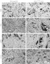
Anomalous accumulation of glycoproteins in the endocrine spongiotrophoblast cells of the Jz in the Eif2s1tm1RjK placenta. Biotinylated lectins, ConA (concanavalin A), e-PHA (Phaseolus vulgaris, erythrohaemagglutinin), DSA (Datura stramonium agglutinin), and STA (Solanum tuberosum agglutinin) were used to stain the placental sections. Note that intensely dark staining accumulations in the rounded spongiotrophoblast cells (arrowed) are more common in the mutants. The cells must be distinguished from the irregularly shaped blood channels (asterisks), which also stain positively. All images taken at 20× magnification. tm1RjKm
None
Eif2s1tm1RjK MEFs exhibit higher ER stress, slower cell proliferation rate, and lower Akt–mTOR signalling than wild-type controls. (A, C) Both wild-type and Eif2s1tm1RjK (A/A) MEFs were cultured in serum-free medium for 24 h. ER stress markers were investigated using antibodies against P-Perk, Perk, and Grp78, while Akt–mTOR signalling was examined using specific antibodies against P-Akt(Ser473), P-Akt(Thr308), Akt, P-4E-BP1 (Ser65), and 4E-BP1. Ponceau S staining was used as the loading control. For Xbp-1 mRNA splicing, total RNA was isolated and RT-PCR was used to detect the spliced variants. Tunicamycin (Tm; 400 ng/ml) treatment for 24 h was used as a positive control. (B) MEFs were seeded at the same density and cultured for 4 days. The number of cells was counted using a haemocytometer. Data are presented as mean ± SEM, n = 4, relative to wild-type (S/S), which is presented as 100%. **pp≤0.01
None
Increased ER stress in Eif2s1tm1RjK (A/A) MEFs modulates the bioactivity of secreted proteins. (A) Alteration of protein glycosylation profile (arrows). Both Eif2s1tm1RjK and wild-type MEFs, with or without 5 nm thapsigargin (Tg) or 10 ng/ml tunicamycin (Tm), were cultured in serum-free medium for 48 h in the presence of EGF. Conditioned media (CM) were concentrated and glycoproteins isolated using concanavalin A (ConA) lectin columns, followed by resolving by SDS-PAGE and silver staining. (B) Conditioned medium from Eif2s1tm1RjK MEFs failed to maintain trophoblast stem cells in a multipotent state but this could be reversed by exogenous supplementation with FGF4 and heparin. Conditioned media prepared from wild-type and Eif2s1tm1RjK MEFs were used to culture TS cells in the absence or presence of FGF4 and heparin. Total TS cell RNA was isolated, and qRT-PCR was used to assess the relative levels of different nuclear regulators of differentiation including the stem cell markers Cdx2 and Esrrb, the early differentiation marker Psx1, the ectoplacental cone/spongiotrophoblast marker Tpbpa, and the giant cell markers Pl1 and Pl2. Sdha and Gapdh were used as housekeeping genes. Data are presented as mean ± SEM from three independent experiments. (C) Altered processing of Igf2 in the Eif2s1tm1RjK MEFs. Immunoblotting of conditioned media or cell lysate prepared from both Eif2s1tm1RjK and wild-type MEFs. Antibodies specific for Pro-Igf2 were used to detect the proteins in both CM and cell lysates. β-Actin and Ponceau S staining were used to show equal loading. Left panel: three blots of pro-Igf2 in CM and cell lysates are presented to indicate the variation between experiments. The intensities of the lowest band and of the upper few bands were quantified separately to show the mobility shifting of pro-Igf2 due to changes in glycosylation. Right panel: data are presented as mean ± SEM from four independent experiments. *p < 0.05
None
Down-regulation of Pdk1–Akt–mTOR signalling in the Eif2s1tm1RjK placenta. (A) Western blotting was used to measure the phosphorylation and total level of P-Pdk1 (Ser241), Pdk1, P-Akt(Ser473), P-Akt(Thr308), Akt, P-4E-BP1 (Thr70), and 4E-BP1 using specific antibodies in both the Jz and Lz. (B) Densitometry of band intensity is expressed relative to wild-type (100%). Phosphorylation status is presented as the ratio between phosphorylated and total protein. *p≤0.05; **p≤0.01. (C) Real-time quantitative RT-PCR was used to measure the expression level of three Akt transcripts. Data are presented as mean ± SEM; n = 6. ΔCt was calculated from the cycle difference between target genes and the housekeeping gene, 18S
None
ER stress disrupts trophoblast stem cell differentiation and placental morphogenesis. (A, B) The absolute volumes and relative ratios of Lz and chorionic plate, but not of decidua and Jz, were reduced in Eif2s1tm1RjK placentas. Stereological analysis was used to estimate the absolute volume of different layers. Results are presented in a table (A) and as a relative ratio in graph (B). A typical section of a placenta after H&E staining and used for stereological analysis. D = decidua; Jz = junctional zone; Lz = labyrinth zone; Cp = chorionic plate. Data are expressed as mean ± SEM from four wild-type and five Eif2s1tm1RjK placentas. (C) Alteration of early trophoblast differentiation in Eif2s1tm1RjK placentas. E9.5 placentas were collected and total RNA was isolated. The levels of the pan-trophoblast marker Keratin-18, the syncytiotrophoblast marker Gcm1, Tpbpa, Pl1, and Pl2 were measured. Data are presented as mean ± SEM from five placentas in each group from two litters. All data were normalized with Keratin-18 mRNA before expressed relative to wild-type (S/S, 100%). *p≤0.05

References

    1. Hafner E, Metzenbauer M, Hofinger D, et al. Placental growth from the first to the second trimester of pregnancy in SGA-foetuses and pre-eclamptic pregnancies compared to normal foetuses. Placenta. 2003;24:336–342. - PubMed
    1. Thame M, Osmond C, Bennett F, et al. Fetal growth is directly related to maternal anthropometry and placental volume. Eur J Clin Nutr. 2004;58:894–900. - PubMed
    1. Constancia M, Hemberger M, Hughes J, et al. Placental-specific IGF-II is a major modulator of placental and fetal growth. Nature. 2002;417:945–948. - PubMed
    1. Benton SJ, Hu Y, Xie F, et al. Can placental growth factor in maternal circulation identify fetuses with placental intrauterine growth restriction? Am J Obstet Gynecol. 2012;206:163.e1–7. - PubMed
    1. Yung HW, Calabrese S, Hynx D, et al. Evidence of placental translation inhibition and endoplasmic reticulum stress in the etiology of human intrauterine growth restriction. Am J Pathol. 2008;173:451–462. - PMC - PubMed

Publication types

MeSH terms

Substances