. 2012 Jul 10;109(28):11105-9.
doi: 10.1073/pnas.1209288109. Epub 2012 Jun 25.

On-chip manipulation of single microparticles, cells, and organisms using surface acoustic waves

Affiliations

On-chip manipulation of single microparticles, cells, and organisms using surface acoustic waves

Xiaoyun Ding et al. Proc Natl Acad Sci U S A. .

Abstract

Techniques that can dexterously manipulate single particles, cells, and organisms are invaluable for many applications in biology, chemistry, engineering, and physics. Here, we demonstrate standing surface acoustic wave based "acoustic tweezers" that can trap and manipulate single microparticles, cells, and entire organisms (i.e., Caenorhabditis elegans) in a single-layer microfluidic chip. Our acoustic tweezers utilize the wide resonance band of chirped interdigital transducers to achieve real-time control of a standing surface acoustic wave field, which enables flexible manipulation of most known microparticles. The power density required by our acoustic device is significantly lower than its optical counterparts (10,000,000 times less than optical tweezers and 100 times less than optoelectronic tweezers), which renders the technique more biocompatible and amenable to miniaturization. Cell-viability tests were conducted to verify the tweezers' compatibility with biological objects. With its advantages in biocompatibility, miniaturization, and versatility, the acoustic tweezers presented here will become a powerful tool for many disciplines of science and engineering.


Conflict of interest statement

The authors declare no conflict of interest.

Figures

Fig. 1.
Fig. 1.
Device structure and working mechanism of the acoustic tweezers. (A) Schematic illustrating a microfluidic device with orthogonal pairs of chirped IDTs for generating standing SAW. An optical image of the device can be seen in Fig. S2. (B) A standing SAW field generated by driving chirped IDTs at frequency f1 and f2. When particles are trapped at the nth pressure node, they can be translated a distance of (Δλ/2)n by switching from f1 to f2. This relationship indicates that the particle displacement can be tuned by varying the pressure node where the particle is trapped.
Fig. 2.
Fig. 2.
Quantitative analysis of the acoustic tweezers. (A) Simulated pressure field between adjacent pressure anti-nodes. (B) One-dimensional particle motion induced by a constant frequency change at varying applied acoustic power (experimental results). (C) Velocity plots corresponding to the displacement curves in B. Inset (smoothed with a moving-average filter of five data points) shows that a velocity of 30 μm/s is achieved at the power input of 11 dBm. (D) Experimentally measured acoustic radiation force (ARF) on the particles as a function of distance from the nearest pressure node (discrete points) at different input power levels. The fitted curves are shown in solid lines. (E) Demonstration of reproducible particle motion. Here x-direction particle motion is repetitively shown between two stationary frequencies to show reproducibility. (F) Demonstration of continuous particle translation along the x direction in well defined steps, while holding stationary in the y direction.
Fig. 3.
Fig. 3.
Independent two-dimensional single particle and cell manipulation. (A) Stacked images used to demonstrate independent motion in x and y using a 10-μm fluorescent polystyrene bead to write the word “PNAS.” (B) Stacked images showing dynamic control of a bovine red blood cell to trace the letters “PSU.” The diameter of bovine red blood cell is about 6 μm.
Fig. 4.
Fig. 4.
Experimental results for cell viability and proliferation tests. HeLa cells were incubated for 20 h after being treated in SAW field for 6 s, 1 min, and 10 min, respectively, under the input power of 23 dBm, and then (A) metabolic activity was measured at 450 nm after 2 h BrdU labeling and following 2 h reagent WST-1 reincubation, to verify the cell viability. Subsequently, (B) DNA synthesis was determined using Cell Proliferation ELISA to verify the cell viability. As control experiments, cells were examined without SAW treatment and at 65 °C for 1 h. The culture medium with no cell was also measured as comparison. Each group was tested five times.
Fig. 5.
Fig. 5.
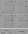
Single C. elegans manipulation. One single C. elegans was (A) trapped, (B) moved in y direction, (C) moved in x direction, and (D) moved in y direction again and released, with the average velocity of approximately 40 μm/s. An optical image of C. elegans (E) before and (F) after being fully stretched.

Comment in

  • Moved by sound.
    Marx V. Marx V. Nat Methods. 2012 Sep;9(9):867. doi: 10.1038/nmeth.2150. Nat Methods. 2012. PMID: 23097785 No abstract available.

References

    1. Hui EE, Bhatia SN. Micromechanical control of cell–cell interactions. Proc Natl Acad Sci USA. 2007;104:5722–5726. - PMC - PubMed
    1. Chronis N, Zimmer M, Bargmann CI. Microfluidics for in vivo imaging of neuronal and behavioral activity in Caenorhabditis elegans. Nat Methods. 2007;4:727–731. - PubMed
    1. Rohde CB, Zeng F, Gonzalez-Rubio R, Angel M, Yanik MF. Microfluidic system for on-chip high-throughput whole-animal sorting and screening at subcellular resolution. Proc Natl Acad Sci USA. 2007;104:13891–13895. - PMC - PubMed
    1. Ashkin A, Dziedzic JM, Bjorkholm JE, Chu S. Observation of a single-beam gradient force optical trap for dielectric particles. Opt Lett. 1986;11:288–290. - PubMed
    1. Grier DG. A revolution in optical manipulation. Nature. 2003;424:810–816. - PubMed

Publication types

Substances