Determination of solution structures of proteins up to 40 kDa using CS-Rosetta with sparse NMR data from deuterated samples
- PMID: 22733734
- PMCID: PMC3390869
- DOI: 10.1073/pnas.1203013109
Determination of solution structures of proteins up to 40 kDa using CS-Rosetta with sparse NMR data from deuterated samples
Abstract
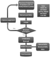
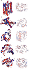
We have developed an approach for determining NMR structures of proteins over 20 kDa that utilizes sparse distance restraints obtained using transverse relaxation optimized spectroscopy experiments on perdeuterated samples to guide RASREC Rosetta NMR structure calculations. The method was tested on 11 proteins ranging from 15 to 40 kDa, seven of which were previously unsolved. The RASREC Rosetta models were in good agreement with models obtained using traditional NMR methods with larger restraint sets. In five cases X-ray structures were determined or were available, allowing comparison of the accuracy of the Rosetta models and conventional NMR models. In all five cases, the Rosetta models were more similar to the X-ray structures over both the backbone and side-chain conformations than the "best effort" structures determined by conventional methods. The incorporation of sparse distance restraints into RASREC Rosetta allows routine determination of high-quality solution NMR structures for proteins up to 40 kDa, and should be broadly useful in structural biology.
Conflict of interest statement
The authors declare no conflict of interest.
Figures


References
-
- Markley JL, Putter I, Jardetzky O. High-resolution nuclear magnetic resonance spectra of selectively deuterated staphylococcal nuclease. Science. 1968;161:1249–1251. - PubMed
-
- Tjandra N, Bax A. Direct measurement of distances and angles in biomolecules by NMR in a dilute liquid crystalline medium. Science. 1997;278:1111–1114. - PubMed
-
- Mueller GA, et al. Global folds of proteins with low densities of NOEs using residual dipolar couplings: Application to the 370-residue maltodextrin-binding protein. J Mol Biol. 2000;300:197–212. - PubMed
Publication types
MeSH terms
Substances
Associated data
- Actions
- Actions
- Actions
- Actions
- Actions
- Actions
Grants and funding
LinkOut - more resources
Full Text Sources
Molecular Biology Databases

