. 2012 Jul 10;109(28):11408-12.
doi: 10.1073/pnas.1118704109. Epub 2012 Jun 25.

Gonads directly regulate growth in teleosts

Affiliations

Gonads directly regulate growth in teleosts

Sandip Bhatta et al. Proc Natl Acad Sci U S A. .

Abstract

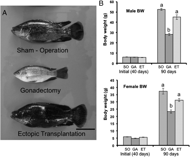
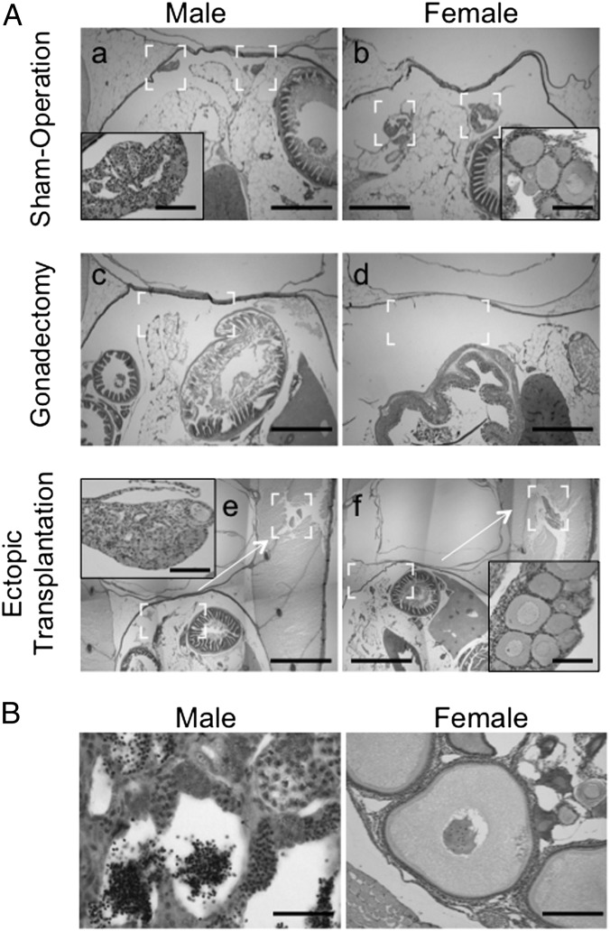
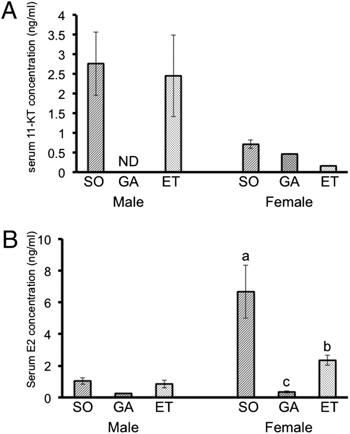
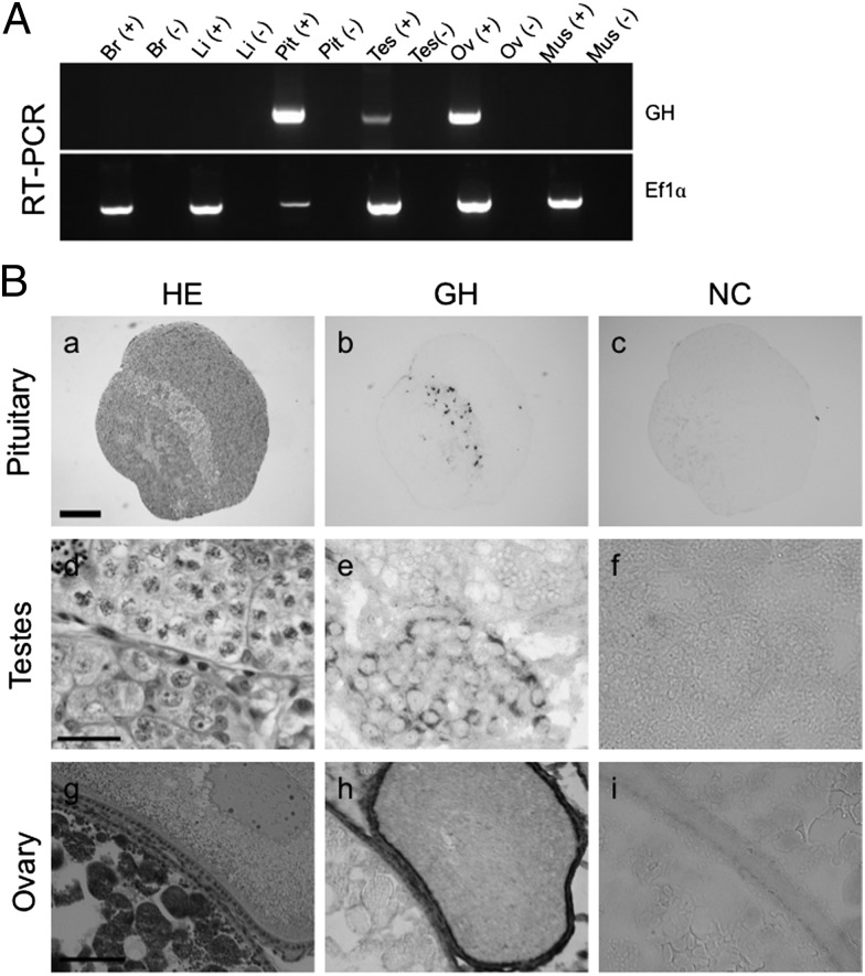
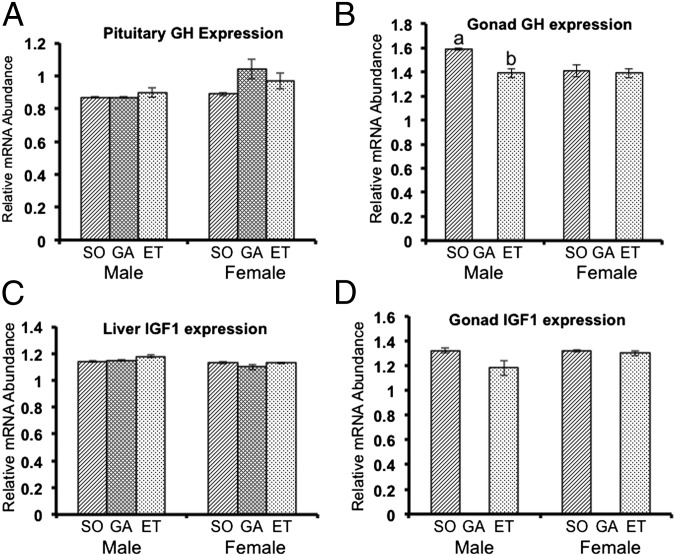
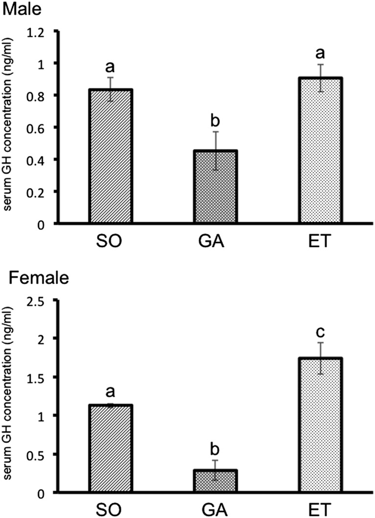
In general, there is a relationship between growth and reproduction, and gonads are known to be important organs for growth, but direct evidence for their role is lacking. Here, using a fish model, we report direct evidence that gonads are endocrine organs equal to the pituitary in controlling body growth. Gonadal loss of function, gain of function, and rescue of growth were investigated in tilapia. Gonadectomy experiments were carried out in juvenile males and females. Gonadectomy significantly retarded growth compared with controls; however, this retardation was rescued by the implantation of extirpated gonads. Because gonads express growth hormone, it is possible that gonads control body growth through the secretion of growth hormone and/or other endocrine factors. We propose that gonads are integral players in the dynamic regulation of growth in teleosts.


Conflict of interest statement

The authors declare no conflict of interest.

Figures

Fig. 1.
Fig. 1.
Gonads promote the growth of tilapia. (A) Photographs of male tilapia at 50 d after sham operation, gonadectomy, or ectopic transplantation. (Scale bar: 2 cm.) (B) Comparison of body weight among the experimental groups. At 40 d (initial, before surgery), the average body weight was no different among groups. At 90 d (50 d after surgery), gonadectomized fish of both sexes (GA) exhibited growth retardation compared with sham-operated fish (SO). Fish that received the ectopic gonad transplant (ET) did not differ in weight compared with SO fish. Results are mean ± SEM calculated for each group. Values with different letters are significantly different (P < 0.05; n ≥14).
Fig. 2.
Fig. 2.
Histological examination of fish. (A) The complete fish was fixed at 2 d after surgery. All sections were stained with H&E. The insets in a, b, e, and f are enlarged in the brackets of figures. The sham-operated fish appeared normal with no damage (a and b). The gonads were completely removed in gonadectomized fish (c and d). Sections of ectopic-transplanted fish showed that the gonads were completely removed from the normal location, and the transplanted gonads were located in the muscle (e and f). (Scale bars: 200 μm in a, b, c, and d; 0.5 mm in e and f; 20 μm in Insets in a, b, e, and f.) (B) Ectopic gonads were fixed at 50 d after operation, and all sections were stained with H&E. Male gonads were completely mature, and spermatozoa were observed (male). Mature eggs were not observed in ectopic female gonads (female), but ovarian follicles at the early vitellogenic stage were present. (Scale bars: 20 μm for males, 50 μm for females.)
Fig. 3.
Fig. 3.
Measurement of steroid hormone levels. (A) Serum 11-KT levels. Levels were similar in sham-operated (SO) and ectopically transplanted (ET) male fish. 11-KT was not detected in serum of male gonadectomized fish (GA). In females, serum 11-KT levels were very low compared those detected in male fish. (B) Serum E2 levels. ET fish had higher levels of E2 compared with GA fish, but unlike 11-KT in males, E2 levels did not recover to the same level in ET fish as in SO fish. This result was in agreement with the findings of histological analysis. Given that E2 level is dependent on the developmental stage of the ovary, SO and ET female fish have differing E2 levels. Transplanted male gonads developed into mature testis containing spermatozoa, but female gonads were not completely mature and contained early vitellogenic ovarian follicles. Results are given as mean ± SEM. Value with different letters are significantly different (P < 0.05; n ≥ 6).
Fig. 4.
Fig. 4.
Expression of GH in tilapia. (A) GH mRNA expression in tilapia as detected by RT-PCR analysis of various tissues, including brain (Br), liver (Li), pituitary (Pit), testis (Tes), ovary (Ov), and muscle (Mus). (−) indicates a reverse-transcription reaction without reverse transcriptase. EF1α served as the internal control. (B) Immunohistochemical analysis of GH in tilapia pituitary, testis, and ovary. Each adjacent section of the tissues was stained with H&E (HE; a, d, and g), immunostained with anti-GH antibody and AP-conjugated secondary antibody (GH; b, e, and h), or immunostained without primary antibody (NC; c, f, and i). Strong signals (black) were detected in GH cells of pituitary (b). No signal was detected without primary antibody (c). Signals were detected in the Sertoli cells around the spermatogonia of testis (e) and in the granulosa and fibroblast (thecal) cells in the ovary (h). (Scale bars: 200 μm in a, 20 μm in d, 50 μm in g.)
Fig. 5.
Fig. 5.
Quantitative real-time PCR analysis of expression of GH and IGF-I mRNAs in various tissues, using the EF1α gene as an internal control. (A) GH expression in the pituitary. (B) GH expression in the gonads. (C) IGF-I expression in the liver. (D) IGF-I expression in the gonads. No significant differences were seen among treatments. Results are given as mean ± SEM. Values with different letters are significantly different (P < 0.05; n ≥6). ET, ectopically transplanted; GA, gonadectomized; SO, sham-operated.
Fig. 6.
Fig. 6.
Measurement of serum GH levels by ELISA. At 90 d, serum levels were significantly lower in male gonadectomized (GA) fish compared with the other two male groups. In females, serum levels were higher in ectopically transplanted (ET) fish compared with sham-operated (SO) fish. Results are given as mean ± SEM. Values with different letters are significantly different (P < 0.05; n ≥ 6).

Similar articles

Cited by

References

    1. Fairbair D-J. Allometry for sexual size dimorphism: Pattern and process in the coevolution of body size in males and females. Annu Rev Ecol Syst. 1997;28:659–687.
    1. Norman J-R. A Systematic Monograph of the Flatfishes (Heterosomata), 1: Psettodidae, Bothidae, Pleuronectidae. London: British Museum; 1934.
    1. Kobelkowsky A. Sexual dimorphism of the flounder Bothus robinsi (Pisces: Bothidae) J Morphol. 2004;260:165–171. - PubMed
    1. Lowe-McConnel R-H. Ecological Studies in Tropical Fish Communities. Cambridge, UK: Cambridge Univ Press; 1987.
    1. Zakes Z, Demska-Zakes K. Effect of diets on growth and reproductive development of juvenile pikeperch, Stizostedion lucioperca (L.), reared under intensive culture conditions. Aquacult Res. 1996;27:841–845.

Publication types

LinkOut - more resources