. 2012 Jul 10;109(28):11437-42.
doi: 10.1073/pnas.1119173109. Epub 2012 Jun 25.

Evolutionarily conserved glycan signal to degrade aberrant brassinosteroid receptors in Arabidopsis

Affiliations

Evolutionarily conserved glycan signal to degrade aberrant brassinosteroid receptors in Arabidopsis

Zhi Hong et al. Proc Natl Acad Sci U S A. .

Abstract

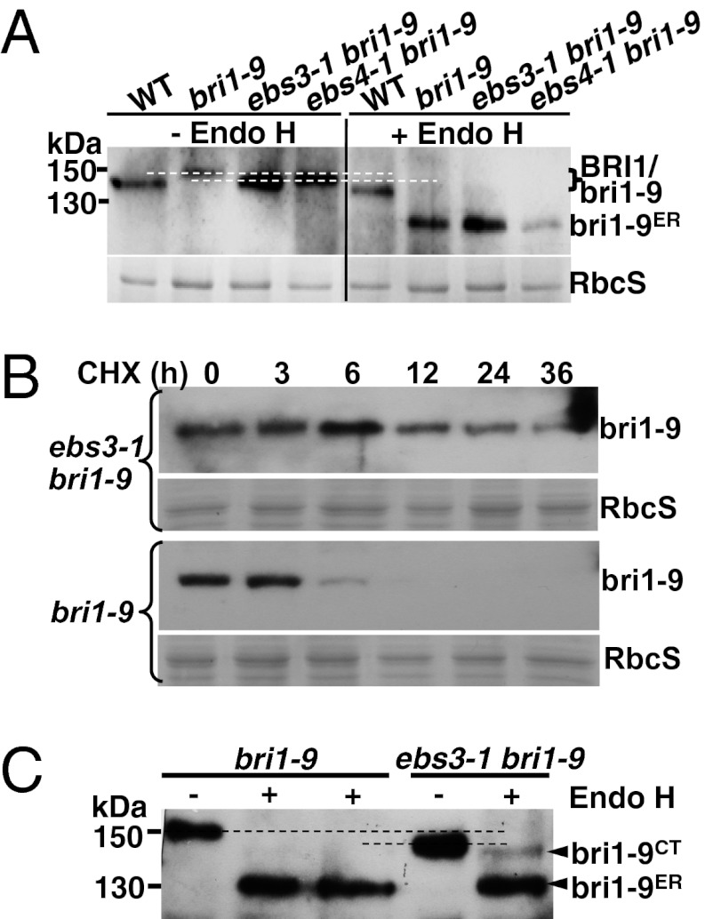
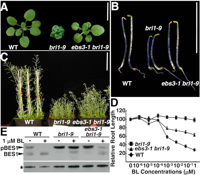
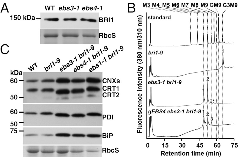
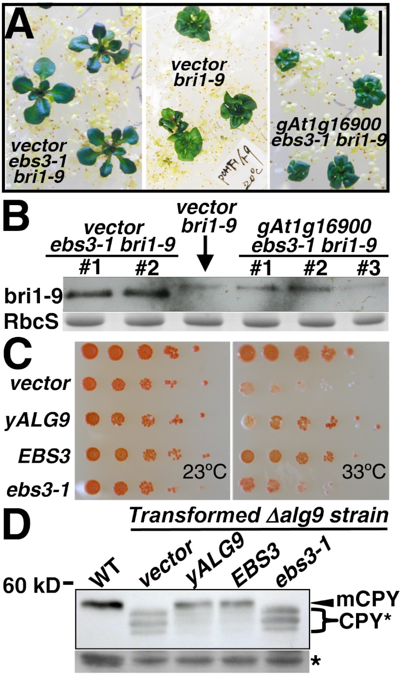
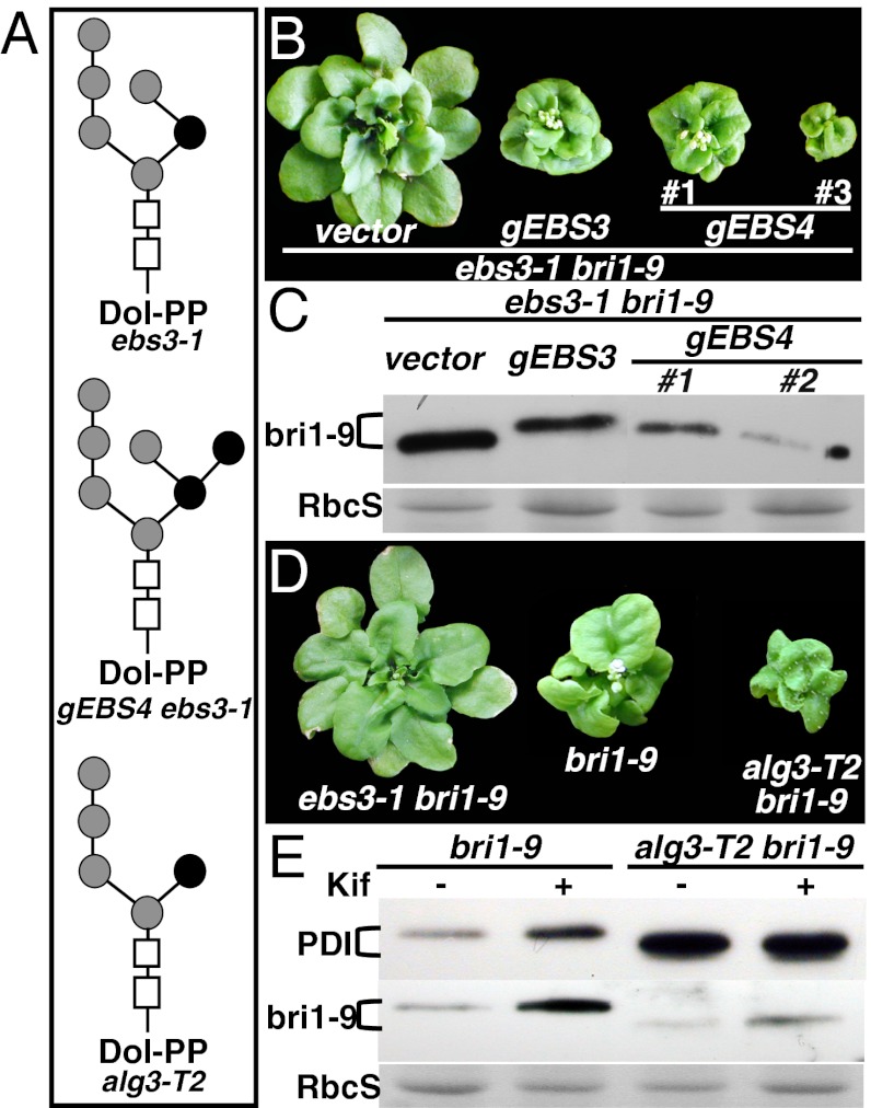
Asparagine-linked glycans (N-glycans) are crucial signals for protein folding, quality control, and endoplasmic reticulum (ER)-associated degradation (ERAD) in yeast and mammals. Although similar ERAD processes were reported in plants, little is known about their biochemical mechanisms, especially their relationships with N-glycans. Here, we show that a missense mutation in the Arabidopsis EMS-mutagenized bri1 suppressor 3 (EBS3) gene suppresses a dwarf mutant, bri1-9, the phenotypes of which are caused by ER retention and ERAD of a brassinosteroid receptor, BRASSINOSTEROID-INSENSITIVE 1 (BR1). EBS3 encodes the Arabidopsis ortholog of the yeast asparagine-linked glycosylation 9 (ALG9), which catalyzes the ER luminal addition of two terminal α1,2 mannose (Man) residues in assembling the three-branched N-glycan precursor [glucose(Glc)](3)(Man)(9)[N-acetylglucosamine(GlcNAc)](2). Consistent with recent discoveries revealing the importance of the Glc(3)Man(9)GlcNAc(2) C-branch in generating an ERAD signal, the ebs3-1 mutation prevents the Glc(3)Man(9)GlcNAc(2) assembly and inhibits the ERAD of bri1-9. By contrast, overexpression of EBS4 in ebs3-1 bri1-9, which encodes the Arabidopsis ortholog of the yeast ALG12 catalyzing the ER luminal α1,6 Man addition, adds an α1,6 Man to the truncated N-glycan precursor accumulated in ebs3-1 bri1-9, promotes the bri1-9 ERAD, and neutralizes the ebs3-1 suppressor phenotype. Furthermore, a transfer (T)-DNA insertional alg3-T2 mutation, which causes accumulation of an even smaller N-glycan precursor carrying a different exposed α1,6 Man, promotes the ERAD of bri1-9 and enhances its dwarfism. Taken together, our results strongly suggest that the glycan signal to mark an ERAD client in Arabidopsis is likely conserved to be an α1,6 Man-exposed N-glycan.


Conflict of interest statement

The authors declare no conflict of interest.

Figures

Fig. 1.
Fig. 1.
ebs3-1 mutation inhibits the ERAD of bri1-9. (A) Immunoblot analysis of bri1-9 in ebs3-1 bri1-9. (B) Immunoblot analysis of bri1-9 stability in bri1-9 and ebs3-1 bri1-9. (C) Endo H analysis of bri1-9 in bri1-9 and ebs3-1 bri1-9. For A and C, total proteins from 4-wk-old leaves were treated with or without Endo H, separated by SDS/PAGE, and analyzed by immunoblot with anti-BRI1 antibody. Equal amounts of total proteins were used in A, whereas five times more proteins in bri1-9 than ebs3-1 bri1-9 were used in C, which also contains technical duplicates of Endo H-treated bri1-9 samples. For B, 3-wk-old seedlings were transferred from 1/2 MS-agar medium into liquid 1/2 MS medium containing 180 μM CHX. Equal amounts of seedlings were removed at different time points to extract total proteins into 2× SDS sample buffer, which were subsequently separated by SDS/PAGE and analyzed by immunoblot with anti-BRI1 antibody. The numbers on the left in A and C indicate molecular mass, bri1-9ER denotes Endo H-sensitive form, and bri1-9CT represents bri1-9 carrying C-type N-glycans. Dashed lines in A and C show the mobility difference between the bri1-9 band in bri1-9 and that of ebs3-1 bri1-9. Coomassie blue staining of the small subunit of Rubisco (RbcS) serves as the loading control.
Fig. 2.
Fig. 2.
ebs3-1 is a weak suppressor of the bri1-9 mutant. (AC) Images of 3-wk-old soil-grown plants (A), 5-d-old etiolated seedlings (B), and 7-wk-old soil-grown mature plants (C) of WT, bri1-9, and ebs3-1 bri1-9. [Scale bars: 1 cm (A and B) and 10 cm (C).] (D) Quantitative analysis of BR sensitivity. Root length of 10-d-old seedlings grown on BL-containing medium were measured and presented as the relative value of average root length of BL-treated seedlings to that of mock-treated seedlings. Each data point represents the average of ∼40 seedlings of duplicated experiments. Error bars denote SE. (E) Immunoblot analysis of BL-induced BES1 dephosphorylation. Total proteins were extracted in 2× SDS buffer from 2-wk-old seedlings treated with or without 1 μM BL for 1 h, separated by SDS/PAGE, and analyzed by immunoblot using an anti-BES1 antibody (27). Star indicates a nonspecific band for loading control.
Fig. 3.
Fig. 3.
ebs3-1 likely affects assembly of the N-glycan precursor. (A) Immunoblot analysis of BRI1 in WT and ebs3-1 BRI1+. (B) Analysis of LLOs in bri1-9, ebs3-1 bri1-9, and a transgenic gEBS4 ebs3-1 bri1-9 line. LLOs of mature plants were extracted, acid-hydrolyzed, fluorescently labeled with PA, and analyzed using SF-HPLC by comparing their elution profiles with that of PA-sugar chain standards (Mn for MannGlcNAc2-PA and G3M9 for Glc3Man9GlcNAc2-PA). Asterisks indicate minor contaminants derived from the labeling process. (C) ebs3-1 affects the electrophoretic mobility of several ER-localized glycoproteins. For A and C, equal amounts of total proteins from 4-wk-old leaves were separated by SDS/PAGE and analyzed by immunoblot with antibodies against BRI1, maize CRT, PDI, or BiP. Numbers on the left indicate molecular mass. Coomassie blue staining of RbcS served as a loading control.
Fig. 4.
Fig. 4.
EBS3 encodes the Arabidopsis ortholog of yALG9. (A) Images of 3-wk-old transgenic bri1-9 lines carrying an empty vector and transgenic ebs3-1 bri1-9 seedlings carrying a genomic EBS3 transgene (gEBS3) or an empty vector. (Scale bar: 1 cm.) (B) Immunoblot analysis of bri1-9. The numbers on top of the gel panel indicate different transgenic lines. (C) EBS3 complemented the yeast Δalg9 mutation. Growth efficiency of WT or Δalg9 wbp1-2 cells transformed with the vector or an expression plasmid of yALG9, EBS3 or ebs3-1 cDNA. Transformants were spotted in 10-fold serial dilution on synthetic medium and grown for 3 d at 23 °C or 33 °C. (D) Immunoblot analysis of CPY of the WT yeast strain and Δalg9 strain transformed with indicated plasmids. For B and D, equal amounts of total proteins extracted from 4-wk-old soil-grown seedlings (B) or yeast cells of midlog growth phase (D) were separated by SDS/PAGE and analyzed by immunoblot using an anti-BRI1 (B) or anti-CPY (D) antibody. Coomassie blue staining of RbcS served as a loading control in (B). mCPY indicates the mature CPY, CPY*s represent three isoforms of CPY carrying different numbers of truncated N-glycan in (D), and star denotes a cross-reacting band used for loading control.
Fig. 5.
Fig. 5.
An exposed α1,6 Man residue is likely the glycan signal for bri1-9 ERAD. (A) Schematic structures of suspected major LLOs in ebs3-1 bri1-9, alg3-T2 bri1-9, or transgenic gEBS4 ebs3-1 bri1-9 line. Rectangles denote the two GlcNAc residues and circles represent Man residues with black circles indicating α1,6-linked Man residues. (B) Images of 5-wk-old soil-grown transgenic ebs3-1 bri1-9 mutant containing pPZP212, a gEBS3, or gEBS4 genomic transgene. (C) Immunoblot analysis of the bri1-9 abundance. Numbers in B and C indicate independent transgenic lines. (D) Images of 5-wk-old soil-grown mutants of ebs3-1 bri1-9, bri1-9, and alg3-T2 bri1-9. (E) Immunoblot analysis of PDI and bri1-9. For C and E, equal amounts of total proteins extracted in 2× SDS buffer from 4-wk-old leaves were treated with or without 10 μM Kif, separated by SDS/PAGE, and analyzed by immunoblot with anti-PDI or anti-BRI1 antibody. Coomassie blue staining of RbcS served as a loading control.

References

    1. Abu-Qarn M, Eichler J, Sharon N. Not just for Eukarya anymore: Protein glycosylation in Bacteria and Archaea. Curr Opin Struct Biol. 2008;18:544–550. - PubMed
    1. Kelleher DJ, Gilmore R. An evolving view of the eukaryotic oligosaccharyltransferase. Glycobiology. 2006;16:47R–62R. - PubMed
    1. Helenius A, Aebi M. Roles of N-linked glycans in the endoplasmic reticulum. Annu Rev Biochem. 2004;73:1019–1049. - PubMed
    1. Molinari M. N-glycan structure dictates extension of protein folding or onset of disposal. Nat Chem Biol. 2007;3:313–320. - PubMed
    1. Caramelo JJ, Parodi AJ. How sugars convey information on protein conformation in the endoplasmic reticulum. Semin Cell Dev Biol. 2007;18:732–742. - PMC - PubMed

Publication types

MeSH terms