. 2012 Sep 7;287(37):31155-64.
doi: 10.1074/jbc.M112.362632. Epub 2012 Jun 25.

MicroRNA-30d induces insulin transcription factor MafA and insulin production by targeting mitogen-activated protein 4 kinase 4 (MAP4K4) in pancreatic β-cells

Affiliations

MicroRNA-30d induces insulin transcription factor MafA and insulin production by targeting mitogen-activated protein 4 kinase 4 (MAP4K4) in pancreatic β-cells

Xiaomin Zhao et al. J Biol Chem. .

Abstract

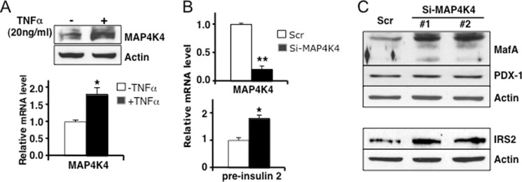
MicroRNAs (miRNAs) represent small noncoding RNAs that play a role in many diseases, including diabetes. miRNAs target genes important for pancreas development, β-cell proliferation, insulin secretion, and exocytosis. Previously, we documented that microRNA-30d (miR-30d), one of miRNAs up-regulated by glucose, induces insulin gene expression in pancreatic β-cells. Here, we found that the induction of insulin production by overexpression of miR-30d is associated with increased expression of MafA, a β-cell-specific transcription factor. Of interest, overexpression of miR-30d prevented the reduction in both MafA and insulin receptor substrate 2 (IRS2) with TNF-α exposure. Moreover, we identified that mitogen-activated protein 4 kinase 4 (MAP4K4), a TNF-α-activated kinase, is a direct target of miR-30d. Overexpression of miR-30d protected β-cells against TNF-α suppression on both insulin transcription and insulin secretion through the down-regulation of MAP4K4 by the miR-30d. A decrease of miR-30d expression was observed in the islets of diabetic db/db mice, in which MAP4K4 expression level was elevated. Our data support the notion that miR-30d plays multiple roles in activating insulin transcription and protecting β-cell functions from impaired by proinflammatory cytokines and underscore the concept that miR-30d may represent a novel pharmacological target for diabetes intervention.


Figures

FIGURE 1.
FIGURE 1.
Decreased expression of miR-30d in the islets of diabetic mice. A, in situ hybridization of pancreatic sections of 10-week-old heterozygous control mice (control, left panels) and diabetic mice (db/db, right panels) using digoxigenin-labeled probes for miR-30d and let-7b control. B, real-time PCR confirmed that miR-30d expression is decreased in freshly isolated islets of db/db mice at the age of 10 weeks when compared with control mice. The data are shown as mean ± S.D. of three independent experiments. *, p < 0.05, **, p < 0.01.
FIGURE 2.
FIGURE 2.
Insulin promoter activity was activated by miR-30d. A, schematic representation of the rat insulin 1 gene promoter-luciferase construct (pRIP-Luc) in pGL3. Major important cis-regulatory elements and transcription factors that bind to these elements are indicated. B, the pRIP-Luc reporter construct was co-transfected into MIN6 cells along with pre-miR-30d, pre-control, anti-miR-30d, or anti-control oligonucleotides, respectively. The insulin promoter activities were normalized by the co-transfected pRLTK Renilla luciferase activity. Data are expressed as relative luciferase activities to the level in control. RIP, rat insulin promoter. C, the change of intracellular expression level of miR-30d was detected by real-time PCR after transfection of pre-miR-30d and anti-miR-30d. Data represent means ± S.D. of three independent experiments. **, p < 0.01.
FIGURE 3.
FIGURE 3.
Overexpression of miR-30d activates the expression of MafA in both MIN6 and islets. A, MIN6 cells were transfected with pre-miR-30d or pre-control. After 48 h, cells were incubated with 1 or 25 mm glucose in DMEM for 16 h. The expressions of MafA were measured in both protein level and mRNA level. B, miR30d-GFP or control GFP plasmids were transfected into Min6 cells. MafA protein level was markedly activated in cells (bright red cells marked by arrows) with overexpressed miR30d-GFP, but not with GFP control. MafA was immunostained with anti-MafA and shown in red, and miR-30d transfected cells are shown in green through the GFP signal. C, expression of miR-30d using recombinant adenovirus (Ad-miR-30d) activates the expression of MafA and mouse insulin 2 in isolated islets. Ad-GFP (control) or Ad-miR-30d was used to infect freshly isolated islets. After 48 h, total RNA was extracted to analyze the expression of miR-30d (top), MafA, and insulin 2 by real-time PCR (bottom left). Protein lysates were harvested for immunoblots of MafA (bottom right). The presented data are the average of three independent experiments ± S.D. *, p < 0.05, **, p < 0.01. normal. to U6, normalized to U6.
FIGURE 4.
FIGURE 4.
miR-30d expression partially recovered the TNF-α-induced suppression of insulin gene transcription (shown in increased MafA level and insulin content), insulin signaling (shown in increased IRS2 level), and insulin secretion. A and B, 48 h after transfection with pre-miR-30d, pre-control, anti-miR-30d, and anti-control oligonucleotides, MIN6 cells were incubated with or without 20 ng/ml TNF-α for an additional 24 h. Cell lysates were harvested for Western blot analysis to detect the expression level of MafA, PDX-1, IRS2, and actin. Protein levels for MafA (A) and IRS2 (B) were quantified using the ImageJ software and normalized to actin loading control. C and D, miR-30d overexpression partially restores the TNF-α-suppressed insulin secretion (C) and insulin content (D). Insulin content and secreted insulin levels in the medium were quantified using mouse insulin ELISA and normalized to total DNA. The presented data are the average of three independent experiments ± S.D. *, p < 0.05, **, p < 0.01 versus pre-control or anti-control.
FIGURE 5.
FIGURE 5.
MAP4K4 is the target of miR-30d in pancreatic β-cells. A, bioinformatic prediction of the interaction between miR-30d and the 3′-UTRs of Map4k4s of various species. Mmu, mouse; Rno, rat; Has, human; Cfa, dog; and Ptr, chimpanzee. The predicted free energy of the hybrid is indicated. The mutant sequence (Map4k4-mut) is identical to the Map4k4-WT construct except for the seven point mutations (indicated in lowercase). B, miR-30d down-regulates MAP4K4 protein expression level. 48 h after transfection with pre-miR-30d or pre-control, Min6 cells were incubated with 20 ng/ml TNF-α for an additional 24 h. C, luciferase assay confirmed that miR-30d inhibited the luciferase reporter activity, in which the Map4k4 3′-UTR (Map4k4-WT) was fused with a Renilla luciferase reporter gene in pRLTK. The reporter constructs containing either Map4k4-WT or mutant (Map4k4-mut) were co-transfected into Min6 cells with either pre-miR-30d or anti-miR-30d. The pGL3 firefly luciferase plasmid was co-transfected for detection of transfection efficiency. 48 h after transfection, luciferase activity was assayed using Dual-Luciferase reporter assay kit. Data represent mean ± S.D. of three independent experiments. **, p < 0.01.
FIGURE 6.
FIGURE 6.
TNF-α-induced MAP4K4 inhibits insulin gene transcription and insulin signaling in pancreatic β-cells. A, MAP4K4 was activated by TNF-α (20 ng/ml) after a 24-h incubation. The expression of MAP4K4 was measured at both the protein level and the mRNA level. B, silencing of MAP4K4 activated insulin gene transcription. Min6 cells were transfected with siRNAs against MAP4K4 (si-MAP4K4) or scrambled siRNA (Scr). After 48 h, total RNAs were extracted for measuring MAP4K4 and pre-insulin mRNA levels by real-time PCR. C, silencing of MAP4K4 activated the expression of MafA and IRS2, but not PDX-1. 48 h after transfection with siRNA against MAP4K4 (si-MAP4K4-1 and si-MAP4K4-2) or scrambled siRNA, cells were incubated with or without TNF-α for an additional 24 h, and cell lysates were prepared for Western blots to detect the expression of MafA, PDX-1, IRS2, and actin. Lanes #1 and #2 show two different MAP4K4 siRNAs. Data represent three independent experiments ± S.D. *, p < 0.05, **, p < 0.01.
FIGURE 7.
FIGURE 7.
Altered expression levels of MAP4K4 in diabetic islets. A, in situ hybridization of pancreatic sections of 10-week-old normal control (left panels) and diabetic db/db (right panels) mice using digoxigenin-labeled probes for MAP4K4 and miR-30d. B, the expression of MAP4K4 was analyzed in freshly isolated islets of the control and db/db mice at the age of 10 weeks by using Western blots (left) and real-time PCR (right). Values shown are mean ± S.D. of three different experiments. *, p < 0.05. normal. to Actin, normalized to actin.
FIGURE 8.
FIGURE 8.
A proposed mechanism for the functions of miR-30d and other interacting components in pancreatic β-cells. miR-30d plays a key role in stimulating insulin transcription (Fig. 2) and secretion (Fig. 4C) by activating MafA and IRS2 through targeting MAP4K4 and other unknown genes (red indicates a newly discovered regulatory pathway by this study). Under normal conditions, high glucose induced the production of miR-30d (based on our previous publication (24)), which further stimulates MafA (Figs. 3 and 4A), but not PDX-1 (Fig. 4A), likely by targeting unknown negative regulators of MafA. In contrast, under stress conditions, TNF-α is activated, a process that not only inhibits miR-30d, but also activates MAP4K4 (Fig. 6), the negative regulator of MafA and IRS2. At this point, miR-30d directly targets MAP4K4 and promotes the production of MafA and IRS2 (Figs. 5 and 6) and consequently insulin transcription and secretion, partially negating the reverse effects of TNF-α on β-cell functions. The function of miR-30d in activating insulin secretion is undermined by TNF-α-induced negative miRNAs miR-21, -34a, and -146 (based on previous publications) (20).

Similar articles

Cited by

References

    1. Andrali S. S., Sampley M. L., Vanderford N. L., Ozcan S. (2008) Glucose regulation of insulin gene expression in pancreatic β-cells. Biochem. J. 415, 1–10 - PubMed
    1. Pinney S. E., Simmons R. A. (2010) Epigenetic mechanisms in the development of type 2 diabetes. Trends Endocrinol. Metab. 21, 223–229 - PMC - PubMed
    1. Ohneda K., Ee H., German M. (2000) Regulation of insulin gene transcription. Semin. Cell Dev. Biol. 11, 227–233 - PubMed
    1. Babu D. A., Chakrabarti S. K., Garmey J. C., Mirmira R. G. (2008) Pdx1 and β2/NeuroD1 participate in a transcriptional complex that mediates short-range DNA looping at the insulin gene. J. Biol. Chem. 283, 8164–8172 - PMC - PubMed
    1. Hang Y., Stein R. (2011) MafA and MafB activity in pancreatic β-cells. Trends Endocrinol. Metab. 22, 364–373 - PMC - PubMed

Publication types

MeSH terms