Review
. 2012 Jul;199(1):W35-42.
doi: 10.2214/AJR.11.8134.

Rectal imaging: part 1, High-resolution MRI of carcinoma of the rectum at 3 T

Affiliations
Review

Rectal imaging: part 1, High-resolution MRI of carcinoma of the rectum at 3 T

Vivek Gowdra Halappa et al. AJR Am J Roentgenol. 2012 Jul.

Abstract

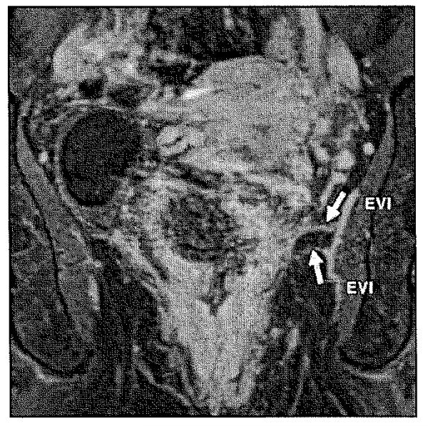
Objective: MRI is currently the imaging modality of choice for the detection, characterization, and staging of rectal cancer. A variety of examinations have been used for preoperative staging of rectal cancer, including digital rectal examination, endorectal (endoscopic) ultrasound, CT, and MRI. Endoscopic ultrasound is the imaging modality of choice for small and small superficial tumors. MRI is superior to CT for assessing invasion to adjacent organs and structures, especially low tumors that carry a high risk of recurrence.

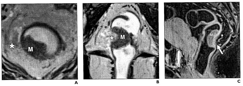
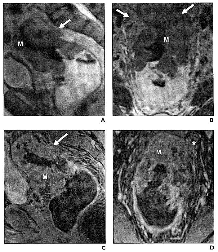
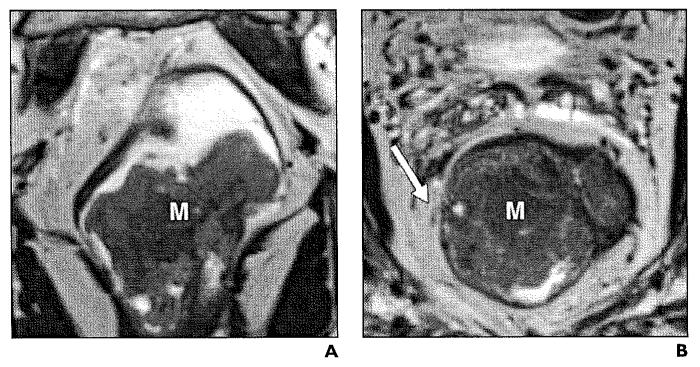
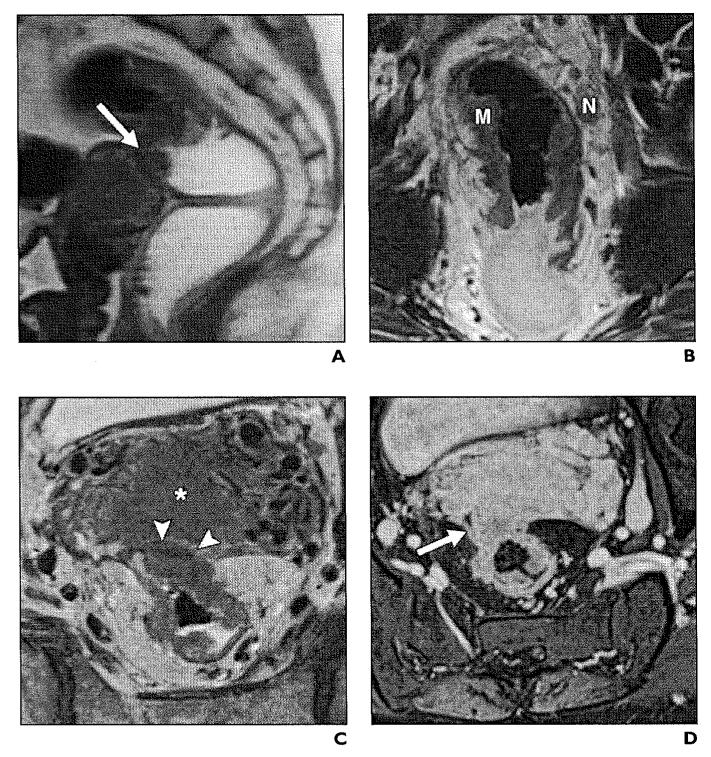
Conclusion: High-resolution MRI is an accurate and sensitive imaging method delineating tumoral margins, mesorectal involvement, nodes, and distant metastasis. In this article, we will review the utility of rectal MRI in local staging, preoperative evaluation, and surgical planning. MRI at 3 T can accurately delineate the mesorectal fascia involvement, which is one of the main decision points in planning treatment.


Figures

Fig. 1
Fig. 1
Normal anatomy of mesorectum. A, T2-weighted axial image shows hyperintense mesorectal fat (asterisk) surrounded by mesorectal fascia, which is represented by thin hypointense line (solid arrows). Submucosa is represented by hyperintense layer (arrowheads), whereas muscularis propria is hypointense layer (dashed arrows). B, Coronal T2-weighted high-resolution MRI shows normal anatomy of levator ani muscle (arrows) and puborectal muscle (asterisk).
Fig. 2
Fig. 2
Diagram of total mesorectal excision, which involves en bloc resection of tumor (M) with mesorectal fat and lymph nodes (N) with intact mesorectal fascia. (Drawing by Corona Villalobos CP)
Fig. 3
Fig. 3
54-year-old woman with stage T1 rectal adenocarcinoma. A and B, Axial (A) and coronal (B) T2-weighted high-resolution images show exophytic hypointense mass (M) on left lateral rectal wall. Muscular layer (arrows) appears to be spared of tumor. Mesorectal fat and mesorectal fascia (asterisk, A) are intact.
Fig. 4
Fig. 4
Stage T2 rectal cancer. A and B, Axial (A) and coronal (B) T2-weighted high-resolution images show asymmetric thickening of rectal wall (arrows) involving mucosa, submucosa, and muscularis propria (M, A). Boundary between muscularis and submucosa are ill defined at right lateral wall (line, B). Mesorectal fat (asterisk, A) is not involved.
Fig. 5
Fig. 5
44-year-old woman with stage T3 moderately differentiated rectal adenocarcinoma. A and B, Axial (A) and coronal (B) T2-weighted high-resolution images show exophytic mass (M) with transmural involvement of rectal layers infiltrating mesorectal fat (asterisk). C, Sagittal T1-weighted fat-saturated image shows enhancing rectal tumor (M) along posterior wall with edema (asterisk) and enhancement of mesorectal fat (arrow).
Fig. 6
Fig. 6
45-year-old man with stage T3 rectal adenocarcinoma. A and B, Sagittal (A) and coronal (B) T2-weighted images show asymmetrical circumferential wall thickening (M) obliterating rectal lumen. There is mesorectal fat involvement (arrows). C and D, Contrast-enhanced sagittal (C) and coronal (D) T1-weighted images show heterogeneous enhancement (arrow, C) of tumor (M) and mesorectal fat (asterisk, D).
Fig. 7
Fig. 7
50-year-old man with stage T3 rectal adenocarcinoma arising in association with tubulovillous adenoma with high-grade dysplasia. A and B, Coronal (A) and axial (B) T2-weighted high-resolution images show polypoid mass (M) along anterior wall of rectum 3.5 cm superior to anal verge. Tumor (arrow, B) invades mesorectal fat and fascia anteriorly.
Fig. 8
Fig. 8
51-year-old woman with stage T4 infiltrating adenocarcinoma. A, Sagittal T2-weighted image illustrates large mass (arrow) causing thickening of rectal wall, disruption of mesorectal fascia, and infiltration of uterus. B, High-resolution coronal T2-weighted image shows circumferential mass (M) in distal sigmoid rectum, with associated suspicious mesorectal node (N). C, In axial plane, there is tumor extension to fundus of uterus (asterisk) and no visible fat plane (arrowheads). D, Axial gadolinium-enhanced image clearly demonstrates invasion of tumor (arrow) to retroverted uterus.
Fig. 9
Fig. 9
Stage IIIB (T3, Nl) rectal carcinoma with tumor invasion into mesorectal fat. Axial T2-weighted image illustrates asymmetrical thickening (asterisk) of left lateral rectal wall with metastatic mesorectal lymphadenopathy (N) measuring 1.1 cm in size.
Fig. 10
Fig. 10
Stage IIIC (T3, N2) rectal carcinoma. Sagittal T2-weighted image shows enhancement of thickened rectal wall (asterisk) with multiple metastatic mesorectallymphadenopathy (N).
Fig. 11
Fig. 11
Rectal cancer. Axial T2-weighted image shows polypoid tumor (M) with less than 1 mm invasion of mesorectal fat (double arrow). Circumferential resection margin is preserved with intact meso rectal fascia.
Fig. 12
Fig. 12
Rectal cancer. A and B, Axial (A) and coronal (B) T2-weighted high-resolution images show significant infiltration to meso rectal fat (arrows) threatening circumferential resection margin.
Fig. 13
Fig. 13
Rectal cancer. Enhanced coronal T1-weighted MRI shows vascular tumor invasion of middle rectal artery branches (arrows). EVI = extramural vascular invasion.

References

    1. American Cancer Society . Cancer facts & figures 2011. American Cancer Society; Atlanta, GA: 2011.
    1. Shihab OC, Moran BJ, Heald RJ, Quirke P, Brown G. MRI staging of low rectal cancer. Eur Radial. 2009;19:643–650. - PubMed
    1. MERCURY Study Group Diagnostic accuracy of preoperative magnetic resonance imaging in predicting curative resection of rectal cancer: prospective observational study. BMJ. 2006;333:779. - PMC - PubMed
    1. Vliegen RF, Beets GL, Lammering G, et al. Mesorectal fascia invasion after neoadjuvant chemotherapy and radiation therapy for locally advanced rectal cancer: accuracy of MR imaging for prediction. Radiology. 2008;246:454–462. - PubMed
    1. Williamson PR, Hellinger MD, Larach SW, Ferrara A. Endorectal ultrasound of T3 and T4 rectal cancers after preoperative chemoradiation. Dis Colon Rectum. 1996;39:45–49. - PubMed

MeSH terms